Abstract
Introduction:
Painter first described painful periarticular soft-tissue calcium deposits in 1907. Further research has led to a variety of nomenclature, including calcareous tendinitis, pseudopodagra, and rheumatism. This report details the journey of a patient with acute calcific periarthritis (ACP) and explores issues concerning diagnosis, management, and provides possible preventative strategies.
Case Report:
A 39-year-old female presented to the emergency department after developing severe thumb metacarpophalangeal joint pain and swelling in her right dominant hand. A slight pain over the radial aspect of the first metacarpal was noticed 5 months before her acute hospital attendance.
Conclusion:
ACP of the thumb is commonly misdiagnosed due to its comparably low prevalence and clinical features which mimic more common conditions. Our case highlights the need for radiological imaging in patients with acute atraumatic thumb pain and evidence of a connection between chronic behavioral patterns and the development of ACP.
Keywords: Acute calcific periarthritis, behavioral injury, monoarticular arthritis, atraumatic
Learning Point of the Article:
To consider calcific etiology in patients presenting with acute joint pain and an atraumatic history.
Introduction
This report details the journey of a patient with acute calcific periarthritis (ACP) and explores issues concerning diagnosis, management, and preventative strategies for patients. Although ACP is a relatively uncommon condition its clinical presentation can mimic that of other more common conditions. Accurate diagnosis requires the evaluation of radiological findings, clinical presentation, and blood tests to prevent misdiagnoses. Friedman et al. discussed the limitations of ultrasound-guided techniques to diagnose ACP and confirmed the need for radiographic investigations in patients where ACP is suspected. Doumas et al. [1] also suggested that ACP must be considered in patients with a previous history of trauma. Both factors are key to preventing patients from receiving avoidable surgical and antibiotic treatments.
Case Report
A 39-year-old female general practitioner presented to the emergency department (ED) after developing severe thumb metacarpophalangeal pain and swelling in her right dominant hand. A slight pain over the radial aspect of the first metacarpal was noticed 5 months before her acute hospital attendance. The patient initially suspected it to be a repetitive strain injury and managed by avoiding thumb use and movement. Five days before presenting, the patient reported severe pain and swelling over the base of the first metacarpal when using her keyboard and performing mother–baby checks at work. There was no history of trauma and symptoms worsened significantly over the 5 days.
On examination, she was apyrexial. She had swelling and tenderness at the base of the thumb. Mild erythema was detected around the thenar eminence and in the webspace between the first and second metacarpals on the palmar side of the right hand. All ranges of motion of her thumb were reduced and reproduced severe pain. She was neurovascularly intact distally. All other metacarpals had a full range of motion without pain.
Blood tests undertaken acutely were normal (full blood count [FBC] and C-reactive protein [CRP]), with a low concern of infection. The subsequent X-ray was ordered upon her presentation to the ED and was used to discern the cause of her presenting pain.
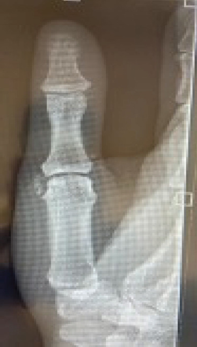
Our patient’s X-ray (Fig. 1) shows calcific periarthritis and juxta-articular deposits of formless calcium hydroxyapatite, on the radial side of the thumb 1st MCP joint in the right hand (thumb). The patient has hypermobility of the thumb, which has led to poor positioning of the joint during functional tasks, such as typing and writing. This is evident from the presence of a writer’s callus on the 2nd finger of this patient’s right hand.
Figure 1.

Anteroposterior plain radiograph of the right hand taken upon presentation to the emergency department. The 1st metacarpophalangeal joint (MCP) shows a well-circumscribed ovoid calcification on the radial side of the 1st MCP joint.
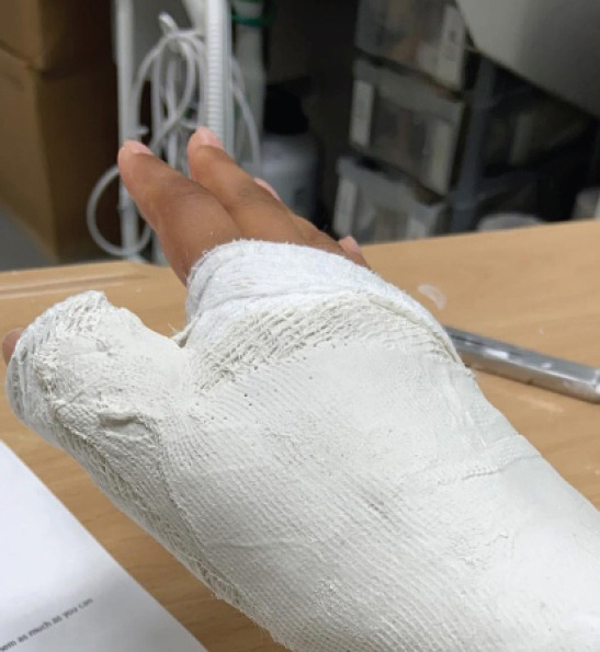
Due to this patient’s occupation, activity levels and manageable symptoms, conservative treatment was the mainstay of management. The patient’s hand was immobilized in a plaster of Paris for 10 days (Fig. 2) after presenting to the ED. She then progressed to the removal of her cast and tried gradually progressive therapeutic hand exercises with soft putty. These exercises aimed to change behavior patterns and prevent future flare-ups of pain.
Figure 2.

Plaster of Paris placed around the right thumb and wrist of the patient upon presentation to the emergency department. Worn for 10 days after admission to ensure complete immobilization of the affected 1st metacarpophalangeal joint.
After 4 weeks of hand therapy, the patient self-reported increased function from 10% to 85% in abduction, adduction, flexion, and opposition. She now maintains a range of 85–95% function by reducing thumb use when she feels any worsening of symptoms. She has been discharged by the orthopedic department and follows a hand therapy exercise program from home.
Discussion
Painter first described painful periarticular soft-tissue calcium deposits in 1907 [2]. Further research has led to a variety of nomenclature, including calcareous tendinitis, pseudopodagra, and rheumatism [1, 3, 4]. Cases tend to be monoarticular and self-limiting with tendons in the shoulder being most frequently affected. ACP of the hand and wrist have been described in adult [3-5] and pediatric [6] populations, with the most common mechanism of injury involving flexor carpi ulnaris when in close proximity to the pisiform bone.
Our case highlights a connection between chronic behavioral patterns and the development of ACP, emphasizing the need to consider ACP as a possible diagnosis in patients with these behavioral patterns [7]. ACP of the thumb is commonly misdiagnosed due to its comparably low prevalence in the hand and clinical features that mimic more common conditions, such as various infections. Diagnosis of ACP in this case required radiographic evidence to rule out other conditions that should be considered from this clinical presentation such as cellulitis plus reactive arthritis, septic arthritis, deep thenar space infection, gout, heterotopic ossification (HO), and pseudogout (calcium pyrophosphate deposition disease (CPPD) [2, 8, 9].
When diagnosing ACP, both clinical and radiological findings must be considered together. Based on the clinical findings alone infection would be the most probable diagnosis [8]. However, normal blood tests (FBC and CRP) and the presence of periarticular calcification on the radiograph, would make this less likely [10].
Radiographs may show calcification in both gout and ACP. However, gout most commonly affects the first metatarsophalangeal joint. Furthermore, periarticular erosions may occur in calcific periarthritis [4], resulting in ACP as the most likely diagnosis.
The cortex and internal trabeculations that are typically seen in HO are not present in cases of ACP, and the calcification caused by ACP is not consistent with that of the typical chondrocalcinosis seen in CPPD [4].
Calcific periarthritis is usually managed conservatively using non-steroidal anti-inflammatory drugs, rest, immobilization of the joint, physiotherapy, and local steroid injections [1].
If conservative management fails, needle aspiration/”dry needling,” shock-wave therapy and in some rare cases surgical treatment can be considered [1, 2].
Conservative management is the first-line treatment and patients commonly find reduced symptoms 4–7 days after the acute presentation of pain [1, 3]. Interventions must strive to sustain or advance the patient’s quality of life with the condition.
Conclusion
ACP of the thumb is commonly misdiagnosed due to its comparably low prevalence and clinical features which mimic more common conditions. Our case highlights the need for radiological imaging in patients with acute atraumatic thumb pain and evidence of a connection between chronic behavioral patterns and the development of ACP.
Clinical Message.
This article hopes to highlight the need for early radiological investigations in patients presenting with acute joint pain and an atraumatic history, particularly to rule out or diagnose calcific etiologies.
Biography


Footnotes
Conflict of Interest: Nil
Source of Support: Nil
Consent: The authors confirm that informed consent was obtained from the patient for publication of this case report
References
- 1.Doumas C, Vazirani RM, Clifford PD, Owens P. Acute calcific periarthritis of the hand and wrist:A series and review of the literature. Emerg Radiol. 2007;14:199–203. doi: 10.1007/s10140-007-0626-9. [DOI] [PubMed] [Google Scholar]
- 2.Painter CF. Subdeltoid bursitis. Boston Med Surg J. 1907;156:345–9. [Google Scholar]
- 3.Sandstrom C. Peritendinitis calcarea:A common disease of middle life:Its diagnosis, pathology, and treatment. Am J Roentgenol. 1938;40:1–21. [Google Scholar]
- 4.McCarty DJ, Gatter RA. Recurrent acute inflammation associated with focal apatite crystal deposition. Arthritis Rheum. 1966;9:804–19. doi: 10.1002/art.1780090608. [DOI] [PubMed] [Google Scholar]
- 5.Hayes CW, Conway WF. Calcium hydroxyapatite deposition disease. Radiographics. 1990;10:1031–48. doi: 10.1148/radiographics.10.6.2175444. [DOI] [PubMed] [Google Scholar]
- 6.Walocko FM, Sando IC, Haase SC, Kozlow JH. Acute calcific tendinitis of the index finger in a child. Hand (N Y) 2017;12:NP84–7. doi: 10.1177/1558944716675146. [DOI] [PMC free article] [PubMed] [Google Scholar]
- 7.Lee KB, Song KJ, Kwak HS, Lee SY. Acute calcific periarthritis of proximal interphalangeal joint in a professional golfer's hand. J Korean Med Sci. 2004;19:904–6. doi: 10.3346/jkms.2004.19.6.904. [DOI] [PMC free article] [PubMed] [Google Scholar]
- 8.Friedman SN, Margau R, Friedman L. Acute calcific periarthritis of the thumb:Correlated sonographic and radiographic findings. Radiol Case Rep. 2018;13:205–7. doi: 10.1016/j.radcr.2017.08.015. [DOI] [PMC free article] [PubMed] [Google Scholar]
- 9.Yosipovitch G, Yosipovitch Z. Acute calcific periarthritis of the hand and elbows in women. A study and review of the literature. J Rheumatol. 1993;20:1533–8. [PubMed] [Google Scholar]
- 10.Dimmick S, Hayter C, Linklater J. Acute calcific periarthritis-a commonly misdiagnosed pathology. Skeletal Radiol. 2022;51:1553–61. doi: 10.1007/s00256-022-04006-8. [DOI] [PMC free article] [PubMed] [Google Scholar]


