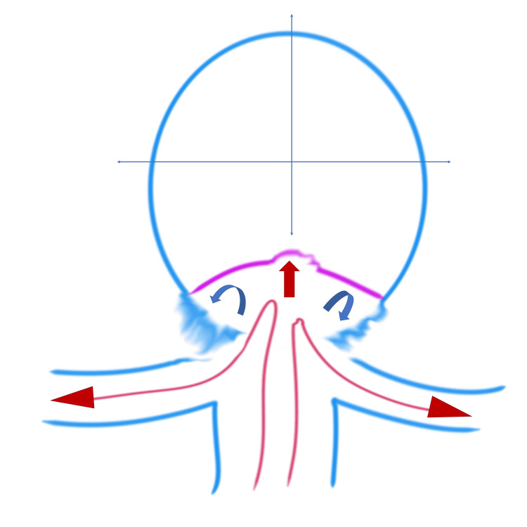
Figure 4. Figure 4: Illustration of the hemodynamic mechanism of RC enlargement.
The basilar artery blood flow (long red arrows) enters the RC, with the main blood flow (red arrow) within the RC impacting the newly formed intimal surface at the center of the RC. Note the detached blood flow (curved blue arrows) on both sides of the RC.
RC: residual cavity
Image Credits: Toru Satoh

