Abstract
Purpose
Psoriasis is a complex inflammatory skin disorder that is closely associated with metabolic syndrome (MetS). Limited information is available on skin metabolic changes in psoriasis; the effect of concurrent MetS on psoriatic skin metabolite levels is unknown. We aimed to expand this information through skin metabolomic analysis.
Patients and Methods
Untargeted metabolomics was conducted using skin samples from 38 patients with psoriasis vulgaris with MetS (PVMS), 23 patients with psoriasis vulgaris without MetS (PVNMS), and 10 healthy controls (HC). Data analyses, including multivariate statistical analysis, KEGG pathway enrichment analysis, correlation analysis, and receiver operating characteristic curve analysis, were performed.
Results
Significant discrepancies were found between skin metabolites in the HC and PVNMS groups, particularly those involved in nucleotide and glycerophospholipid metabolism. Fifteen of these metabolites were positively correlated with psoriasis severity. Furthermore, MetS was found to affect the metabolic profiles of patients with psoriasis. There were some metabolites with consistent alterations in both the PVNMS/HC and PVMS/PVNMS comparisons.
Conclusion
This study may provide new insights into the link between skin metabolism and psoriatic inflammation and the mechanism underlying the interaction between psoriasis and MetS.
Keywords: psoriasis, metabolic syndrome, metabolomics, nucleotide metabolism, glycerophospholipid metabolism
Introduction
Psoriasis manifests as a complex systemic inflammatory skin disorder.1 Millions of people worldwide are afflicted with this disease.2 Psoriasis vulgaris is the most frequent type of psoriasis, with classic presentation of chronic well-defined scaly plaque.3 There are close associations between psoriasis and metabolic and cardiovascular comorbidities, specifically metabolic syndrome (MetS).4,5 Given that high recurrence rates of psoriasis,6 in addition to the aggravated inflammation and a reduced efficacy response in psoriasis with concomitant MetS,7,8 a deeper understanding of pathophysiology of psoriasis and psoriasis with concomitant MetS is warranted.
Metabolites, which are involved in endogenous metabolism, also reflect exogenous exposures, thus representing the interactions between gene and protein function and environmental factors.9 Therefore, metabolomics, the systemic collection of metabolites,10 is an effective tool for exploring the molecular basis and pathophysiology that drive disease. A series of studies investigated metabolic signature within blood sample from patients with psoriasis;11 however, it has been reported that skin metabolites are more reliable than blood metabolites in reflecting core metabolic alterations in psoriasis.12 At present, there is limited research regarding the metabolomic analysis of skin samples from patients with psoriasis, with most of the studies having a sample size not exceeding ten participants.13–15 Both psoriasis and MetS are closely related to systemic inflammation,1,16 in which metabolic disturbances involving various pathways, such as amino-acid, carbohydrate, and lipid metabolisms, were recognized, respectively.11,17 Moreover, significant alterations in serum levels of lipids were found between psoriatic patients with and without metabolic disorders.18 To the best of our knowledge, metabolomics has not been applied for detecting skin metabolic changes in psoriasis complicated with MetS to date.
In this study, we used an untargeted metabolomic method based on liquid chromatography–tandem mass spectrometry (LC–MS/MS) to characterize the metabolic signatures of skin lesions from patients with psoriasis vulgaris, with or without MetS, to provide insights into the pathogenesis of psoriasis and the interaction between psoriasis and MetS.
Material and Methods
Study Population and Sample Collection
In total, 38 patients with psoriasis vulgaris with MetS (PVMS), 23 patients with psoriasis vulgaris without MetS (PVNMS), and 10 healthy controls (HC) were consecutively recruited from the First Medical Center of Chinese PLA General Hospital. The inclusion criteria for recruiting participants with psoriasis vulgaris were adulthood, no topical therapy within 2 weeks and systemic therapy within 4 weeks prior to skin sample collection, and meeting at least three (PVMS) or none (PVNMS) of the diagnostic criteria for MetS.19 Patients with pregnancy, liver or kidney dysfunction, or cancer were excluded from the study. Informed consent was obtained from all participants. This study was approved by the Hospital Ethics Committee.
Skin samples from typical lesional skin of patients with psoriasis vulgaris and normal skin of HC were collected under local anesthesia. After washing with saline, skin tissues were placed in cryogenic vials and stored in liquid nitrogen until analysis. The baseline characteristics of all subjects were presented in our previous work.20
Untargeted Metabolomics
Sample Preparation and LC–MS/MS Analysis
Metabolomic sample preparation and LC–MS/MS analysis were performed referring to previously described methods.21 The skin specimens were thawed on ice, chopped and homogenized. Subsequently, 400 μL 70% methanol was added and 200 μL of supernatant was collected after centrifugation (12000 rpm, 10 min, 4°C) for analysis. A mixture of equal amounts of the supernatant obtained from each skin tissue was prepared as the quality control (QC) sample. One QC sample was injected after running 10 samples to monitor the repeatability of the analytical process.
LC–MS/MS analysis was performed using a Nexera UHPLC LC-30A (Shimadzu Corporation, Kyoto, Japan) system equipped with an ACQUITY Premier HSS T3 Column (1.8 µm, 2.1 mm × 100 mm) (Waters, Milford, MA, USA) plus a TripleTOF 6600 mass spectrometer (SCIEX, Foster City, CA, USA). The chromatographic conditions included 40°C, 0.1% aqueous formic acid (mobile phase A) and acetonitrile containing 0.1% formic acid (mobile phase B), 0.4 mL/min (flow rate), and 4 μL (injection volume). The elution gradients proceeded as follows: 0–2 min, 95% A; 2–5 min, 80% solvent A; 5–6 min, 40% solvent A; 6–7.5 min, 1% A; 7.5–10 min, 95% A. MS was performed with the following parameter: electrospray ionization source; spray voltage set at 5000 V (positive mode) or −4000 V (negative mode); sheath gas, 50 psi; auxiliary gas, 60 psi; curtain gas, 35 psi; collision energy spread, 15V; MS1 TOF masses, 50~1000 Da; MS2 TOF masses, 25~1000 Da; MS1 accumulation time 0.2 s; MS2 accumulation time, 0.04 s.
Data Processing and Analysis
ProteoWizard and R software (version 4.2.2) were used for data processing including format conversion, peak extraction, peak alignment, retention time correction, peak area correction, missing value filling, normalization, data merging in positive and negative ion modes, and metabolite identification. Subsequently, the datasets were imported into SIMCA software (version 14.1, Umetrics, Umea, Sweden) for multivariate statistical analyses, including principal component analysis (PCA) and orthogonal partial least squares discrimination analysis (OPLS-DA). Differential metabolites were screened based on the P-value (< 0.05, Student’s t-test) and variable importance in the projection (VIP > 1). KEGG pathway enrichment analysis and receiver operating characteristic curve (ROC) analysis were performed; heatmaps and volcano plots were generated using R software. The Spearman rank correlation test was used for the correlation analysis between the psoriasis area and severity index (PASI) and significant differential metabolites.
Results
The Overview of Total Metabolites and the PCA
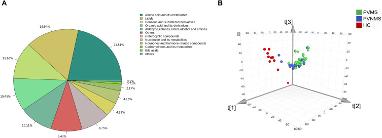
A total of 3224 metabolites (1628 and 1596 metabolites in the positive and negative ion modes, respectively) were identified in all skin samples. The metabolites were categorized into 12 classes (Figure 1A). PCA revealed separation tendencies among the HC, PVNMS, and PVMS groups (Figure 1B).
Figure 1.
Component composition of metabolites and PCA 3D plot of the entire metabolomics data set. (A) Classification and proportion of the identified metabolites from all skin samples. (B) PCA score scatter 3D plot comparing the HC, PVNMS, and PVMS groups (R2Y, 0.617; Q2Y, 0.349).
Skin Metabolic Profile Analysis of Psoriasis Vulgaris
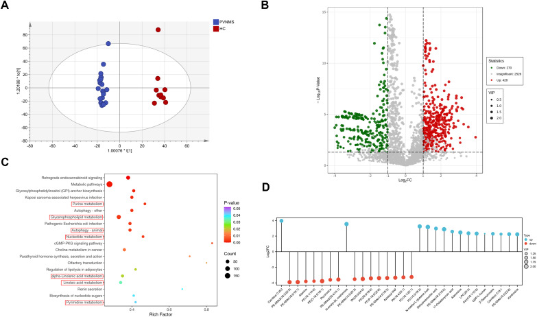
The OPLS-DA model revealed a distinct separation between skin metabolites in the PVNMS and HC groups (Figure 2A), which was validated using a permutation test (Supplementary Figure S1A). Based on the VIP and P-value, 1113 differential metabolites were screened. For higher accuracy, fold-change (FC, ≥ 2 or ≤ 0.5) was added to the screening criteria. We identified 428 upregulated and 270 downregulated metabolites (Figure 2B). These metabolites were enriched for certain pathways, including nucleotide metabolism (purine metabolism and pyrimidine metabolism), autophagy, glycerophospholipid metabolism, α-linolenic metabolism, and linoleic acid metabolism, which might be relevant to the pathogenesis of psoriasis (Figure 2C).
Figure 2.
The Metabolic profile of the PVNMS group compared with the HC group. (A) OPLS-DA score scatter plot of the two groups (R2Y, 0.99; Q2Y, 0.857). (B) Volcano plot illustrating differential metabolites in the PVNMS/HC comparison. (C) KEGG pathway analysis plot. (D) The 30 most significantly altered skin metabolites. FC, fold change.
Figure 2D shows the 30 most significantly altered skin metabolites in the PVNMS/HC comparison, consisting of 5 nucleotide metabolites, 17 glycerophospholipids, 3 fatty acyls, 2 sphingolipids, dehydroascorbic acid, N-acetyl-DL-methionine, and N-methyl-L-glutamic acid.
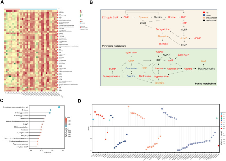
We further analyzed nucleotide and glycerophospholipid metabolites in psoriatic skin lesions. The heatmap describing the levels of important nucleotide metabolites showed that most of important nucleotide metabolites were upregulated in the skin lesions of patients with PVNMS compared to HC (Figure 3A). The levels of many key metabolites involved in purine and pyrimidine metabolism, such as uridine, UMP, UDP, CMP, thymine, adenosine, AMP, inosine, hypoxanthine, and dGMP, were significantly higher in the PVNMS group (Figure 3B). These results revealed enhanced nucleotide metabolism in skin lesions of psoriasis vulgaris. Moreover, the levels of seven nucleotide metabolites, including orotidine, 3’-deoxyguanosine, 8-hydroxyguanosine, 3’-UMP, 2’,3’-cyclic CMP, flavin mononucleotide, and 2-hydroxy-dAMP, were positively correlated with PASI in the PVNMS group (Figure 3C).
Figure 3.
Disturbances in the nucleotide and glycerophospholipid metabolism and correlation analysis of differential metabolites levels and PASI in the PVNMS group. (A) Heatmap of important nucleotide metabolites. (B) Schematic diagram of nucleotide metabolism pathway changes. (C) Levels of 15 differential metabolites positively correlated to PASI. (D) A lollipop plot showing dysregulation of glycerophospholipid metabolites.
The differential glycerophospholipids in the PVNMS/HC comparison included lysophosphatidic acid (LPA), lysophosphatidylcholine (LPC), lysophosphatidylethanolamine (LPE), phosphatidic acid (PA), phosphatidylcholine (PC), phosphatidylethanolamine (PE), phosphatidylglycerol (PG), and phosphatidylserine (PS). The levels of LPA (18:3) were upregulated in the PVNMS group (Figure 3D), and positively correlated with PASI (Figure 3C). The expression levels of LPE and PG were elevated and accompanied by the downregulation of PS. Six of the eight LPCs, including LPC(20:4), LPC(22:6), LPC(18:1), LPC(22:5), LPC(20:3), and LPC(20:5), were upregulated, whereas the majority of PAs were downregulated. In addition, abundant metabolites in the PC and PE classes were significantly altered (Figure 3D).
In addition to the levels of seven nucleotide metabolites and LPA (18:3), the levels of five fatty acyls (linoleic acid, methyl 15-cyanopentadecanoate, mead acid, cis-8,11,14,17-eicosatetraenoic acid, and 2-hydroxyhexadecanoic acid), D-fructose 6-phosphate-disodium salt, and 5-methoxytryptophan, were positively correlated with PASI (Figure 3C).
MetS Affects the Skin Metabolic Profile of Psoriasis Vulgaris
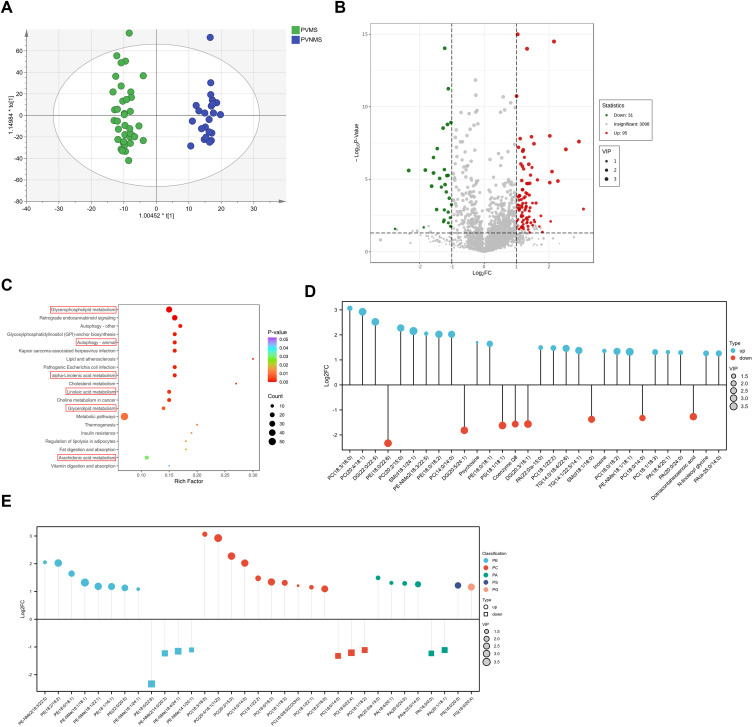
In the OPLS-DA, a clear separation was observed between the PVNMS and PVMS groups (Figure 4A and Supplementary Figure S1B). A total of 676 differential metabolites were screened for the PVMS/PVNMS comparison. Taking FC (≥ 2 or ≤ 0.5) into consideration, we further identified 95 upregulated and 31 downregulated differential metabolites (Figure 4B). These metabolites were involved in glycerophospholipid metabolism, glycerolipid metabolism, autophagy, α-linolenic metabolism, linoleic acid metabolism, arachidonic acid metabolism, etc. (Figure 4C), indicating dysregulated lipid metabolism in the skin lesions of patients with PVMS. As shown in Figure 4D, the 30 skin metabolites with the greatest FC variation were glycerophospholipids, followed by glycerolipids, sphingolipids, fatty acyls, inosine, and coenzyme Q8. In terms of glycerophospholipid metabolites, the PVMS group exhibited higher levels of 8 PEs, 10 PCs, 4 PAs, PS(18:0/20:0), and PG(16:0/20:4), in addition to lower levels of 4 PEs, 3 PCs, and 2 PAs than the PVNMS group (Figure 4E).
Figure 4.
The metabolic profile of the PVMS group compared with the PVNMS group. (A) OPLS-DA score scatter plot of the two groups (R2Y, 0.967; Q2Y, 0.74). (B) Volcano plot elucidating differential metabolites in the PVMS/PVNMS comparison. (C) KEGG pathway analysis plot. (D) The top 30 skin metabolites with the greatest fold change (FC) variation. (E) A lollipop plot displaying changes of differential glycerophospholipids.
Metabolites Showing Consistent Alterations in Both the PVNMS/HC and PVMS/PVNMS Comparisons
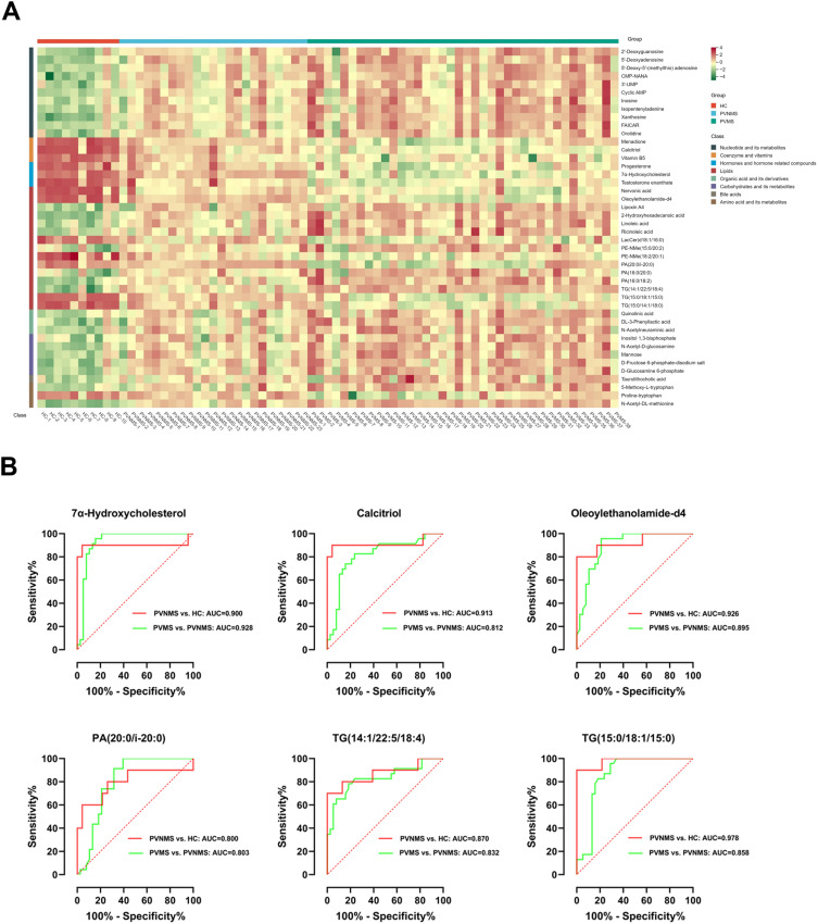
We compared the differential metabolites (based on VIP and P-value) in the PVNMS/HC and PVMS/PVNMS comparisons and found 44 metabolites with consistent alterations. Notably, a higher abundance of 11 nucleotide metabolites was observed in the PVMS group than in the PVNMS group. Disturbances in the levels of 15 lipids, including 6 glycerophospholipids, 6 fatty acyls, and 3 triglycerides (TGs), were enhanced in patients with PVMS compared with those with PVNMS (Figure 5A).
Figure 5.
The overlaps of metabolites and potential biomarkers in the PVNMS/HC and PVMS/PVNMS comparisons. (A) Heatmap of 44 metabolites with consistent alterations. (B) ROC curve plots of six metabolites with high AUC values (≥ 0.8) in both the PVNMS/HC and PVMS/PVNMS distinguishment.
Univariate ROC analysis of these 44 metabolites was performed to assess biomarker performance for distinguishing psoriatic skin lesions from normal skin and psoriatic skin lesions in patients with MetS from those in patients without MetS. The area under the curve (AUC) values of six metabolites, including 7α-hydroxycholesterol, calcitriol, oleoylethanolamide-d4, PA(20:0/i-20:0), TG(14:1/22:5/18:4), and TG(15:0/18:1/15:0), in both the PVNMS/HC and PVMS/PVNMS distinguishment were higher than 0.8, suggesting that these metabolites could be considered shared potential biomarkers and might play important roles in linking MetS with psoriasis.
Discussion
Metabolic regulation plays a pivotal role in the functions of keratinocytes and immune cells in psoriasis.22,23 Skin metabolomic analysis provides a snapshot of the metabolic perturbations in psoriatic lesions. To date, limited information is available on this issue. Considering that psoriasis and MetS have been identified as a risk factor of each other,24,25 there may be shared metabolic pathways and molecular networks involved in both diseases. It has been reported that comorbid metabolic disorders are complicated by dysregulated serum lipid metabolism in psoriasis.18 However, the impact of Mets on the metabolism of psoriatic skin remains unknown.
In this study, we analyzed the metabolic profile of the psoriatic skin and explored its implications. Numerous differential metabolites were observed in the PVNMS group compared with normal skin, particularly elevated levels of multiple nucleotide metabolites and eight dysregulated types of glycerophospholipids. Fifteen of these metabolites positively correlated with psoriasis severity. Various differential metabolites were found between the PVMS and PVNMS groups, most of which were involved in lipid metabolism. Moreover, disturbances in the levels of nucleotide metabolites and lipids were enhanced in the PVMS group compared with those in the PVNMS group. Six metabolites were screened for good discriminative abilities in both patient groups and in the PVNMS and HC groups.
Nucleotide Metabolism
Our results showed enhanced nucleotide metabolism in psoriatic skin tissues. Xiong et al also reported increased levels of inosine in psoriatic lesional skin compared to those in non-lesional skin and observed a positive correlation between inosine levels and PASI.26 In addition, plasma metabolomic analysis showed elevations in 5′-ADP, xanthosine, and 5.6-dihydrothymine in psoriatic plasma in comparison to HC plasma.14 Furthermore, higher levels of inosine monophosphate (IMP) in the urine were found in patients with psoriasis than in HC, which was reduced after treatment.27 Nucleotide metabolism, providing necessary substrates for many biological processes, is vital for cell growth and proliferation.28 Its enhancement can meet the demand of excessive proliferation of keratinocytes in psoriasis. Moreover, purinergic signaling is involved in the pathophysiology of psoriasis via inflammasome and immune cell activation.29–31 Elevated purine nucleotide levels can induce the expression of MHC class I related chain A and subsequently affect the function of immune cells expressing its receptor,32 such as γδ17 cells and group 3 innate lymphoid cells, which play roles in psoriasis.33,34 Additionally, recent research has shown that pyrimidine metabolism may have a non-proliferative role.35 Notably, in the PVMS/PVNMS comparison, the levels of 11 nucleotide metabolites were significantly higher. Few studies have investigated the implication of MetS and its components on nucleotide metabolism. Dai et al reported that obesity-related factors indirectly enhanced pyrimidine metabolism in endometrial cancer.36 Furthermore, the levels of three nucleotide metabolites in the urine were found to be increased in hyperlipidemic rats.37
Glycerophospholipid Metabolism
Recent research has noted that lipid metabolism, especially glycerophospholipid metabolism, is significantly altered in psoriatic blood, and its dysregulation can be improved after treatment.18,38–40 Our findings highlight the obvious dysregulation of glycerophospholipid metabolism in psoriatic skin. Significant changes in glycerophospholipid profiles, notably LPA, LPC, PC, PE, and PA, were found in psoriatic skin compared with normal skin. Pohla et al reported elevated levels of LPC and PC in psoriatic lesional skin compared to those in non-lesional and healthy skin.41 Consistent with the skin profiles in this study, upregulated LPA and LPC38,39 and dysregulated PC and PE18 were observed in blood samples from patients with psoriasis. However, downregulation of two types of PA in psoriatic blood were reported in prior research,38 whereas the levels of most PAs were found to be decreased in psoriatic skin in our study.
LPC, generated from the cleavage of PC, is a ligand for the G-protein coupled receptors, G2A and GPR4, and is regarded as an inflammatory bioactive lipid.42 LPC promoted psoriasiform inflammation by inducing the production of inflammatory mediators in keratinocytes and facilitating Th1 and Th17 differentiation.43 LPA originates from the hydrolysis of LPC and PA.44 Recent studies have indicated that LPA activates keratinocytes and macrophages by engaging its receptors, contributing to the pathogenesis of psoriasis.45–47 PC and PE are two primary components of membrane phospholipids. PC also serves as an essential substrates of LPA, PA, diacylglycerol, and arachidonic acid, the latter two of which participate in the cellular signal transduction and prostaglandin pathways, respectively.48 Our results showed that some types of PC were upregulated, while others were downregulated, in psoriatic skin. This may be attributed to the hyperproliferation of keratinocytes and the different functions of different PC isoforms.48 PE, a multifunctional phospholipid, participates in autophagosome formation and navigation to ferroptosis.49,50 The complicated roles of autophagy and ferroptosis in the pathological processes of psoriasis51,52 might partly explain the dysregulated PE in psoriatic skin. As a lipid secondary messenger, PA performs diverse functions.53 Dysregulated PA signaling might be associated with cancer, inflammation, and neurodegenerative diseases.54
Previous studies have revealed the associations between PC, PE, and PC/PE and MetS-related features.55 Diacylglycerol kinase, which is responsible for the balance of diacylglycerol and PA, is intimately related to metabolic diseases.56 Our findings add new evidence that glycerophospholipids are altered in the skin of patients with psoriasis and that concurrent MetS intensified glycerophospholipid metabolic disturbances in psoriatic skin.
Other Metabolites
Positive correlations were observed between several fatty acyls and PASI in our study. Methyl 15-cyanopentadecanoate and 2-hydroxyhexadecanoic acid are the derivatives of pentadecanoic acid and palmitic acid, respectively. Pentadecanoic acid was identified as a protective odd-chain fatty acid against coronary heart disease, insulin resistance, and diabetes,57 while methyl 15-cyanopentadecanoate positively correlated with psoriasis severity in our study. Exogenous palmitic acid amplified the expression of inflammatory mediators and indirectly promoted Th1/Th17-immune responses.58,59 Linoleic acid and eicosatetraenoic acid belong to the ω-6 family of polyunsaturated fatty acids. Eicosatetraenoic acid, which is synthesized from linoleic acid, is a major precursor of eicosanoids, including prostaglandins and leukotrienes. These eicosanoids play a key role in the inflammation of psoriasis.60 A scaly and hyperproliferative dermatosis was induced in mice by applying mead acid;61 however, recent reports have indicated that mead acid inhibits the production of leukotrienes and thereby curbs the infiltration and directional migration of neutrophils.62
Our previous study demonstrated that increased glycolysis levels are associated with psoriasis.20 Fructose 6-phosphate, an intermediate of glycolysis, might be implicated in the elevated level of D-fructose 6-phosphate-disodium salt fructose.
5-Methoxytryptophan is regarded as an anti-inflammatory molecule with an inhibitory function on cyclooxygenase-2 and macrophage activation.63 Its positive correlation with PASI in this study raises the possibility that it is involved in the negative regulation of psoriatic inflammation.
Some differential metabolites in the PVNMS/HC and PVMS/PVNMS comparisons were both enriched in α-linolenic metabolism. α-linolenic acid, as an essential ω-3 polyunsaturated fatty acid, were found to be anti-inflammatory and antioxidative.64,65 It improved the hyperproliferation and abnormal differentiation of keratinocytes and inhibited the secretion of inflammatory mediators in the psoriatic skin model.66 A diet enriched in marine ω-3 polyunsaturated fatty acids was associated with low rates of psoriasis.67 A meta-analysis suggested that ω-3 supplementation improved skin lesions in psoriatic patients.68
High levels of total TGs in the blood are one of defining features in MetS. Elevated total TGs levels are associated with a higher risk of psoriasis.69,70 However, recent research has highlighted the heterogeneity of individual TG species and indicated that specific individual TG molecules are more relevant to obesity and insulin resistance.71,72 Furthermore, TGs containing different fatty acid chains have different variation trends in skin tissues from weight-controlled mice and human hepatocellular carcinoma.73,74 Here, we screened out two types of TGs that distinguished the PVNMS group from controls and the PVMS group from the PVNMS group, while one TG was upregulated and another downregulated in both the PVNMS/HC and PVMS/PVNMS. Therefore, the physiological functions of individual TG molecules in psoriasis and MetS require further investigation.
7α-hydroxycholesterol, one of the cholesterol oxides, induces secretion of chemokines in monocytic cells and triggers the migration of Th1 lymphocytes in atherosclerosis.75,76 However, we observed downregulated 7α-hydroxycholesterol in psoriatic skin lesions. Calcitriol, a form of active vitamin D, exerts a variety of immunomodulatory effects; its low level is implicated in the onset of psoriasis.77 Topical application of calcitriol has been reported to improve psoriatic lesions.78 Here, we found decreased levels of calcitriol in psoriatic lesions and lower levels in the PVMS group relative to the PVNMS group, which underlines vitamin D management in patients with psoriasis, particularly those with MetS.
Oleoylethanolamide, an endogenous peroxisome proliferator-activated receptor (PPAR)-alpha agonist, is considered an anti-inflammatory mediator and protective lipid metabolism regulator.79 In the present study, low level of oleoylethanolamide was a good discriminator in the PVNMS/HC and PVMS/PVNMS comparisons. Mehta et al also reported significantly decreased PPAR-α gene expression in psoriatic lesions and PPAR-α activation product in psoriatic serum.80
This study indicates distinct metabolic profiles in psoriasis and the condition complicated by MetS. Our findings will help develop useful biomarkers and establish strategies for personalized interventions in psoriasis and comorbidities.
This study has some limitations. First, the results were not verified in an independent cohort or animal experiments. Future studies with a larger sample size are needed to confirm these findings and screen critical metabolic changes among them. Second, other factors related to skin metabolites, such as hydration level, physical activity, dietary habits, prior treatment, and pharmaco-genomics, were not considered in this study. Third, more studies should be conducted to explore the roles of the observed changes in psoriasis with or without MetS and the underlying mechanisms. Fourth, the integration of the metabolomics results with other omics studies will benefit bench-to-bedside translation into diagnostic and therapeutic strategies for psoriasis and psoriasis with MetS.
Conclusion
Metabolomics revealed new clues regarding the pathophysiology of psoriasis and the implications of comorbidities. This study described the skin metabolic signatures of psoriasis and psoriasis complicated by MetS. Our findings may contribute to a deeper exploration of the link between skin metabolism and psoriatic inflammation and provide new insights into the interaction between psoriasis and MetS from the perspective of skin metabolites.
Funding Statement
This work was supported by funds from the National Natural Science Foundation of China (82273530).
Abbreviations
MetS, metabolic syndrome; PVMS, psoriasis vulgaris with MetS; PVNMS, psoriasis vulgaris without MetS; HC, healthy controls; LC–MS/MS, liquid chromatography–tandem mass spectrometry; QC, quality control; PCA, principal component analysis; OPLS-DA, orthogonal partial least squares discrimination analysis; VIP, variable importance in the projection; ROC, receiver operating characteristic curve; PASI, the psoriasis area and severity index; FC, fold-change; LPA, lysophosphatidic acid; LPC, lysophosphatidylcholine; LPE, lysophosphatidylethanolamine; PA, phosphatidic acid; PC, phosphatidylcholine; PE, phosphatidylethanolamine; PG, phosphatidylglycerol; PS, phosphatidylserine; TG, triglyceride; AUC, the area under the curve; IMP, inosine monophosphate.
Ethics Statement
This study was complied with the Declaration of Helsinki and approved by the Ethics Committee of the Chinese PLA General Hospital (S2023-742-01). All participants signed an informed consent form.
Disclosure
The authors report no conflicts of interest in this work.
References
- 1.Yamanaka K, Yamamoto O, Honda T. Pathophysiology of psoriasis: a review. J Dermatol. 2021;48(6):722–731. doi: 10.1111/1346-8138.15913 [DOI] [PubMed] [Google Scholar]
- 2.Michalek IM, Loring B, John SM, World Health O. Global report on psoriasis. World Health Organization. 2016:44. [Google Scholar]
- 3.Griffiths CEM, Armstrong AW, Gudjonsson JE, Barker J. Psoriasis. Lancet. 2021;397(10281):1301–1315. doi: 10.1016/s0140-6736(20)32549-6 [DOI] [PubMed] [Google Scholar]
- 4.Egeberg A, Gisondi P, Carrascosa JM, Warren RB, Mrowietz U. The role of the interleukin-23/Th17 pathway in cardiometabolic comorbidity associated with psoriasis. J Eur Acad Dermatol Venereol. 2020;34(8):1695–1706. doi: 10.1111/jdv.16273 [DOI] [PMC free article] [PubMed] [Google Scholar]
- 5.Wu JJ, Kavanaugh A, Lebwohl MG, Gniadecki R, Merola JF. Psoriasis and metabolic syndrome: implications for the management and treatment of psoriasis. J Eur Acad Dermatol Venereol. 2022;36(6):797–806. doi: 10.1111/jdv.18044 [DOI] [PMC free article] [PubMed] [Google Scholar]
- 6.Liu N, Qin H, Cai Y, et al. Dynamic trafficking patterns of IL-17-producing γδ T cells are linked to the recurrence of skin inflammation in psoriasis-like dermatitis. Ebiomedicine. 2022;82:104136. doi: 10.1016/j.ebiom.2022.104136 [DOI] [PMC free article] [PubMed] [Google Scholar]
- 7.Singh S, Young P, Armstrong AW. An update on psoriasis and metabolic syndrome: a meta-analysis of observational studies. PLoS One. 2017;12(7):e0181039. doi: 10.1371/journal.pone.0181039 [DOI] [PMC free article] [PubMed] [Google Scholar]
- 8.Merola JF, Kavanaugh A, Lebwohl MG, Gniadecki R, Wu JJ. Clinical Efficacy and Safety of Psoriasis Treatments in Patients with Concomitant Metabolic Syndrome: a Narrative Review. Dermatol Ther. 2022;12(10):2201–2216. doi: 10.1007/s13555-022-00790-2 [DOI] [Google Scholar]
- 9.González-Domínguez Á, González-Domínguez R. How far are we from reliable metabolomics-based biomarkers? The often-overlooked importance of addressing inter-individual variability factors. Biochim Biophys Acta Mol Basis Dis. 2024;1870(1):166910. doi: 10.1016/j.bbadis.2023.166910 [DOI] [PubMed] [Google Scholar]
- 10.Tweeddale H, Notley-McRobb L, Ferenci T. Effect of slow growth on metabolism of Escherichia coli, as revealed by global metabolite pool (”metabolome”) analysis. J Bacteriol. 1998;180(19):5109–5116. doi: 10.1128/JB.180.19.5109-5116.1998 [DOI] [PMC free article] [PubMed] [Google Scholar]
- 11.Guo L, Jin H. Research progress of metabolomics in psoriasis. Chin Med J. 2023;136(15):1805–1816. doi: 10.1097/CM9.0000000000002504 [DOI] [PMC free article] [PubMed] [Google Scholar]
- 12.Dutkiewicz EP, Hsieh KT, Urban PL, Chiu HY. Temporal Correlations of Skin and Blood Metabolites with Clinical Outcomes of Biologic Therapy in Psoriasis. J Appl Lab Med. 2020;5(5):877–888. doi: 10.1093/jalm/jfaa009 [DOI] [PubMed] [Google Scholar]
- 13.Sorokin AV, Norris PC, English JT, et al. Identification of proresolving and inflammatory lipid mediators in human psoriasis. J Clin Lipidol. 2018;12(4):1047–1060. doi: 10.1016/j.jacl.2018.03.091 [DOI] [PMC free article] [PubMed] [Google Scholar]
- 14.Sorokin AV, Domenichiello AF, Dey AK, et al. Bioactive Lipid Mediator Profiles in Human Psoriasis Skin and Blood. J Invest Dermatol. 2018;138(7):1518–1528. doi: 10.1016/j.jid.2018.02.003 [DOI] [PMC free article] [PubMed] [Google Scholar]
- 15.Tarentini E, Odorici G, Righi V, et al. Integrated metabolomic analysis and cytokine profiling define clusters of immuno-metabolic correlation in new-onset psoriasis. Sci Rep. 2021;11(1):10472. doi: 10.1038/s41598-021-89925-7 [DOI] [PMC free article] [PubMed] [Google Scholar]
- 16.Zhao Y, Shao W, Zhu Q, et al. Association between systemic immune-inflammation index and metabolic syndrome and its components: results from the National Health and Nutrition Examination Survey 2011-2016. J Transl Med. 2023;21(1):691. doi: 10.1186/s12967-023-04491-y [DOI] [PMC free article] [PubMed] [Google Scholar]
- 17.Comte B, Monnerie S, Brandolini-Bunlon M, et al. Multiplatform metabolomics for an integrative exploration of metabolic syndrome in older men. Ebiomedicine. 2021;69:103440. doi: 10.1016/j.ebiom.2021.103440 [DOI] [PMC free article] [PubMed] [Google Scholar]
- 18.Dai D, He C, Wang S, Wang M, Guo N, Song P. Toward Personalized Interventions for Psoriasis Vulgaris: molecular Subtyping of Patients by Using a Metabolomics Approach. Front Mol Biosci. 2022;9:945917. doi: 10.3389/fmolb.2022.945917 [DOI] [PMC free article] [PubMed] [Google Scholar]
- 19.Yan L, Yu C, Zhao Z, Zhang Y, Wang R, Li C. Higher IL-9 Level is Associated with Psoriasis Vulgaris Complicated by Metabolic Syndrome. Clin Cosmet Invest Dermatol. 2023;16:2297–2307. doi: 10.2147/CCID.S422355 [DOI] [Google Scholar]
- 20.Yan L, Wang W, Qiu Y, Yu C, Wang R, Li C. Role of glucose metabolism reprogramming in keratinocytes in the link between psoriasis and metabolic syndrome. Int Immunopharmacol. 2024;139:112704. doi: 10.1016/j.intimp.2024.112704 [DOI] [PubMed] [Google Scholar]
- 21.Deng Y, Shuai P, Wang H, et al. Untargeted metabolomics for uncovering plasma biological markers of wet age-related macular degeneration. Aging (Albany NY). 2021;13(10):13968–14000. doi: 10.18632/aging.203006 [DOI] [PMC free article] [PubMed] [Google Scholar]
- 22.Dhamija B, Marathe S, Sawant V, et al. IL-17A Orchestrates Reactive Oxygen Species/HIF1α-Mediated Metabolic Reprogramming in Psoriasis. J Immunol. 2024;212(2):302–316. doi: 10.4049/jimmunol.2300319 [DOI] [PMC free article] [PubMed] [Google Scholar]
- 23.Kanno T, Nakajima T, Miyako K, Endo Y. Lipid metabolism in Th17 cell function. Pharmacol Ther. 2023;245:108411. doi: 10.1016/j.pharmthera.2023.108411 [DOI] [PubMed] [Google Scholar]
- 24.Snekvik I, Nilsen TIL, Romundstad PR, Saunes M. Metabolic syndrome and risk of incident psoriasis: prospective data from the HUNT Study, Norway. Br J Dermatol. 2019;180(1):94–99. doi: 10.1111/bjd.16885 [DOI] [PubMed] [Google Scholar]
- 25.Langan SM, Seminara NM, Shin DB, et al. Prevalence of metabolic syndrome in patients with psoriasis: a population-based study in the United Kingdom. J Invest Dermatol. 2012;132(3 Pt 1):556–562. doi: 10.1038/jid.2011.365 [DOI] [PMC free article] [PubMed] [Google Scholar]
- 26.Xiong Q, Zhong D, Li Q, et al. LC-MS metabolomics reveal skin metabolic signature of psoriasis vulgaris. Exp Dermatol. 2023;32(6):889–899. doi: 10.1111/exd.14796 [DOI] [PubMed] [Google Scholar]
- 27.Lu C, Deng J, Li L, Wang D, Li G. Application of metabolomics on diagnosis and treatment of patients with psoriasis in traditional Chinese medicine. Biochim Biophys Acta. 2014;1844(1 Pt B):280–288. doi: 10.1016/j.bbapap.2013.05.019 [DOI] [PubMed] [Google Scholar]
- 28.Mullen NJ, Singh PK. Nucleotide metabolism: a pan-cancer metabolic dependency. Nat Rev Cancer. 2023;23(5):275–294. doi: 10.1038/s41568-023-00557-7 [DOI] [PMC free article] [PubMed] [Google Scholar]
- 29.Ferrari D, Casciano F, Secchiero P, Reali E. Purinergic Signaling and Inflammasome Activation in Psoriasis Pathogenesis. Int J Mol Sci. 2021;22(17):9449. doi: 10.3390/ijms22179449 [DOI] [PMC free article] [PubMed] [Google Scholar]
- 30.Cronstein BN, Sitkovsky M. Adenosine and adenosine receptors in the pathogenesis and treatment of rheumatic diseases. Nat Rev Rheumatol. 2017;13(1):41–51. doi: 10.1038/nrrheum.2016.178 [DOI] [PMC free article] [PubMed] [Google Scholar]
- 31.Yin L, Zhang E, Mao T, et al. Macrophage P2Y6R activation aggravates psoriatic inflammation through IL-27-mediated Th1 responses. Acta Pharm Sin B. 2024. doi: 10.1016/j.apsb.2024.06.008 [DOI] [Google Scholar]
- 32.McCarthy MT, Moncayo G, Hiron TK, et al. Purine nucleotide metabolism regulates expression of the human immune ligand MICA. J Biol Chem. 2018;293(11):3913–3924. doi: 10.1074/jbc.M117.809459 [DOI] [PMC free article] [PubMed] [Google Scholar]
- 33.Dyring-Andersen B, Geisler C, Agerbeck C, et al. Increased number and frequency of group 3 innate lymphoid cells in nonlesional psoriatic skin. Br J Dermatol. 2014;170(3):609–616. doi: 10.1111/bjd.12658 [DOI] [PubMed] [Google Scholar]
- 34.Akitsu A, Iwakura Y. Interleukin-17-producing γδ T (γδ17) cells in inflammatory diseases. Immunology. 2018;155(4):418–426. doi: 10.1111/imm.12993 [DOI] [PMC free article] [PubMed] [Google Scholar]
- 35.Siddiqui A, Ceppi P. A non-proliferative role of pyrimidine metabolism in cancer. Mol Metab. 2020;35:100962. doi: 10.1016/j.molmet.2020.02.005 [DOI] [PMC free article] [PubMed] [Google Scholar]
- 36.Dai M, Yang B, Chen J, et al. Nuclear-translocation of ACLY induced by obesity-related factors enhances pyrimidine metabolism through regulating histone acetylation in endometrial cancer. Cancer Lett. 2021;513:36–49. doi: 10.1016/j.canlet.2021.04.024 [DOI] [PubMed] [Google Scholar]
- 37.Wu Q, Zhang H, Dong X, et al. UPLC-Q-TOF/MS based metabolomic profiling of serum and urine of hyperlipidemic rats induced by high fat diet. J Pharm Anal. 2014;4(6):360–367. doi: 10.1016/j.jpha.2014.04.002 [DOI] [PMC free article] [PubMed] [Google Scholar]
- 38.Zeng C, Wen B, Hou G, et al. Lipidomics profiling reveals the role of glycerophospholipid metabolism in psoriasis. Gigascience. 2017;6(10):1–11. doi: 10.1093/gigascience/gix087 [DOI] [Google Scholar]
- 39.Cao H, Su S, Yang Q, et al. Metabolic profiling reveals interleukin-17A monoclonal antibody treatment ameliorate lipids metabolism with the potentiality to reduce cardiovascular risk in psoriasis patients. Lipids Health Dis. 2021;20(1):16. doi: 10.1186/s12944-021-01441-9 [DOI] [PMC free article] [PubMed] [Google Scholar]
- 40.Ottas A, Fishman D, Okas TL, Kingo K, Soomets U. The metabolic analysis of psoriasis identifies the associated metabolites while providing computational models for the monitoring of the disease. Arch Dermatol Res. 2017;309(7):519–528. doi: 10.1007/s00403-017-1760-1 [DOI] [PMC free article] [PubMed] [Google Scholar]
- 41.Pohla L, Ottas A, Kaldvee B, et al. Hyperproliferation is the main driver of metabolomic changes in psoriasis lesional skin. Sci Rep. 2020;10(1):3081. doi: 10.1038/s41598-020-59996-z [DOI] [PMC free article] [PubMed] [Google Scholar]
- 42.Ismaeel S, Qadri A. ATP Release Drives Inflammation with Lysophosphatidylcholine. Immunohorizons. 2021;5(4):219–233. doi: 10.4049/immunohorizons.2100023 [DOI] [PubMed] [Google Scholar]
- 43.Liu P, Zhou Y, Chen C, et al. Lysophosphatidylcholine facilitates the pathogenesis of psoriasis through activating keratinocytes and T cells differentiation via glycolysis. J Eur Acad Dermatol Venereol. 2023;37(7):1344–1360. doi: 10.1111/jdv.19088 [DOI] [PubMed] [Google Scholar]
- 44.Sheng X, Yung YC, Chen A, Chun J. Lysophosphatidic acid signalling in development. Development. 2015;142(8):1390–1395. doi: 10.1242/dev.121723 [DOI] [PMC free article] [PubMed] [Google Scholar]
- 45.Lei L, Yan B, Liu P, et al. Lysophosphatidic acid mediates the pathogenesis of psoriasis by activating keratinocytes through LPAR5. Signal Transduct Target Ther. 2021;6(1):19. doi: 10.1038/s41392-020-00379-1 [DOI] [PMC free article] [PubMed] [Google Scholar]
- 46.Kim D, Khin PP, Lim OK, Jun HS. LPA/LPAR1 signaling induces PGAM1 expression via AKT/mTOR/HIF-1alpha pathway and increases aerobic glycolysis, contributing to keratinocyte proliferation. Life Sci. 2022;311(Pt B):121201. doi: 10.1016/j.lfs.2022.121201 [DOI] [PubMed] [Google Scholar]
- 47.Gaire BP, Lee CH, Kim W, Sapkota A, Lee DY, Choi JW. Lysophosphatidic Acid Receptor 5 Contributes to Imiquimod-Induced Psoriasis-Like Lesions through NLRP3 Inflammasome Activation in Macrophages. Cells-Basel. 2020;9(8):1753. doi: 10.3390/cells9081753 [DOI] [Google Scholar]
- 48.Gibellini F, Smith TK. The Kennedy pathway--De novo synthesis of phosphatidylethanolamine and phosphatidylcholine. Iubmb Life. 2010;62(6):414–428. doi: 10.1002/iub.337 [DOI] [PubMed] [Google Scholar]
- 49.Calzada E, Onguka O, Claypool SM. Phosphatidylethanolamine Metabolism in Health and Disease. Int Rev Cell Mol Biol. 2016;321:29–88. doi: 10.1016/bs.ircmb.2015.10.001 [DOI] [PMC free article] [PubMed] [Google Scholar]
- 50.Kagan VE, Mao G, Qu F, et al. Oxidized arachidonic and adrenic PEs navigate cells to ferroptosis. Nat Chem Biol. 2017;13(1):81–90. doi: 10.1038/nchembio.2238 [DOI] [PMC free article] [PubMed] [Google Scholar]
- 51.Wu X, Song J, Zhang Y, et al. Exploring the role of autophagy in psoriasis pathogenesis: insights into sustained inflammation and dysfunctional keratinocyte differentiation. Int Immunopharmacol. 2024;135:112244. doi: 10.1016/j.intimp.2024.112244 [DOI] [PubMed] [Google Scholar]
- 52.Le J, Meng Y, Wang Y, et al. Molecular and therapeutic landscape of ferroptosis in skin diseases. Chin Med J. 2024;137(15):1777–1789. doi: 10.1097/cm9.0000000000003164 [DOI] [PubMed] [Google Scholar]
- 53.Zhou H, Huo Y, Yang N, Wei T. Phosphatidic acid: from biophysical properties to diverse functions. Febs J. 2024;291(9):1870–1885. doi: 10.1111/febs.16809 [DOI] [PubMed] [Google Scholar]
- 54.Thakur R, Naik A, Panda A, Raghu P. Regulation of Membrane Turnover by Phosphatidic Acid: cellular Functions and Disease Implications. Front Cell Dev Biol. 2019;7:83. doi: 10.3389/fcell.2019.00083 [DOI] [PMC free article] [PubMed] [Google Scholar]
- 55.Chen S, Wu Q, Zhu L, et al. Plasma glycerophospholipid profile, erythrocyte n-3 PUFAs, and metabolic syndrome incidence: a prospective study in Chinese men and women. Am J Clin Nutr. 2021;114(1):143–153. doi: 10.1093/ajcn/nqab050 [DOI] [PubMed] [Google Scholar]
- 56.Nakano T, Goto K. Diacylglycerol Kinase ε in Adipose Tissues: a Crosstalk Between Signal Transduction and Energy Metabolism. Front Physiol. 2022;13:815085. doi: 10.3389/fphys.2022.815085 [DOI] [PMC free article] [PubMed] [Google Scholar]
- 57.Jenkins B, West JA, Koulman A. A review of odd-chain fatty acid metabolism and the role of pentadecanoic Acid (c15:0) and heptadecanoic Acid (c17:0) in health and disease. Molecules. 2015;20(2):2425–2444. doi: 10.3390/molecules20022425 [DOI] [PMC free article] [PubMed] [Google Scholar]
- 58.Ikeda K, Morizane S, Akagi T, et al. Obesity and Dyslipidemia Synergistically Exacerbate Psoriatic Skin Inflammation. Int J Mol Sci. 2022;23(8):4312. doi: 10.3390/ijms23084312 [DOI] [PMC free article] [PubMed] [Google Scholar]
- 59.Stelzner K, Herbert D, Popkova Y, et al. Free fatty acids sensitize dendritic cells to amplify TH1/TH17-immune responses. Eur J Immunol. 2016;46(8):2043–2053. doi: 10.1002/eji.201546263 [DOI] [PubMed] [Google Scholar]
- 60.Peng L, Chen L, Wan J, Liu W, Lou S, Shen Z. Single-cell transcriptomic landscape of immunometabolism reveals intervention candidates of ascorbate and aldarate metabolism, fatty-acid degradation and PUFA metabolism of T-cell subsets in healthy controls, psoriasis and psoriatic arthritis. Front Immunol. 2023;14:1179877. doi: 10.3389/fimmu.2023.1179877 [DOI] [PMC free article] [PubMed] [Google Scholar]
- 61.Nguyen TT, Ziboh VA, Uematsu S, McCullough JL, Weinstein G. New model of a scaling dermatosis: induction of hyperproliferation in hairless mice with eicosa-5,8,11-trienoic acid. J Invest Dermatol. 1981;76(5):384–387. doi: 10.1111/1523-1747.ep12520900 [DOI] [PubMed] [Google Scholar]
- 62.Kawashima H, Yoshizawa K. The physiological and pathological properties of Mead acid, an endogenous multifunctional n-9 polyunsaturated fatty acid. Lipids Health Dis. 2023;22(1):172. doi: 10.1186/s12944-023-01937-6 [DOI] [PMC free article] [PubMed] [Google Scholar]
- 63.Wu KK. Control of Tissue Fibrosis by 5-Methoxytryptophan, an Innate Anti-Inflammatory Metabolite. Front Pharmacol. 2021;12:759199. doi: 10.3389/fphar.2021.759199 [DOI] [PMC free article] [PubMed] [Google Scholar]
- 64.Hassan A, Ibrahim A, Mbodji K, et al. An α-linolenic acid-rich formula reduces oxidative stress and inflammation by regulating NF-κB in rats with TNBS-induced colitis. J Nutr. 2010;140(10):1714–1721. doi: 10.3945/jn.109.119768 [DOI] [PubMed] [Google Scholar]
- 65.Leung KS, Galano JM, Oger C, Durand T, Lee JC. Enrichment of alpha-linolenic acid in rodent diet reduced oxidative stress and inflammation during myocardial infarction. Free Radic Biol Med. 2021;162:53–64. doi: 10.1016/j.freeradbiomed.2020.11.025 [DOI] [PubMed] [Google Scholar]
- 66.Morin S, Simard M, Rioux G, Julien P, Pouliot R. Alpha-Linolenic Acid Modulates T Cell Incorporation in a 3D Tissue-Engineered Psoriatic Skin Model. Cells-Basel. 2022;11(9). doi: 10.3390/cells11091513 [DOI] [Google Scholar]
- 67.Bang HO. Lipid research in Greenland. Preventive and therapeutic consequences. Scand J Soc Med. 1990;18(1):53–57. doi: 10.1177/140349489001800108 [DOI] [PubMed] [Google Scholar]
- 68.Clark CCT, Taghizadeh M, Nahavandi M, Jafarnejad S. Efficacy of ω-3 supplementation in patients with psoriasis: a meta-analysis of randomized controlled trials. Clin Rheumatol. 2019;38(4):977–988. doi: 10.1007/s10067-019-04456-x [DOI] [PubMed] [Google Scholar]
- 69.Greve AM, Wulff AB, Bojesen SE, Nordestgaard BG. Elevated plasma triglycerides increase the risk of psoriasis: a cohort and Mendelian randomization study. Br J Dermatol. 2024;191(2):209–215. doi: 10.1093/bjd/ljae089 [DOI] [PubMed] [Google Scholar]
- 70.Xiao Y, Jing D, Tang Z, et al. Serum Lipids and Risk of Incident Psoriasis: a Prospective Cohort Study from the UK Biobank Study and Mendelian Randomization Analysis. J Invest Dermatol. 2022;142(12):3192–3199e12. doi: 10.1016/j.jid.2022.06.015 [DOI] [PubMed] [Google Scholar]
- 71.Sanders F, McNally B, Griffin JL. Blood triacylglycerols: a lipidomic window on diet and disease. Biochem Soc Trans. 2016;44(2):638–644. doi: 10.1042/bst20150235 [DOI] [PubMed] [Google Scholar]
- 72.Kotronen A, Velagapudi VR, Yetukuri L, et al. Serum saturated fatty acids containing triacylglycerols are better markers of insulin resistance than total serum triacylglycerol concentrations. Diabetologia. 2009;52(4):684–690. doi: 10.1007/s00125-009-1282-2 [DOI] [PubMed] [Google Scholar]
- 73.King BS, Lu L, Yu M, et al. Lipidomic profiling of di- and tri-acylglycerol species in weight-controlled mice. PLoS One. 2015;10(2):e0116398. doi: 10.1371/journal.pone.0116398 [DOI] [PMC free article] [PubMed] [Google Scholar]
- 74.Guan M, Dai D, Li L, et al. Comprehensive qualification and quantification of triacylglycerols with specific fatty acid chain composition in horse adipose tissue, human plasma and liver tissue. Talanta. 2017;172:206–214. doi: 10.1016/j.talanta.2017.05.042 [DOI] [PubMed] [Google Scholar]
- 75.Kim SM, Kim BY, Lee SA, et al. 27-Hydroxycholesterol and 7alpha-hydroxycholesterol trigger a sequence of events leading to migration of CCR5-expressing Th1 lymphocytes. Toxicol Appl Pharmacol. 2014;274(3):462–470. doi: 10.1016/j.taap.2013.12.007 [DOI] [PubMed] [Google Scholar]
- 76.Son Y, Kim BY, Kim M, Kim J, Kwon RJ, Kim K. Glucocorticoids Impair the 7α-Hydroxycholesterol-Enhanced Innate Immune Response. Immune Netw. 2023;23(5):e40. doi: 10.4110/in.2023.23.e40 [DOI] [PMC free article] [PubMed] [Google Scholar]
- 77.Sîrbe C, Rednic S, Grama A, Pop TL. An Update on the Effects of Vitamin D on the Immune System and Autoimmune Diseases. Int J Mol Sci. 2022;23(17). doi: 10.3390/ijms23179784 [DOI] [Google Scholar]
- 78.Chakraborty D, Aggarwal K. Comparative evaluation of efficacy and safety of calcipotriol versus calcitriol ointment, both in combination with narrow-band ultraviolet B phototherapy in the treatment of stable plaque psoriasis. Photodermatol Photoimmunol Photomed. 2023;39(5):512–519. doi: 10.1111/phpp.12893 [DOI] [PubMed] [Google Scholar]
- 79.Comella F, Lama A, Pirozzi C, et al. Oleoylethanolamide attenuates acute-to-chronic kidney injury: in vivo and in vitro evidence of PPAR-α involvement. Biomed Pharmacother. 2024;171:116094. doi: 10.1016/j.biopha.2023.116094 [DOI] [PubMed] [Google Scholar]
- 80.Mehta NN, Li K, Szapary P, Krueger J, Brodmerkel C. Modulation of cardiometabolic pathways in skin and serum from patients with psoriasis. J Transl Med. 2013;11:194. doi: 10.1186/1479-5876-11-194 [DOI] [PMC free article] [PubMed] [Google Scholar]


