



Abstract
Objective:
Atherosclerosis underlies the most common etiologies of mortality worldwide, resulting in nearly 10 million deaths annually. In atherosclerosis, inflammation, metabolic factors, and hemodynamics cause the accumulation of extracellular lipids and the formation of plaques in the tunica intima of specific arteries. Atherosclerotic plaques primarily form in the coronary and carotid arteries, the aorta, and the peripheral arteries of the lower extremities. Although a common conceptual model of atherogenesis across these arteries has evolved over decades, there is a limited understanding of the important differences in regional atherosclerotic disease.
Methods:
This review summarizes clinical studies, meta-analyses, and case reports to compare and contrast the impact, risk, plaque features, and clinical management of carotid, coronary, and femoral atherosclerosis in humans.
Results:
Common risk factors, such as smoking and diabetes, influence disease risk differently across vascular beds. In addition, biological variables demonstrate a region-specific relationship with disease as peripheral atherosclerosis is most heritable, and male sex increases the risk of coronary and carotid, but not peripheral artery disease. The pathology of atherosclerotic lesions also varies between vascular territories. Specifically, carotid plaques are primarily lipid rich, whereas coronary plaques more commonly include fibrotic components with lipid-rich features, and femoral plaques are predominantly fibrocalcific. Clinically, interventional outcomes are worst in the carotid arteries and response to medical therapies, particularly statins, is not consistent across diseased regions, even within individual patients.
Conclusions:
Atherosclerosis manifests in site-specific ways with regional differences in susceptibility and treatment response. Despite advances in the scientific understanding and clinical management of atherosclerosis, little is known about the mechanisms determining vessel-specific disease patterns and risk. Further research is needed urgently to delineate factors controlling plaque initiation and progression specific to vascular beds.
Keywords: Atherosclerosis, Plaque, Coronary artery disease, Peripheral arterial disease, Carotid stenosis
Atherosclerosis is the localized thickening of artery walls caused by intimal lipid deposits, immune and vascular cell types, and extracellular matrix proteins.1 These lesions form plaques, which can create fixed, hemodynamically significant stenoses or occlusions that limit blood flow to distal arterial beds, leading to angina and transient ischemic attacks. Moreover, plaques can rupture, thereby causing ischemia in tissues such as the heart and brain, resulting in myocardial infarction and stroke, respectively. Atherosclerosis initiation is driven by low-density lipoprotein cholesterol (LDL-C) exposure, leading to deposition and oxidation of LDL-C particles in the arterial wall. Further injury and inflammation of the arterial wall via risk factors, such as cigarette smoking, support plaque development and immune cell recruitment. Hemodynamics contribute to the regional vascular susceptibility of atherosclerosis,2 with the disease concentrating in the coronary arteries, carotid arteries, peripheral lower extremity arteries, and the aorta.
The stages of plaque development have unique morphological and cellular features.3,4 In coronary arteries, macrophages are present in pathological intimal thickening and fatty streaks whereas extracellular lipid pools accumulate. Later stage plaques form fibroatheromas, lipid-rich plaques with necrotic cores covered by a fibrous cap and characterized by abundant macrophages and T cells in the shoulder areas bordering the acellular core. In advanced lesions, macrophages and lymphocytes are present in the cap and adventitia, respectively. Atherosclerosis of the aorta demonstrates a similar process of lymphocyte infiltration.4 Importantly, not all vascular sites prone to atherosclerosis have similar manifestations of plaque progression or symptomatic disease.
Atherosclerosis initiates at an early age as minimal, regional fatty streaks in the aorta that are detectable in all humans from age 7, and fibrous plaques appear in aortas during the fourth decade of life.5 The coronary and carotid arteries develop lesions before the femoral arteries.6 However, in adults, plaques are found more commonly in femoral arteries (76%) than coronary (45%) or carotid (35%) arteries.7
The purpose of this review is to examine and discuss the similarities and unique features associated with atherosclerotic cardiovascular disease (ASCVD) in carotid, coronary, and peripheral arteries. Although a unified model for atherogenesis is widely accepted, this process is not consistent throughout the body.
EPIDEMIOLOGY OF ATHEROSCLEROSIS IN CAROTID, CORONARY, AND PERIPHERAL ARTERY DISEASE
ASCVD contributes to the two leading causes of death worldwide, ischemic (coronary) heart disease (IHD) and ischemic stroke in addition to peripheral artery disease (PAD) (Table I). Each year, IHD accounts for 9.4 million global deaths, and ischemic stroke causes 3.9 million deaths.8 A relatively low percent (11.5%) of ischemic strokes stem from carotid artery stenosis or occlusion.9 Carotid stenosis is present in 2% of adults >50 years of age, which leads to stroke in 0.9% of patients per year, causing approximately 60,000 strokes owing to carotid stenosis or occlusion in the United States annually.9–11 In the United States, IHD has an incidence of almost 1 million people per year with a prevalence between 8 and 20 million.8,12 Although iliofemoral atherosclerosis is the most common manifestation of ASCVD, a recent Lancet Global Burden of Disease study estimates PAD to be less prevalent than IHD8; however, other studies estimate a higher global prevalence (≤236 million) of PAD.13 PAD in the United States affects 12.4 million patients with an incidence of 1 million per year.8 Current estimates of ASCVD prevalence reflect clinical disease that typically becomes apparent to patients and physicians after years of undetected progression. Although atherosclerosis can initiate in arteries with the formation of fatty streaks during childhood and adolescence, these stages of atherogenesis are not tested for or studied closely. Therefore, the true prevalence of atherosclerotic disease is likely underestimated by current sources, and current estimates may not reflect differences in early-stage plaque development across vascular beds.
Table I.
Global epidemiology of disease from carotid, coronary, and lower extremity atherosclerosis
| Global statistics | Coronary heart disease8,14 | Carotid stenosis8,15 | PAD8,16 |
|---|---|---|---|
| Disease prevalence | 197,219,000 | 57,790,000 | 154,700,000 |
| Annual incidence | 21,203,000 | Not available | 10,504,000 |
| Annual mortality | 9,440,000 | 445,000a | 71,200 |
| Changes since 1990 | Prevalence +75% | Prevalence +59%, | Prevalence +72%, |
| Changes since 2010 | Prevalence +29.8%, age-standardized prevalence +0.4% | Prevalence +31.8%, age-standardized prevalence +4.3% | Prevalence +25.9%, age-standardized prevalence −5.0% |
Cardiovascular disease prevalence is increasing largely owing to aging of the global population. From 2010 to 2019, the global age-standardized prevalence of IHD was stable, increased 4% for ischemic stroke, and decreased 5% for PAD.8 Overall the prevalence of IHD, ischemic stroke, and PAD increased approximately 30% during this time interval and increased >50% for all three conditions since 1990.14,15,17 However, the rate of increase of ASCVD prevalence has slowed since 2009, likely owing to the development of improved management strategies.16 A number of factors, many of which are preventable, drive ASCVD progression and cardiovascular events,18 and these risk factors display selective effects in carotid, coronary, and femoral vascular territories (Table II). Although subclinical atherosclerosis is associated with age, sex, dyslipidemia, smoking, hypertension, and diabetes, these relationships differ between carotid and femoral disease.19 In PAD, proximal disease is strongly associated with cigarette smoking and dyslipidemia, whereas distal disease is associated with diabetes and advanced age.20,21
Table II.
Comparison of risk factors for carotid, coronary, and lower extremity atherosclerosis
| Coronary | Carotid | Peripheral (proximal/distal) | |
|---|---|---|---|
| Age | ++ | ++ | ++/+++ |
| Male sex | ++ | ++ | −/+ |
| Dyslipidemia/cholesterol | + | + | ++/+ |
| Smoking | ++ | + | +++/++ |
| Hypertension | ++ | ++ | ++ |
| Diabetes | ++ | + | ++/+++ |
| Family history | ++ | + | ++ |
| Heritability | ~40% | ~50% | ~60% |
Approximate impact of cardiovascular risk factors estimated ranging from – (protective effect)/0 (no effect)/+ (mild risk)/++ (moderate risk)/+++ (strong risk).
Lifestyle and biological factors share roles in the development of ASCVD. High genetic risk increases risk of coronary events by 91%, and favorable lifestyle lowers risk by 46%.22 Atherosclerosis increases significantly with age.5,6,23 Severe carotid stenosis prevalence increases sharply at ages 50 and 60,10 and the incidence of IHD increases progressively with age.24,25 Similarly, age is a risk factor for PAD prevalence.13 Stratification by sex/gender demonstrates differences of atherosclerosis in specific vascular territories. In men, IHD is the most common first clinical manifestation of cardiovascular disease, whereas in women, cardiovascular disease generally manifests at older ages after menopause, often presenting as pathology of the brain vasculature.26,27 Coronary atherosclerosis and myocardial infarction are delayed about 10 years in women compared with men, and female sex is associated with a decreased risk of carotid plaque14,28,29; however, women have an increased prevalence of PAD.13
Modifiable risk factors show different patterns for regional atherosclerotic disease (Table II). In the carotid arteries, hypercholesterolemia increases risk of atherosclerosis to a similar degree as changes in LDL-C and high-density lipoprotein cholesterol increase risk for IHD.30,31 Hypercholesterolemia and LDL-C have a stronger associated risk for PAD, comparatively.13 Similarly, cigarette smoking increases the risk of PAD to a greater extent than IHD or carotid disease.32 Recent meta-analyses show that smoking significantly increases the risk of carotid atherosclerosis, and smoking has the strongest associated risk of any modifiable risk factor for IHD and PAD.15,30,33 Unlike hypercholesterolemia and smoking, hypertension has a similarly associated risk for IHD, carotid atherosclerosis, and PAD.13,30,34 Although diabetes is significantly associated with carotid atherosclerosis risk, the corresponding risk is likely higher for IHD and PAD.14,30,33
Last, genetic and hereditary factors play an important role in regional atherosclerosis. A family history of stroke, IHD, and PAD are associated with increased risk of carotid stenosis, IHD, and PAD, respectively.35–37 Heritability varies between areas of ASCVD: it is lowest in the coronaries and highest in the femoral arteries.38–41
CORONARY ARTERY DISEASE
In the coronary arteries, the development of atherosclerosis causes IHD and leads to angina and myocardial infarction. Coronary atherosclerosis is most common (in decreasing order) in the left anterior descending, the right coronary, and the left circumflex arteries.28 Revascularization is recommended for symptomatic patients with high-grade stenosis (≥70%) owing to atherosclerotic disease.42
Coronary artery atherosclerotic plaques can undergo acute changes leading to thrombosis that obstructs blood flow to cardiac muscle, causing myocardial infarction and chest pain. Chronic atherosclerosis without thrombosis can also impair heart function owing to ischemia from decreased perfusion. Classically, thrombotic occlusions follow plaque rupture, in which the fibrous cap breaks, exposing the thrombogenic, lipid-rich core of the plaque to platelets and red blood cells and resulting in thrombosis.43 Inflammation and proteolytic enzymes from immune cells, particularly macrophages, are key contributors to plaque rupture. Lymphocytes and macrophages are absent in healthy coronary arteries and most abundant in coronary atherosclerosis with a high lipid content.3,44 Coronary plaques with a cap thickness of ≤65 μm are considered vulnerable to rupture.45 In stable angina, plaques have a cap thickness of approximately 180 μm.46
Although plaque rupture underlies approximately 70% of acute coronary thrombi, approximately 30% of acute coronary thrombi result from plaque erosion.47 In acute coronary syndromes (ie, unstable angina and myocardial infarction) where coronary thrombi are not always present, plaque rupture and erosion cause 50% and 40% of events, respectively.48 Although plaque rupture can lead to coronary thrombosis, not all plaque ruptures lead to clinically significant outcomes. Indeed, evidence of prior plaque rupture that did not lead to acute events is present in one-quarter of diseased arteries.49 In culprit coronary arteries in acute coronary syndrome, approximately 50% of coronary plaques are lipid-rich fibroatheromas, and 30% have a more stable, fibrocalcific morphology.50 For diseased coronary arteries not causing acute symptoms, 10% are fibrocalcific, 30% are fibroatheromas, 45% are calcified fibroatheromas, and 15% are pathological intimal thickening.51 Coronary plaque features are important determinants of cardiovascular event risk. Plaque rupture is associated with increased necrotic and lipid-rich core size and macrophage content, as well as decreased cap thickness.52 Cap thickness and plaque rupture have not as consistently been shown to associate with lumen diameter and percent stenosis.52,53 Moreover, adverse cardiovascular events can be associated with high-risk coronary plaque features independent of stenosis severity.54 Overall, coronary plaques have a primarily fibroatheromatous structure and, in the case of decreased cap thickness and increased necrotic core size, are especially susceptible to rupture and erosion leading to clinical events.
CAROTID ARTERY DISEASE
Atherosclerosis of the carotid arteries is most common in the carotid bulb along the bifurcation in the common carotid artery and internal carotid artery (ICA).55 Although disease of the carotid arteries is relatively common, many patients with carotid atherosclerosis and stenosis are asymptomatic. Current guidelines suggest avoiding intervention for asymptomatic patients with stenosis <70%.56 Symptomatic patients experiencing complete occlusion, high-grade stenosis, or atheroembolic events can present clinically with an ischemic stroke or transient ischemic attack. Approximately 25% of ischemic strokes are caused by large vessel occlusion, with stenosis or occlusion of the ICA responsible for one-half of these cases.9,57 In patients with ischemic cerebrovascular events, intervention to treat carotid plaque is recommended for stenosis ≥50%.56 However, some studies suggest that carotid atherosclerosis causing <50% stenosis can be the source of cerebrovascular events because ipsilateral carotid atherosclerosis is associated with cryptogenic stroke,58 and one-quarter of ischemic strokes have no determined cause.57
Plaque features in the carotid artery contribute to clinical manifestations of stroke and transient ischemic attack. Carotid plaques typically have a large fibrous cap with a thickness of approximately 400 μm, which may decrease the risk of rupture and thromboembolic occlusion relative to coronary plaques, which have thinner caps.59 Carotid plaques are more commonly lipid rich than coronary plaques; 70% to 90% of carotid plaques contain a lipid-rich structure and only 20% have a fibrocalcific structure.6,60,61 In general, symptomatic ICA disease is associated with plaque rupture, a thinner fibrous cap, lipid-rich necrotic cores, and intraplaque hemorrhage.62 In symptomatic carotid atherosclerosis, vulnerable plaque features are common; 58% of plaques show rupture and 64% show intraplaque hemorrhage.63 Thrombotically active carotid plaques are caused by rupture in 90% of cases, but can also be caused by erosion.64 Intraplaque hemorrhage is less common in asymptomatic disease, where it is present in approximately 20% of patients.65 There is evidence that structural elements, such as intraplaque hemorrhage, mediate the risk of clinical events in carotid stenosis, suggesting that plaque features may be more important than plaque burden for cerebrovascular events.65,66 Carotid plaque features also influence outcomes of surgery because greater lipid core size and macrophage content are associated with a decreased risk of restenosis after endarterectomy.67
PAD
PAD in the lower extremities is the most common manifestation of atherosclerosis. In PAD, atherosclerosis restricts blood flow to the lower extremities. This flow restriction causes severe decreases in systolic blood pressure in the legs relative to that in the arms, occasionally causing complete flow obstruction. Approximately one-half of patients meeting the criteria for PAD are asymptomatic.68 PAD lesions are most commonly in the superficial femoral artery (SFA) and second most commonly in the popliteal artery,69 although many patients also have aortoiliac and/or infrapopliteal lesions.21,60 The anatomy of the SFA is thought to contribute to its clinical importance as the adductor canal around the SFA restricts outward remodeling, resulting in plaque-induced flow restriction.70 Additional biological and lifestyle factors are important for PAD localization. Hypercholesterolemia and smoking are associated with large artery (eg, iliac) atherosclerosis in PAD. Conversely, age and diabetes increase the risk of infrapopliteal atherosclerosis.21
PAD can present clinically with buttocks or thigh cramping, calf claudication and coldness, skin changes, and hair loss of the lower extremities owing to chronic arterial obstruction. Anatomical assessment and intervention are not recommended for asymptomatic patients, but patients with disabling symptoms despite exercise and/or medical therapy are candidates for endovascular or surgical intervention. In addition to chronic obstruction, PAD can present rarely with acute atheroembolic events resulting in acute ischemic changes, such as blue toe syndrome.71
Although femoral atherosclerotic plaques can rupture, these lesions are classically calcified and fibrotic with collagen-rich material unlike those in the coronary and carotid arteries.72 Atheroembolic and thrombotic events are less common in PAD, likely owing to the more fibrocalcific features of iliofemoral plaques. Approximately 70% to 80% of femoral plaques are calcified and >50% are fibrotic, and foam cells are rarely present.6,73 There is limited evidence of the relationship between plaque structure and PAD progression. Lipid-rich necrotic cores in the SFA have been associated with lower extremity vascular events; however, overall plaque quantity and lumen area, but not lipid-rich necrotic cores, have been linked to risk of mobility loss in PAD.74,75 In patients undergoing intervention, restenosis after endarterectomy is associated with greater plaque collagen content.76
Comparison of plaque features across carotid, coronary, and peripheral arteries.
Studies comparing plaque features in depth across arterial beds are lacking. The progression of proximal intimal thickening and fatty streaks giving rise to fibroatheromas that become vulnerable and rupture as soft plaques or calcify and harden as fibrocalcific structures has been suspected for over 60 years.5 However, this depiction does not fully reflect the manifestations of chronic atherosclerosis throughout the body, because lipid-rich plaques with thin caps are rare in the peripheral arteries and only a minority of carotid and coronary plaques are fibrocalcific. Although patients often experience atherosclerosis in multiple vascular beds, the differences and causative factors in plaque architecture, development, and symptomatology are not appreciated fully. There are clear differences in atherosclerosis pathology in the carotid, coronary, and femoral arteries (Figure), but the basis of these differences is unknown. Coronary and carotid plaques have similar phenotypes and are believed to form before femoral plaques based on autopsy data.6,53,77 Both coronary and carotid lesions commonly contain lipid cores, but foam cell-rich lesions are most common in the carotid artery.6 Carotid and lower extremity plaques show contrasting features; femoral and iliac plaques have more calcification and frequent fibrotic phenotypes while carotid plaques have greater lipid content and lower collagen content.50,78,79 There may be important similarities in regional plaque initiation and progression that are not detectable macroscopically or histologically. For example, whereas carotid and coronary plaques are more similar in morphology, PAD is associated more strongly with coronary events than carotid stenosis.80,81 Intrapatient data in multisite atherosclerosis is limited, but Helck et al82 have shown that carotid plaques have greater necrotic cores than femoral plaques in individual patients, consistent with the reports measuring lipid cores discussed elsewhere in the current article.6,78,79
Figure.
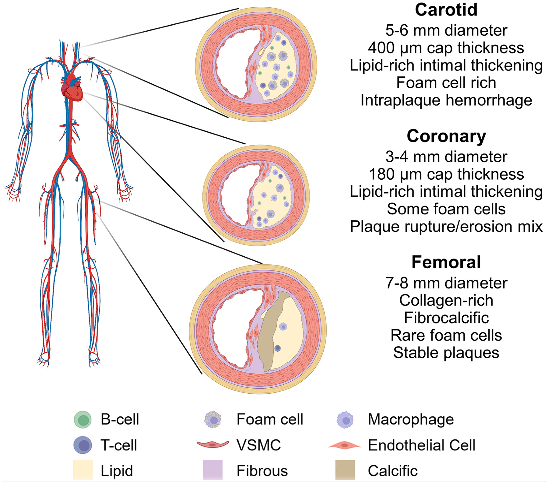
Plaque features in carotid, coronary, and peripheral atherosclerosis. Characteristics of the vascular territory and atherosclerotic plaque morphology including approximate cell type composition of plaques.
Distinct genetic factors may contribute to disease risk at various vascular locations. A European twin study indicates greater heritability of calcified plaque content compared with a less dense plaque composition.83 Similarly, a distinct study reported that noncalcified coronary plaques are not heritable, whereas calcified plaques have a heritability of 78%.84 This difference may explain partly why femoral plaque, which is largely fibrocalcific, has a greater heritability than carotid or coronary atherosclerosis.
At the transcriptional level, bulk signaling is more distinct among healthy than atherosclerotic arteries in different vascular beds.85 In comparison with smooth muscle cells of other vessels, femoral vascular smooth muscle cells from healthy arteries show increased mineralization potential with elevated Runx2, alkaline phosphatase, and osteopontin expression and upregulated transforming growth factor-β signaling.78 Indeed, both healthy and atherosclerotic femoral arteries have enriched gene expression related to bone development, and osteoid metaplasia is common in femoral plaques.79 These previous studies have exclusively evaluated bulk tissue gene expression and not considered cellular and spatial heterogeneity. Multiple different cell types contribute to atherosclerosis and comprise significant portions of the plaque. Therefore, additional studies are needed to evaluate whether cellular and molecular patterns across plaques are consistent or indicate distinct signaling pathways, which may predict responses to medical therapy or interventions. A better understanding of intrinsic differences in vascular beds could allow for treatment tailored to the type and site of atherosclerosis and secondary prevention accounting for different rates of recurrence after intervention.86
COMPARISON OF ATHEROSCLEROSIS PROGRESSION AND TREATMENT RESPONSE IN CAROTID, CORONARY, AND PERIPHERAL ARTERIES
The progression of atherosclerosis is associated with age, smoking, cholesterol, and hypertension, of which the strongest predictor in patients with ASCVD is smoking and in patients with subclinical (ie, undiagnosed) atherosclerosis is dyslipidemia.87,88 In the carotid arteries, disease progression, as determined by degree of stenosis, occurs in 19% of patients over 5 years and regression occurs in 42%.89 Approximately 15% to 30% of carotid plaques with vulnerable features become lower risk over 3 years.90 Probably reflecting plaque progression, aging is associated with increased carotid plaque calcification and lipid core size, as well as decreased fibrous tissue.91 Coronary atherosclerotic lesions leading to myocardial infarction expand primarily in the year before the event and typically result from plaques that cause only 35% stenosis 1 to 3 years earlier.92 Coronary plaques increase in size over time and typically develop cap thickening if they are fibroatheromas or remain stable as fibrocalcific plaques.93 These changes are sex dependent during aging, with women developing more vulnerable plaque features and men demonstrating no clear trends.94 Femoral artery plaque progression is not typical in symptomatic PAD; however, with aging, femoral plaques have increased calcification and decreased collagen content.95,96
With established medical and interventional strategies available for ASCVD, their influence on plaque progression must be considered (Table III). Statins are efficacious for the management of ASCVD. Biobank studies suggest that increasing use of statins has decreased lipid cores and calcification in carotid and femoral endarterectomy samples.97,98 Conversely, meta-analysis of IHD studies show that statin therapy decreases fibrous volumes and increases dense calcium volumes of coronary plaques without altering necrotic core or fibrofatty volumes.99 These results suggest that statins have differential effects on plaques across vascular territories, decreasing necrotic core volume100 and likely lowering calcification of carotid plaques while increasing calcium volume without altering necrotic core content in coronary plaques.
Table III.
Plaque morphology response to therapy in carotid, coronary, and femoral atherosclerosis
| Carotid | Coronary | PAD/femoral | |
|---|---|---|---|
| Plaque change in disease course | More atheromatous, less fibrous | More vulnerable plaque features in women; some increased calcification in men | More calcification, less collagen |
| Statins | Decrease IMT for LDL-C <70 mg/dL | Decrease plaque size for LDL-C <70 mg/dL | Prevent plaque progression |
| Aspirin | Prevent plaque progression | Not studied | Not studied |
| Cilostazol | Decrease IMT by ~5% | Decrease plaque size by 7% | Not studied |
In the carotid arteries, statin therapy can decrease intima-media thickness (IMT) when LDL-C levels of <70 mg/dL are achieved.101 Similarly, LDL-C lowering below an inflection point of approximately 70 mg/dL induces plaque regression in coronary arteries.92 Two-thirds of patients experience coronary plaque regression with statin therapy, which decreases plaque volume by 1% over 2 years.102 Some studies suggest that this effect may be primarily mediated by anti-inflammatory effects rather than directly by lipid lowering.103 Similarly, in PAD, statin initiation prevents the progression of plaque enlargement, although clear effects on regression have not been shown.104 Notably, in the REGRESS trial, plaque changes and regression with statin treatment across vascular beds in patients did not correlate between coronary, femoral, and carotid arteries, and pravastatin decreased femoral, but not carotid, IMT.105
Alongside statin therapy, antiplatelet agents are used widely in ASCVD. Surprisingly, although aspirin is commonly prescribed for ASCVD, its effect on atherosclerotic disease progression has not been studied widely. In carotid atherosclerosis, aspirin does not change IMT, but prevents the progression of plaque volume without inducing regression.106,107 Definitive studies of the effect of aspirin on plaque in IHD and PAD are lacking. Cilostazol is an antiplatelet medication with important effects on plaque evolution. Cilostazol prevents progression, and in fact increases regression of carotid IMT.108,109 In the coronary arteries, cilostazol causes plaque regression in three-quarters of patients and decreases atheroma volume an average of 7% over 9 months.110
Interventional procedures are widely used in ASCVD. Efficacy rates of interventional treatments likely vary across vascular territories owing to regional plaque features and vascular differences, although differences in procedural technique and risk factor management likely contribute as well. Carotid drug-eluting stents have a primary patency of 72% at 1 year and 59% at 5 years, whereas coronary drug-eluting stents have a patency of 97% at 1 year and 96% at 2 years.111,112 Comparatively, femoral drug-eluting stent placement shows a patency of 84% at one year and 66% at five years.113 In case of endarterectomy, carotid arteries have 79% patency at one year and 70% patency at five years while femoral arteries have a patency of 93% at one year and 89% at five years.111,114,115 These differences are likely mediated by technical factors, such as stent size and plaque length, as well as factors specific to each vascular region. Fibrocalcific plaque features are associated with greater risk of restenosis after endarterectomy, and, conversely, lipid-rich plaques have a reduced risk of restenosis.67,76 It would be expected that vascular territories with commonly fibrocalcific plaques would have inferior patency rates after intervention. However, this expected pattern fails to match the higher rates of restenosis of carotid plaques relative to femoral plaques when considering the typical atheromatous and fibrocalcific structures, respectively. Therefore, since the overall relationship of plaque features and restenosis within arteries is not matched by plaque morphology across arteries, features specific to atherosclerotic disease in the carotid and femoral arteries may influence intervention efficacy more so than plaque structure itself.
CONCLUSIONS AND FUTURE DIRECTIONS
There is a need to understand how coronary, carotid, and femoropopliteal atherosclerosis initiate and progress in relation to each other. Despite advancements in cardiovascular medicine and vascular biology, the factors influencing differential plaque progression and clinical disease across vascular beds are yet to be well-understood. Thus, existing guideline-directed therapy for ASCVD does not consider unique plaque dynamics fully or the remodeling of specific vascular beds. Understanding the mechanisms regulating plaque development across arteries should allow for new strategies to influence risk profile, atherosclerosis progression, and disease recurrence.
The current clinical approach to diagnosing and treating atherosclerosis limits the understanding of heterogeneity in atherogenesis across separate vascular territories. ASCVD initiates early in life, with the formation of fatty streaks and intimal thickening in arteries and in most patients, and only manifests with symptomatic disease multiple decades later. The prolonged progression of ASCVD obfuscates the pathogenic events in the arterial wall because the disease is commonly evaluated only after being detected clinically, thereby limiting our evaluation to differences in atherosclerosis of advanced, clinically significant disease. Further studies of subclinical atherosclerosis initiation and progression have great potential to clarify how ASCVD pathogenesis differs between vascular territories.
Previous work has shown differences in bulk gene expression in plaques from different vascular beds,85 but the conclusions from this analysis are limited owing to cell heterogeneity and structural differences present in carotid, coronary, and femoral plaques. Going forward, it will be important to understand whether plaques in different arteries contain distinct cell types or different mixtures of the same cellular components. Understanding the progression and composition of atherosclerosis in various vascular beds at the single cell level has the potential to identify key signaling pathways determining plaque morphology and maturation. These pathways may allow for therapeutic targeting to influence plaque structure directly. More specifically, there is great potential for using single cell RNA sequencing and assay for transposase-accessible chromatin sequencing studies to assess differences in cellular gene expression and cell types within human plaques across multiple vascular beds. To this end, recent work applying single cell RNA-sequencing identified differences in immune cell polarization and presence in carotid and femoral plaques.116 Extending these approaches with spatial transcriptomics will allow for the comparison of cell identity and gene expression in the context of differential plaque structures observed across vascular beds. Preclinical research in atherosclerosis will benefit from the increased use of larger animal models that can more closely match the anatomy and metabolic conditions of human disease as shown with spatial transcriptomics of porcine coronary atherosclerosis.117
It is important to consider the intrinsic differences in atherosclerosis between patients. Data from humans and animals suggest that a monoclonal or oligoclonal vascular smooth muscle cell expansion can contribute more than one-half of the cells present in advanced plaques.118–120,121 Additionally, animal studies confirm that specific vascular territories have an intrinsic vulnerability to atherogenesis.122,123 Because atherosclerosis risk differs across vessels and patients often develop multiple different types of plaques in separate arteries, unique cellular phenotypes and epigenetics across the vasculature may influence local manifestations of atherosclerosis. In particular, epigenetic differences in regional smooth muscle cells could be crucial in determining large sections of the plaque architecture in atherosclerosis. Biological variables, such as sex and age, are also associated with differences in plaque features and influence ASCVD risk. Building on initial evidence in intrapatient atherosclerosis from Helck et al82 and the REGRESS trial,105 future studies will benefit from further in-depth comparisons of atherosclerosis across vascular beds within individual patients to better understand the cellular and molecular heterogeneity of plaques in different arterial segments.
Clinical medicine can improve the integration of morphological and histological information with clinical decision-making to provide personalized care and therapies. Plaque features are identifiable with imaging modalities, such as duplex and intravascular ultrasound examination, computed tomography scans, and magnetic resonance, and histological plaque samples are available after endarterectomy. Although plaque structure is known to be associated with cerebrovascular events, coronary events, and revascularization, and femoral disease symptoms and progression after intervention, approaches to evaluate plaque structure are not often used in decision-making in patient management. Identifying predictive aspects of plaque features and response to therapies promises to be especially valuable. Atherosclerosis incidence, plaque structure, modifiable risk, and response to intervention vary substantially by vessel location; yet, these disease processes are still considered highly similar. Critical opportunities exist to risk stratify and treat patients based on how and where their atherosclerotic disease manifests.
Acknowledgments
The authors thank house staff and faculty of the Division of Vascular and Endovascular Surgery in the Yale University School of Medicine, including Dr. Ochoa Chaar, for their input. The Figure was created with a licensed account on BioRender.com.
D.G.J. was supported by a Medical Scientist Training Program Grant at Yale University School of Medicine from the National Institutes of Health (T32GM136651). D.M.G. receives funding from the National Institutes of Health (R35HL150766, R01HD110059, R21NS123469) and American Heart Association (Established Investigator Award, 19EIA34660321 and Collaborative Sciences Award, 23CSA1051139).
Footnotes
The editors and reviewers of this article have no relevant financial relationships to disclose per the Journal policy that requires reviewers to decline review of any manuscript for which they may have a conflict of interest.
REFERENCES
- 1.Libby P, Buring JE, Badimon L, et al. Atherosclerosis. Nat Rev Dis Primers. 2019;5:56. [DOI] [PubMed] [Google Scholar]
- 2.Frangos SG, Gahtan V, Sumpio B. Localization of atherosclerosis: role of hemodynamics. Arch Surg. 1999;134:1142–1149. [DOI] [PubMed] [Google Scholar]
- 3.Zuiderwijk M, Geerts M, van Rhijn CJ, et al. Leukocyte dynamics during the evolution of human coronary atherosclerosis: conclusions from a sevenfold, chromogen-based, immunohistochemical evaluation. Am J Pathol. 2018;188:1524–1529. [DOI] [PubMed] [Google Scholar]
- 4.van Dijk RA, Duinisveld AJ, Schaapherder AF, et al. A change in inflammatory footprint precedes plaque instability: a systematic evaluation of cellular aspects of the adaptive immune response in human atherosclerosis. J Am Heart Assoc. 2015;4:e001403. [DOI] [PMC free article] [PubMed] [Google Scholar]
- 5.Holman RL, Mc GH, Strong JP, Geer JC. The natural history of atherosclerosis: the early aortic lesions as seen in New Orleans in the middle of the of the 20th century. Am J Pathol. 1958;34:209–235. [PMC free article] [PubMed] [Google Scholar]
- 6.Dalager S, Paaske WP, Kristensen IB, Laurberg JM, Falk E. Artery-related differences in atherosclerosis expression: implications for atherogenesis and dynamics in intima-media thickness. Stroke. 2007;38:2698–2705. [DOI] [PubMed] [Google Scholar]
- 7.Pan Y, Jing J, Cai X, et al. Prevalence and vascular distribution of multiterritorial atherosclerosis among community-dwelling adults in southeast China. JAMA Netw Open. 2022;5:e2218307. [DOI] [PMC free article] [PubMed] [Google Scholar]
- 8.Diseases GBD, Injuries C. Global burden of 369 diseases and injuries in 204 countries and territories, 1990–2019: a systematic analysis for the Global Burden of Disease Study 2019. Lancet. 2020;396:1204–1222. [DOI] [PMC free article] [PubMed] [Google Scholar]
- 9.Flaherty ML, Kissela B, Khoury JC, et al. Carotid artery stenosis as a cause of stroke. Neuroepidemiology. 2013;40:36–41. [DOI] [PMC free article] [PubMed] [Google Scholar]
- 10.de Weerd M, Greving JP, Hedblad B, et al. Prevalence of asymptomatic carotid artery stenosis in the general population: an individual participant data meta-analysis. Stroke. 2010;41:1294–1297. [DOI] [PMC free article] [PubMed] [Google Scholar]
- 11.Chang RW, Tucker LY, Rothenberg KA, et al. Incidence of ischemic stroke in patients with asymptomatic severe carotid stenosis without surgical intervention. JAMA. 2022;327:1974–1982. [DOI] [PMC free article] [PubMed] [Google Scholar]
- 12.Tsao CW, Aday AW, Almarzooq ZI, et al. Heart disease and stroke statistics-2023 update: a report from the American heart association. Circulation. 2023;147:e93–e621. [DOI] [PMC free article] [PubMed] [Google Scholar]
- 13.Song P, Rudan D, Zhu Y, et al. Global, regional, and national prevalence and risk factors for peripheral artery disease in 2015: an updated systematic review and analysis. Lancet Glob Health. 2019;7:e1020–e1030. [DOI] [PubMed] [Google Scholar]
- 14.Song P, Fang Z, Wang H, et al. Global and regional prevalence, burden, and risk factors for carotid atherosclerosis: a systematic review, meta-analysis, and modelling study. Lancet Glob Health. 2020;8:e721–e729. [DOI] [PubMed] [Google Scholar]
- 15.Eid MA, Mehta K, Barnes JA, et al. The global burden of peripheral artery disease. J Vasc Surg. 2023;77:1119–11126.e1. [DOI] [PubMed] [Google Scholar]
- 16.Vancheri F, Tate AR, Henein M, et al. Time trends in ischaemic heart disease incidence and mortality over three decades (1990–2019) in 20 Western European countries: systematic analysis of the Global Burden of Disease Study 2019. Eur J Prev Cardiol. 2022;29:396–403. [DOI] [PubMed] [Google Scholar]
- 17.Dai H, Much AA, Maor E, et al. Global, regional, and national burden of ischaemic heart disease and its attributable risk factors, 1990–2017: results from the Global Burden of Disease Study 2017. Eur Heart J Qual Care Clin Outcomes. 2022;8:50–60. [DOI] [PMC free article] [PubMed] [Google Scholar]
- 18.Berry JD, Dyer A, Cai X, et al. Lifetime risks of cardiovascular disease. N Engl J Med. 2012;366:321–329. [DOI] [PMC free article] [PubMed] [Google Scholar]
- 19.Lopez-Melgar B, Fernandez-Friera L, Oliva B, et al. Subclinical atherosclerosis burden by 3D ultrasound in mid-life: the PESA study. J Am Coll Cardiol. 2017;70:301–313. [DOI] [PubMed] [Google Scholar]
- 20.Chen Q, Smith CY, Bailey KR, Wennberg PW, Kullo IJ. Disease location is associated with survival in patients with peripheral arterial disease. J Am Heart Assoc. 2013;2:e000304. [DOI] [PMC free article] [PubMed] [Google Scholar]
- 21.Diehm N, Shang A, Silvestro A, et al. Association of cardiovascular risk factors with pattern of lower limb atherosclerosis in 2659 patients undergoing angioplasty. Eur J Vasc Endovasc Surg. 2006;31:59–63. [DOI] [PubMed] [Google Scholar]
- 22.Khera AV, Emdin CA, Drake I, et al. Genetic risk, adherence to a healthy lifestyle, and coronary disease. N Engl J Med. 2016;375:2349–2358. [DOI] [PMC free article] [PubMed] [Google Scholar]
- 23.Natural history of aortic and coronary atherosclerotic lesions in youth. Findings from the PDAY study. Pathobiological determinants of atherosclerosis in youth (PDAY) research group. Arterioscler Thromb. 1993;13:1291–1298. [DOI] [PubMed] [Google Scholar]
- 24.Wang F, Yu Y, Mubarik S, et al. Global burden of ischemic heart disease and attributable risk factors, 1990–2017: a secondary analysis based on the global burden of disease study 2017. Clin Epidemiol. 2021;13:859–870. [DOI] [PMC free article] [PubMed] [Google Scholar]
- 25.McClelland RL, Jorgensen NW, Budoff M, et al. 10-Year coronary heart disease risk prediction using coronary artery calcium and traditional risk factors: derivation in the MESA (Multi-Ethnic study of atherosclerosis) with validation in the HNR (Heinz Nixdorf recall) study and the DHS (dallas heart study). J Am Coll Cardiol. 2015;66:1643–1653. [DOI] [PMC free article] [PubMed] [Google Scholar]
- 26.Leening MJ, Ferket BS, Steyerberg EW, et al. Sex differences in lifetime risk and first manifestation of cardiovascular disease: prospective population based cohort study. BMJ. 2014;349:g5992. [DOI] [PMC free article] [PubMed] [Google Scholar]
- 27.George J, Rapsomaniki E, Pujades-Rodriguez M, et al. How does cardiovascular disease first present in women and men? Incidence of 12 cardiovascular diseases in a contemporary cohort of 1,937,360 people. Circulation. 2015;132:1320–1328. [DOI] [PMC free article] [PubMed] [Google Scholar]
- 28.Bergstrom G, Persson M, Adiels M, et al. Prevalence of subclinical coronary artery atherosclerosis in the general population. Circulation. 2021;144:916–929. [DOI] [PMC free article] [PubMed] [Google Scholar]
- 29.Anand SS, Islam S, Rosengren A, et al. Risk factors for myocardial infarction in women and men: insights from the INTERHEART study. Eur Heart J. 2008;29:932–940. [DOI] [PubMed] [Google Scholar]
- 30.Ji X, Leng XY, Dong Y, et al. Modifiable risk factors for carotid atherosclerosis: a meta-analysis and systematic review. Ann Transl Med. 2019;7:632. [DOI] [PMC free article] [PubMed] [Google Scholar]
- 31.Kinosian B, Glick H, Preiss L, Puder KL. Cholesterol and coronary heart disease: predicting risks in men by changes in levels and ratios. J Investig Med. 1995;43:443–450. [PubMed] [Google Scholar]
- 32.Fowkes FG, Housley E, Riemersma RA, et al. Smoking, lipids, glucose intolerance, and blood pressure as risk factors for peripheral atherosclerosis compared with ischemic heart disease in the Edinburgh Artery Study. Am J Epidemiol. 1992;135:331–340. [DOI] [PubMed] [Google Scholar]
- 33.Pencina MJ, Navar AM, Wojdyla D, et al. Quantifying importance of major risk factors for coronary heart disease. Circulation. 2019;139:1603–1611. [DOI] [PMC free article] [PubMed] [Google Scholar]
- 34.Razo C, Welgan CA, Johnson CO, et al. Effects of elevated systolic blood pressure on ischemic heart disease: a Burden of Proof study. Nat Med. 2022;28:2056–2065. [DOI] [PMC free article] [PubMed] [Google Scholar]
- 35.Khaleghi M, Isseh IN, Jouni H, Sohn S, Bailey KR, Kullo IJ. Family history as a risk factor for carotid artery stenosis. Stroke. 2014;45:2252–2256. [DOI] [PubMed] [Google Scholar]
- 36.Lloyd-Jones DM, Nam BH, D’Agostino RB, et al. Parental cardiovascular disease as a risk factor for cardiovascular disease in middle-aged adults: a prospective study of parents and offspring. JAMA. 2004;291:2204–2211. [DOI] [PubMed] [Google Scholar]
- 37.Khaleghi M, Isseh IN, Bailey KR, Kullo IJ. Family history as a risk factor for peripheral arterial disease. Am J Cardiol. 2014;114:928–932. [DOI] [PMC free article] [PubMed] [Google Scholar]
- 38.Dong C, Beecham A, Slifer S, et al. Genomewide linkage and peakwide association analyses of carotid plaque in Caribbean Hispanics. Stroke. 2010;41:2750–2756. [DOI] [PMC free article] [PubMed] [Google Scholar]
- 39.Peyser PA, Bielak LF, Chu JS, et al. Heritability of coronary artery calcium quantity measured by electron beam computed tomography in asymptomatic adults. Circulation. 2002;106:304–308. [DOI] [PubMed] [Google Scholar]
- 40.Lucatelli P, Fagnani C, Tarnoki AD, et al. Genetic influence on femoral plaque and its relationship with carotid plaque: an international twin study. Int J Cardiovasc Imaging. 2018;34:531–541. [DOI] [PubMed] [Google Scholar]
- 41.van Zuydam NR, Stiby A, Abdalla M, et al. Genome-wide association study of peripheral artery disease. Circ Genom Precis Med. 2021;14:e002862. [DOI] [PMC free article] [PubMed] [Google Scholar]
- 42.Lawton JS, Tamis-Holland JE, Bangalore S, et al. 2021 ACC/AHA/SCAI guideline for coronary artery revascularization: a report of the American college of cardiology/American heart association joint committee on clinical practice guidelines. Circulation. 2022;145:e18–e114. [DOI] [PubMed] [Google Scholar]
- 43.Fernandez-Ortiz A, Badimon JJ, Falk E, et al. Characterization of the relative thrombogenicity of atherosclerotic plaque components: implications for consequences of plaque rupture. J Am Coll Cardiol. 1994;23:1562–1569. [DOI] [PubMed] [Google Scholar]
- 44.Cortenbach KRG, Morales Cano D, Meek J, et al. Topography of immune cell infiltration in different stages of coronary atherosclerosis revealed by multiplex immunohistochemistry. Int J Cardiol Heart Vasc. 2023;44:101111. [DOI] [PMC free article] [PubMed] [Google Scholar]
- 45.Burke AP, Farb A, Malcom GT, Liang YH, Smialek J, Virmani R. Coronary risk factors and plaque morphology in men with coronary disease who died suddenly. N Engl J Med. 1997;336:1276–1282. [DOI] [PubMed] [Google Scholar]
- 46.Kubo T, Imanishi T, Kashiwagi M, et al. Multiple coronary lesion instability in patients with acute myocardial infarction as determined by optical coherence tomography. Am J Cardiol. 2010;105:318–322. [DOI] [PubMed] [Google Scholar]
- 47.Kolodgie FD, Burke AP, Farb A, et al. The thin-cap fibroatheroma: a type of vulnerable plaque: the major precursor lesion to acute coronary syndromes. Curr Opin Cardiol. 2001;16:285–292. [DOI] [PubMed] [Google Scholar]
- 48.Jia H, Abtahian F, Aguirre AD, et al. In vivo diagnosis of plaque erosion and calcified nodule in patients with acute coronary syndrome by intravascular optical coherence tomography. J Am Coll Cardiol. 2013;62:1748–1758. [DOI] [PMC free article] [PubMed] [Google Scholar]
- 49.Yin WJ, Jing J, Zhang YQ, et al. Association between non-culprit healed plaque and plaque progression in acute coronary syndrome patients: an optical coherence tomography study. J Geriatr Cardiol. 2021;18:631–644. [DOI] [PMC free article] [PubMed] [Google Scholar]
- 50.Matsuo Y, Takumi T, Mathew V, et al. Plaque characteristics and arterial remodeling in coronary and peripheral arterial systems. Atherosclerosis. 2012;223:365–371. [DOI] [PMC free article] [PubMed] [Google Scholar]
- 51.Calvert PA, Obaid DR, O’Sullivan M, et al. Association between IVUS findings and adverse outcomes in patients with coronary artery disease: the VIVA (VH-IVUS in Vulnerable Atherosclerosis) Study. JACC Cardiovasc Imaging. 2011;4:894–901. [DOI] [PubMed] [Google Scholar]
- 52.Niccoli G, Montone RA, Di Vito L, et al. Plaque rupture and intact fibrous cap assessed by optical coherence tomography portend different outcomes in patients with acute coronary syndrome. Eur Heart J. 2015;36:1377–1384. [DOI] [PubMed] [Google Scholar]
- 53.Mann JM, Davies MJ. Vulnerable plaque. Relation of characteristics to degree of stenosis in human coronary arteries. Circulation. 1996;94:928–931. [DOI] [PubMed] [Google Scholar]
- 54.Puchner SB, Liu T, Mayrhofer T, et al. High-risk plaque detected on coronary CT angiography predicts acute coronary syndromes independent of significant stenosis in acute chest pain: results from the ROMICAT-II trial. J Am Coll Cardiol. 2014;64:684–692. [DOI] [PMC free article] [PubMed] [Google Scholar]
- 55.Li R, Duncan BB, Metcalf PA, et al. B-mode-detected carotid artery plaque in a general population. Stroke. 1994;25:2377–2383. [DOI] [PubMed] [Google Scholar]
- 56.AbuRahma AF, Avgerinos ED, Chang RW, et al. Society for Vascular Surgery clinical practice guidelines for management of extracranial cerebrovascular disease. J Vasc Surg. 2022;75:4S–22S. [DOI] [PubMed] [Google Scholar]
- 57.Ornello R, Degan D, Tiseo C, et al. Distribution and temporal trends from 1993 to 2015 of ischemic stroke subtypes: a systematic review and meta-analysis. Stroke. 2018;49:814–819. [DOI] [PubMed] [Google Scholar]
- 58.Ospel JM, Singh N, Marko M, et al. Prevalence of ipsilateral non-stenotic carotid plaques on computed tomography angiography in embolic stroke of undetermined source. Stroke. 2020;51:1743–1749. [DOI] [PubMed] [Google Scholar]
- 59.Redgrave JN, Gallagher P, Lovett JK, Rothwell PM. Critical cap thickness and rupture in symptomatic carotid plaques: the oxford plaque study. Stroke. 2008;39:1722–1729. [DOI] [PubMed] [Google Scholar]
- 60.Abbas AE, Zacharias SK, Goldstein JA, Hanson ID, Safian RD. Invasive characterization of atherosclerotic plaque in patients with peripheral arterial disease using near-infrared spectroscopy intravascular ultrasound. Catheter Cardiovasc Interv. 2017;90:461–470. [DOI] [PubMed] [Google Scholar]
- 61.Cai JM, Hatsukami TS, Ferguson MS, Small R, Polissar NL, Yuan C. Classification of human carotid atherosclerotic lesions with in vivo multicontrast magnetic resonance imaging. Circulation. 2002;106:1368–1373. [DOI] [PubMed] [Google Scholar]
- 62.Gupta A, Baradaran H, Schweitzer AD, et al. Carotid plaque MRI and stroke risk: a systematic review and meta-analysis. Stroke. 2013;44:3071–3077. [DOI] [PubMed] [Google Scholar]
- 63.Redgrave JN, Lovett JK, Gallagher PJ, Rothwell PM. Histological assessment of 526 symptomatic carotid plaques in relation to the nature and timing of ischemic symptoms: the Oxford plaque study. Circulation. 2006;113:2320–2328. [DOI] [PubMed] [Google Scholar]
- 64.Spagnoli LG, Mauriello A, Sangiorgi G, et al. Extracranial thrombotically active carotid plaque as a risk factor for ischemic stroke. JAMA. 2004;292:1845–1852. [DOI] [PubMed] [Google Scholar]
- 65.Kamtchum-Tatuene J, Wilman A, Saqqur M, Shuaib A, Jickling GC. Carotid plaque with high-risk features in embolic stroke of undetermined source: systematic review and meta-analysis. Stroke. 2020;51:311–314. [DOI] [PMC free article] [PubMed] [Google Scholar]
- 66.McNally JS, McLaughlin MS, Hinckley PJ, et al. Intraluminal thrombus, intraplaque hemorrhage, plaque thickness, and current smoking optimally predict carotid stroke. Stroke. 2015;46:84–90. [DOI] [PubMed] [Google Scholar]
- 67.Hellings WE, Moll FL, De Vries JP, et al. Atherosclerotic plaque composition and occurrence of restenosis after carotid endarterectomy. JAMA. 2008;299:547–554. [DOI] [PubMed] [Google Scholar]
- 68.Hirsch AT, Criqui MH, Treat-Jacobson D, et al. Peripheral arterial disease detection, awareness, and treatment in primary care. JAMA. 2001;286:1317–1324. [DOI] [PubMed] [Google Scholar]
- 69.Singer A Segmental distribution of peripheral atherosclerosis. Arch Surg. 1963;87:384–390. [DOI] [PubMed] [Google Scholar]
- 70.Chi J, Chiu B, Cao Y, et al. Assessment of femoral artery atherosclerosis at the adductor canal using 3D black-blood MRI. Clin Radiol. 2013;68:e213–e221. [DOI] [PubMed] [Google Scholar]
- 71.Karmody AM, Powers SR, Monaco VJ, Leather RP. “Blue toe” syndrome. An indication for limb salvage surgery. Arch Surg. 1976;111:1263–1268. [DOI] [PubMed] [Google Scholar]
- 72.Cunnane EM, Mulvihill JJE, Barrett HE, Hennessy MM, Kavanagh EG, Walsh MT. Mechanical properties and composition of carotid and femoral atherosclerotic plaques: a comparative study. J Biomech. 2016;49:3697–3704. [DOI] [PubMed] [Google Scholar]
- 73.Narula N, Dannenberg AJ, Olin JW, et al. Pathology of peripheral artery disease in patients with critical limb ischemia. J Am Coll Cardiol. 2018;72:2152–2163. [DOI] [PubMed] [Google Scholar]
- 74.McDermott MM, Kramer CM, Tian L, et al. Plaque composition in the proximal superficial femoral artery and peripheral artery disease events. JACC Cardiovasc Imaging. 2017;10:1003–1012. [DOI] [PMC free article] [PubMed] [Google Scholar]
- 75.McDermott MM, Carroll T, Carr J, et al. Femoral artery plaque characteristics, lower extremity collaterals, and mobility loss in peripheral artery disease. Vasc Med. 2017;22:473–481. [DOI] [PMC free article] [PubMed] [Google Scholar]
- 76.Derksen WJ, de Vries JP, Vink A, et al. Histologic atherosclerotic plaque characteristics are associated with restenosis rates after endarterectomy of the common and superficial femoral arteries. J Vasc Surg. 2010;52:592–599. [DOI] [PubMed] [Google Scholar]
- 77.Dalager S, Falk E, Kristensen IB, Paaske WP. Plaque in superficial femoral arteries indicates generalized atherosclerosis and vulnerability to coronary death: an autopsy study. J Vasc Surg. 2008;47:296–302. [DOI] [PubMed] [Google Scholar]
- 78.Espitia O, Chatelais M, Steenman M, et al. Implication of molecular vascular smooth muscle cell heterogeneity among arterial beds in arterial calcification. PLoS One. 2018;13:e0191976. [DOI] [PMC free article] [PubMed] [Google Scholar]
- 79.Herisson F, Heymann MF, Chetiveaux M, et al. Carotid and femoral atherosclerotic plaques show different morphology. Atherosclerosis. 2011;216:348–354. [DOI] [PubMed] [Google Scholar]
- 80.Laclaustra M, Casasnovas JA, Fernandez-Ortiz A, et al. Femoral and carotid subclinical atherosclerosis association with risk factors and coronary calcium: the AWHS study. J Am Coll Cardiol. 2016;67:1263–1274. [DOI] [PubMed] [Google Scholar]
- 81.Fernandez-Friera L, Penalvo JL, Fernandez-Ortiz A, et al. Prevalence, vascular distribution, and multiterritorial extent of subclinical atherosclerosis in a middle-aged cohort: the PESA (progression of early subclinical atherosclerosis) study. Circulation. 2015;131:2104–2113. [DOI] [PubMed] [Google Scholar]
- 82.Helck A, Bianda N, Canton G, et al. Intra-individual comparison of carotid and femoral atherosclerotic plaque features with in vivo MR plaque imaging. Int J Cardiovasc Imaging. 2015;31:1611–1618. [DOI] [PubMed] [Google Scholar]
- 83.Hernyes A, Piroska M, Fejer B, et al. Overlapping genetic background of coronary artery and carotid/femoral atherosclerotic calcification. Medicina (Kaunas). 2021;57:252. [DOI] [PMC free article] [PubMed] [Google Scholar]
- 84.Drobni ZD, Kolossvary M, Karady J, et al. Heritability of coronary artery disease: insights from a classical twin study. Circ Cardiovasc Imaging. 2022;15:e013348. [DOI] [PMC free article] [PubMed] [Google Scholar]
- 85.Steenman M, Espitia O, Maurel B, et al. Identification of genomic differences among peripheral arterial beds in atherosclerotic and healthy arteries. Sci Rep. 2018;8:3940. [DOI] [PMC free article] [PubMed] [Google Scholar]
- 86.DeBakey ME, Glaeser DH. Patterns of atherosclerosis: effect of risk factors on recurrence and survival-analysis of 11,890 cases with more than 25-year follow-up. Am J Cardiol. 2000;85:1045–1053. [DOI] [PubMed] [Google Scholar]
- 87.van der Meer IM, Iglesias del Sol A, Hak AE, Bots ML, Hofman A, Witteman JC. Risk factors for progression of atherosclerosis measured at multiple sites in the arterial tree: the Rotterdam Study. Stroke. 2003;34:2374–2379. [DOI] [PubMed] [Google Scholar]
- 88.Lopez-Melgar B, Fernandez-Friera L, Oliva B, et al. Short-Term progression of multiterritorial subclinical atherosclerosis. J Am Coll Cardiol. 2020;75:1617–1627. [DOI] [PubMed] [Google Scholar]
- 89.Hogberg D, Bjorck M, Mani K, Svensjo S, Wanhainen A. Five year outcomes in men screened for carotid artery stenosis at 65 Years of age: a population based cohort study. Eur J Vasc Endovasc Surg. 2019;57:759–766. [DOI] [PubMed] [Google Scholar]
- 90.Catalano O, Bendotti G, Aloi TL, et al. Evidence of carotid atherosclerosis vulnerability regression in real life from magnetic resonance imaging: results of the MAGNETIC prospective study. J Am Heart Assoc. 2023;12:e026469. [DOI] [PMC free article] [PubMed] [Google Scholar]
- 91.Redgrave JN, Lovett JK, Rothwell PM. Histological features of symptomatic carotid plaques in relation to age and smoking: the oxford plaque study. Stroke. 2010;41:2288–2294. [DOI] [PubMed] [Google Scholar]
- 92.Ahmadi A, Argulian E, Leipsic J, Newby DE, Narula J. From subclinical atherosclerosis to plaque progression and acute coronary events: JACC state-of-the-art review. J Am Coll Cardiol. 2019;74:1608–1617. [DOI] [PubMed] [Google Scholar]
- 93.Kubo T, Maehara A, Mintz GS, et al. The dynamic nature of coronary artery lesion morphology assessed by serial virtual histology intravascular ultrasound tissue characterization. J Am Coll Cardiol. 2010;55:1590–1597. [DOI] [PubMed] [Google Scholar]
- 94.Seegers LM, Araki M, Nakajima A, et al. Sex differences in culprit plaque characteristics among different age groups in patients with acute coronary syndromes. Circ Cardiovasc Interv. 2022;15:e011612. [DOI] [PubMed] [Google Scholar]
- 95.Russell KS, Yates DP, Kramer CM, et al. A randomized, placebo-controlled trial of canakinumab in patients with peripheral artery disease. Vasc Med. 2019;24:414–421. [DOI] [PMC free article] [PubMed] [Google Scholar]
- 96.de Bakker M, Timmerman N, van Koeverden ID, et al. The age- and sex-specific composition of atherosclerotic plaques in vascular surgery patients. Atherosclerosis. 2020;310:1–10. [DOI] [PubMed] [Google Scholar]
- 97.van Lammeren GW, den Ruijter HM, Vrijenhoek JE, et al. Time-dependent changes in atherosclerotic plaque composition in patients undergoing carotid surgery. Circulation. 2014;129:2269–2276. [DOI] [PubMed] [Google Scholar]
- 98.Haitjema S, van Haelst STW, de Vries JPM, et al. Time-dependent differences in femoral artery plaque characteristics of peripheral arterial disease patients. Atherosclerosis. 2016;255:66–72. [DOI] [PubMed] [Google Scholar]
- 99.Zheng G, Li Y, Huang H, Wang J, Hirayama A, Lin J. The effect of statin therapy on coronary plaque composition using virtual histology intravascular ultrasound: a meta-analysis. PLoS One. 2015;10:e0133433. [DOI] [PMC free article] [PubMed] [Google Scholar]
- 100.Brinjikji W, Lehman VT, Kallmes DF, et al. The effects of statin therapy on carotid plaque composition and volume: a systematic review and meta-analysis. J Neuroradiol. 2017;44:234–240. [DOI] [PubMed] [Google Scholar]
- 101.Amarenco P, Hobeanu C, Labreuche J, et al. Carotid atherosclerosis evolution when targeting a low-density lipoprotein cholesterol concentration <70 mg/dL after an ischemic stroke of atherosclerotic origin. Circulation. 2020;142:748–757. [DOI] [PubMed] [Google Scholar]
- 102.Nicholls SJ, Ballantyne CM, Barter PJ, et al. Effect of two intensive statin regimens on progression of coronary disease. N Engl J Med. 2011;365:2078–2087. [DOI] [PubMed] [Google Scholar]
- 103.Gao DR, Hua R, Jiesisibieke D, et al. C-reactive protein and coronary atheroma regression following statin therapy: a meta-regression of randomized controlled trials. Front Cardiovasc Med. 2022;9:989527. [DOI] [PMC free article] [PubMed] [Google Scholar]
- 104.West AM, Anderson JD, Meyer CH, et al. The effect of ezetimibe on peripheral arterial atherosclerosis depends upon statin use at baseline. Atherosclerosis. 2011;218:156–162. [DOI] [PMC free article] [PubMed] [Google Scholar]
- 105.de Groot E, Jukema JW, van Boven AJ, et al. Effect of pravastatin on progression and regression of coronary atherosclerosis and vessel wall changes in carotid and femoral arteries: a report from the Regression Growth Evaluation Statin Study. Am J Cardiol. 1995;76:40C–46C. [DOI] [PubMed] [Google Scholar]
- 106.Hu X, Hu Y, Sun X, Li Y, Zhu Y. Effect of aspirin in patients with established asymptomatic carotid atherosclerosis: a systematic review and meta-analysis. Front Pharmacol. 2022;13:1041400. [DOI] [PMC free article] [PubMed] [Google Scholar]
- 107.Ranke C, Hecker H, Creutzig A, Alexander K. Dose-dependent effect of aspirin on carotid atherosclerosis. Circulation. 1993;87:1873–1879. [DOI] [PubMed] [Google Scholar]
- 108.Huang R, Mills K, Romero J, et al. Comparative effects of lipid lowering, hypoglycemic, antihypertensive and antiplatelet medications on carotid artery intima-media thickness progression: a network meta-analysis. Cardiovasc Diabetol. 2019;18:14. [DOI] [PMC free article] [PubMed] [Google Scholar]
- 109.Heo SH, Lee JS, Kim BJ, Hwang KJ, Kim JH, Chang DI. Effects of cilostazol against the progression of carotid IMT in symptomatic ischemic stroke patients. J Neurol. 2013;260:122–130. [DOI] [PMC free article] [PubMed] [Google Scholar]
- 110.Ko YG, Choi SH, Chol Kang W, Kwon Lee B, Wook Kim S, Shim WH. Effects of combination therapy with cilostazol and probucol versus monotherapy with cilostazol on coronary plaque, lipid and biomarkers: SECURE study, a double-blind randomized controlled clinical trial. J Atheroscler Thromb. 2014;21:816–830. [DOI] [PubMed] [Google Scholar]
- 111.Bonati LH, Gregson J, Dobson J, et al. Restenosis and risk of stroke after stenting or endarterectomy for symptomatic carotid stenosis in the International Carotid Stenting Study (ICSS): secondary analysis of a randomised trial. Lancet Neurol. 2018;17:587–596. [DOI] [PMC free article] [PubMed] [Google Scholar]
- 112.Sarno G, Lagerqvist B, Frobert O, et al. Lower risk of stent thrombosis and restenosis with unrestricted use of ‘new-generation’ drug-eluting stents: a report from the nationwide Swedish Coronary Angiography and Angioplasty Registry (SCAAR). Eur Heart J. 2012;33:606–613. [DOI] [PubMed] [Google Scholar]
- 113.Dake MD, Ansel GM, Jaff MR, et al. Durable clinical effectiveness with Paclitaxel-eluting stents in the femoropopliteal artery: 5-year results of the zilver PTX randomized trial. Circulation. 2016;133:1472–1483. discussion: 83. [DOI] [PMC free article] [PubMed] [Google Scholar]
- 114.Boufi M, Ejargue M, Gaye M, Boyer L, Alimi Y, Loundou AD. Systematic review and meta-analysis of endovascular versus open repair for common femoral artery atherosclerosis treatment. J Vasc Surg. 2021;73:1445–1455. [DOI] [PubMed] [Google Scholar]
- 115.Cardon A, Aillet S, Jarno P, et al. [Endarteriectomy of the femoral tripod: long-term results and analysis of failure factors]. Ann Chir. 2001;126:777–782. [DOI] [PubMed] [Google Scholar]
- 116.Slysz J, Sinha A, DeBerge M, et al. Single-cell profiling reveals inflammatory polarization of human carotid versus femoral plaque leukocytes. JCI Insight. 2023;8:e171359. [DOI] [PMC free article] [PubMed] [Google Scholar]
- 117.Sukhanov S, Higashi Y, Yoshida T, et al. Insulin-like growth factor 1 reduces coronary atherosclerosis in pigs with familial hypercholesterolemia. JCI Insight. 2023;8:e165713. [DOI] [PMC free article] [PubMed] [Google Scholar]
- 118.Murry CE, Gipaya CT, Bartosek T, Benditt EP, Schwartz SM. Monoclonality of smooth muscle cells in human atherosclerosis. Am J Pathol. 1997;151:697–705. [PMC free article] [PubMed] [Google Scholar]
- 119.Benditt EP, Benditt JM. Evidence for a monoclonal origin of human atherosclerotic plaques. Proc Natl Acad Sci U S A. 1973;70:1753–1756. [DOI] [PMC free article] [PubMed] [Google Scholar]
- 120.Misra A, Feng Z, Chandran RR, et al. Integrin beta3 regulates clonality and fate of smooth muscle-derived atherosclerotic plaque cells. Nat Commun. 2018;9:2073. [DOI] [PMC free article] [PubMed] [Google Scholar]
- 121.Kabir I, Zhang X, Dave JM, et al. Age of bone marrow dictates clonality of smooth muscle cell-derived cells in the atherosclerotic plaque. Nat Aging. 2023;3:64–81. [DOI] [PMC free article] [PubMed] [Google Scholar]
- 122.Haimovici H, Maier N, Strauss L. Fate of aortic homografts in experimental canine atherosclerosis; study of fresh thoracic implants into abdominal aorta. AMA Arch Surg. 1958;76:282–288. [DOI] [PubMed] [Google Scholar]
- 123.Haimovici H, Maier N. Fate of aortic homografts in canine atherosclerosis. 3. Study of fresh abdominal and thoracic aortic implants into thoracic aorta: role of tissue susceptibility in atherogenesis. Arch Surg. 1964;89:961–969. [DOI] [PubMed] [Google Scholar]