Abstract
At present, there is no doubt that RNA recombination is one of the major factors responsible for the generation of new RNA viruses and retroviruses. Numerous experimental systems have been created to investigate this complex phenomenon. Consequently, specific RNA structural motifs mediating recombination have been identified in several viruses. Unfortunately, up till now a unified model of genetic RNA recombination has not been formulated, mainly due to difficulties with the direct comparison of data obtained for different RNA-based viruses. To solve this problem, we have attempted to construct a universal system in which the recombination activity of various RNA sequences could be tested. To this end, we have used brome mosaic virus, a model (+)RNA virus of plants, for which the structural requirements of RNA recombination are well defined. The effectiveness of the new homomolecular system has been proven in an experiment involving two RNA sequences derived from the hepatitis C virus genome. In addition, comparison of the data obtained with the homomolecular system with those generated earlier using the heteromolecular one has provided new evidence that the mechanisms of homologous and non-homologous recombination are different and depend on the virus' mode of replication.
INTRODUCTION
RNA recombination is a very common phenomenon. It has been observed in all types of viruses using RNA as a carrier of genetic information: in positive-sense, single-stranded RNA viruses (1–4), in negative-sense, single-stranded RNA viruses (5,6), in double-stranded RNA viruses (7,8) and in retroviruses (9–11). Moreover, it has been shown that RNA recombination enables the exchange of genetic material not only between the same or similar viruses but also between distinctly different viruses (12). Sometimes it also permits crossovers between viral and host RNA (13–17). Taking into account the structure of viral genomic molecules and the location of crossover sites, three basic types of RNA recombination were distinguished: homologous, aberrant homologous and non-homologous (3,4,18). The former two occur between two identical or similar RNAs (or between molecules displaying local homology), while the latter involves two different molecules. Most of the collected data suggest that RNA recombinants are formed according to a copy choice model (4,18). A viral replication complex starts nascent RNA strand synthesis on one template, called RNA donor and then switches to another template, called RNA acceptor. Accordingly, two main factors are thought to affect RNA recombination: the structure of recombining molecules and the ability of the viral replicase to switch templates.
To gain more knowledge of the mechanism of RNA recombination, several model experimental systems have been created. They provided us with some specific data describing homologous and/or non-homologous recombination in particular viruses, e.g. in poliovirus, (19) mouse hepatitis virus (20,21), brome mosaic virus (BMV) (4,22), turnip crinkle virus (23,24) or tomato bushy stunt virus (25). As a result, the involvement of viral replicase proteins in recombination has been demonstrated (26,27) and a wide spectrum of RNA motifs supporting recombination have been identified (4,23,28–30). In general, the collected data suggest that there exist two major types of RNA structural elements that induce recombination events: (i) universal ones mediating template switching by different viral replicases, e.g. regions of local homology (28,31) or complementarity (32–35) and (ii) virus-specific ones, e.g. promoter-like structures (36,37). Unfortunately, up till now there has been no in vivo recombination system that could be used to test the recombination activity of any given RNA sequence and consequently to verify the above hypothesis and find some general laws governing the studied process.
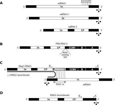
In our studies on genetic RNA recombination we have used the well-characterized in vivo system developed in BMV (30,33). BMV is a model (+)RNA virus of plants (38). Its genome is composed of three segments called RNA1, RNA2 and RNA3. RNA1 and RNA2 encode BMV replicase proteins 1a and 2a, respectively. RNA3 encodes movement (3a) and coat proteins (CP) (38). All three BMV RNAs possess an almost identical 3′-untranslated region (3′-UTR). The first BMV-based recombination system was created by Nagy and Bujarski (33). They constructed a recombinationally active BMV mutant whose genome is composed of wtRNA1, wtRNA2 and modified RNA3 (PN0-RNA3 called the recombination vector, for details see Figure 1). Only 3′-UTR was modified in PN0-RNA3, while its 5′-UTR, intergenic and coding regions were unchanged. Despite the introduced changes, the recombination vector is stable and replicates when used together with wtRNA1 and wtRNA2 to infect plants. It starts to recombine if a recombinationally active sequence (RAS) is introduced just between the CP coding sequence and the modified 3′ region (into the RAS-cloning site). Non-homologous recombination was observed when a 140–60 nt sequence complementary to RNA1 between positions 2856 and 2992 was inserted into PN0-RNA3 (a sequence from the 3′-portion of RNA1 was introduced in antisense orientation) (30,33). Interestingly, the same RNA1 fragment inserted in sense orientation did not support homologous crossovers (33). Non-homologous recombination repaired the RNA3 vector by replacing its highly modified 3′ end with 3′-UTR derived from RNA1. The resultant recombinants replicated and accumulated better than the parental RNA3 molecule, and so the latter was out competed from the infected cells. The above system is extremely efficient, since it employs selection pressure to support the accumulation of RNA3 recombinants. Because RAS is placed in two different segments of the BMV genome, we have proposed to name this system heteromolecular.
Figure 1.
The BMV-based recombination system. White, black and gray boxes represent coding, noncoding and recombinationally active sequences, respectively. The location of the primers (A and B) used for specific RT–PCR amplification of the 3′-portion of BMV RNA3 (parental or recombinant) is indicated by arrows. (A) BMV genome. The BMV genome consists of three RNA segments: RNA1, RNA2 and RNA3. All three BMV RNAs share an almost identical 3′-noncoding region with a tRNA-like structure at the very end. (B) Recombination vector. The PN0-RNA3 vector is a wtRNA3 derivative with a modified 3′-noncoding end [(for details see ref. (33)]. The latter includes (i) the RAS cloning site, (ii) a 197 nt sequence derived from the 3′-noncoding region of cowpea chlorotic mottle virus RNA3 (marked as CCMV), (iii) the sequence of wtRNA3 between nt 7 and 200 (counting from the 3′ end—marked as the region B) and (iv) the last 236 nt from the 3′ end of BMV wtRNA1 (marked as A). (C) Non-homologous recombination. Non-homologous recombination was observed if an ∼140 nt sequence, called RAS1as (shown in A), complementary to wtRNA1 between positions 2856 and 2992 was inserted into the PN0-RNA3 vector. The presence of the RAS1as sequence in RNA3 derivative (called Mag1-RNA3) allows local RNA1-RNA3 hybridization that mediates frequent non-homologous crossovers. It is thought that polymerase starts nascent strand synthesis on the 3′ end of RNA1 and then switches to RNA3 within a local double-stranded region. (D) Non-homologous recombinant. Non-homologous recombination repairs Mag1-RNA3 by replacing its modified 3′ end with the 3′-noncoding fragment coming from RNA1.
Unfortunately, Nagy and Bujarski's BMV-based recombination system has one serious limitation. It was designed in such a manner that viable RNA3 recombinants can easily form only if a sequence derived from the 3′-portion of RNA1 or RNA2 is used as a RAS. Consequently, the heteromolecular system could not be applied for testing the recombination capacity of various RNA motifs. Olsthoorn et al. (39) attempted to solve that problem by inserting examined sequences into the 3′-noncoding region of BMV RNA2 and RNA3. This system was not further developed, since any changes in RNA2, which encodes BMV polymerase, could strongly affect the studied process.
Here, we describe a new BMV-based recombination system. It has been constructed in such a way that both tested RASes are placed in the same segment of the BMV genome (in the modified RNA3 molecule); therefore, we have called this system homomolecular. To prove the usefulness of the homomolecular system, we have employed it to examine the recombination activity of sequences derived from the hepatitis C virus (HCV) genome. The examined sequences have been inserted into RNA3 as direct or inverted repeats. This demonstrated that the 101 nt hypervariable region of HCV efficiently supports both homologous and non-homologous crossovers, while the most conservative 98 nt portion of HCV's 3′-UTR induces only non-homologous recombination events. Moreover, a direct comparison of the hetero- and homomolecular systems revealed crucial differences between the mechanisms of homologous and non-homologous recombination. The former involves preferentially two different segments of the BMV genome and the latter occurs more easily between the same genomic RNAs.
MATERIALS AND METHODS
Materials
Plasmids pB1TP3, pB2TP5 and pPN0-RNA3 containing full-length cDNA of BMV RNA1, RNA2 and modified RNA3 (recombination vector), respectively, were the generous gift from J. J. Bujarski (Northern Illinois University, DeKalb, IL).
Restriction enzymes (EcoRI, SpeI and XbaI) T7 RNA polymerase, RNasine, RQ DNase RNase free, MMLV-reverse transcriptase, Taq polymerase and pUC19 cloning vector were from Promega.
The following primers were used for the construction of pMatNH- pMatH-, pMatNH-HVR-, pMatH-HVR-, pMatNH-X-, pMatH-X-RNA3: (i) 5′-AAGGTACCCATGAGGAGTACTGTTTGGTTGCC-3′; (ii) 5′-AAGAATTCTGGTCTCTTTTAGAGATTACAG-3′; (iii) 5′-AAGGTACCACGCG TGGATCCGATATCATCTTTAATTGTGTTAAGTGATGCGC-3′; (iv) 5′-AATCTAGAC GTTGACGGGGGAACCTATTGT-3′; (v) 5′-AATCTAGAAAGCTGGATTTTCTGGGAT GACCC-3′; (vi) 5′-ATTCTAGATTTCCTTTTCTTTCCTTTGGTGGCTCCATCTTAG-3′; (vii) 5′-AATCTAGAACTTGATCTGCAGAGAGGCC-3′; (viii) 5′-ATACGCGTTTTCCTTTTCT TTCCTTTGGTGGCTC-3′; (ix) 5′-AAGATATCACTTGATCTGCAGAGAGGCC-3′; (x) 5′-AAACGCGTCGTTGACGGGGGAACCTATGTG-3′; (xi) 5′-AAGATATCAAGCTGG ATTTTCTGGGATGACCC-3′. The following primers were used for a specific RT–PCR amplification of the 3′-portion of BMV RNA3 (the region where recombinant junction sites are located): primer A—first strand primer complementary to the 3′ end of all BMV genomic RNAs and introducing the EcoRI restriction site, 5′-CAGTGAATTCTGGTCTCTTTTAGAGATTTACAAG-3′; primer B—second strand primer specific for RNA3, representing RNA3 sequence between positions 1726 and 1751.
METHODS
Plasmid construction
Plasmids pMag1- and pMagH-RNA3 contain full-length cDNA of the RNA3 vector carrying the recombinationally active sequence RAS1 inserted in antisense or sense orientation, respectively. Both plasmids were constructed in the same way: pPN0-RNA3 was linearized with SpeI endonuclease and ligated with SpeI cut RAS1 cDNA. Then plasmids carrying RAS1 in antisense (pMag1-RNA3) and sense (pMagH-RNA3) orientation were identified (30).
To prepare pMatNH-RNA3 and pMatH-RNA3 plasmids (containing full-length cDNA of MatNH-RNA3 and MatH-RNA3), pMag1-RNA3 and pMagH-RNA3 were digested with KpnI and EcoRI endonucleases. Then, the deleted fragment was replaced with a KpnI–EcoRI cut 379 nt cDNA fragment corresponding to the BMV RNA1 3′ end (containing the entire 3′-UTR and RAS1). The latter were obtained by PCR involving primers 1, 2 and pB1TP3 as a template.
To construct pMat0-RNA3, i.e. a plasmid containing cDNA of the universal recombination vector Mat0-RNA3, the following modifications were introduced into pMatNH-RNA3. First, it was digested with SpeI endonuclease and religated. This way 5′RAS1as was removed and the 5′RAS cloning site (including only one restriction site SpeI) was created. Next, the plasmid was cut with KpnI and EcoRI to remove RNA1 3′-UTR and 3′RAS1s. Instead, a 295 nt fragment of RNA1 3′ end (between positions 2940 and 3234) followed by the 3′RAS cloning site (including KpnI, MluI, BamHI and EcoRV restriction sites) was ligated into pMatNH-RNA3. The inserted sequence was obtained by PCR using primers 2, 3 and pB1TP3 as a template and digested with KpnI and EcoRI, prior to ligation.
To test the recombination activity of HCV-derived sequences (hypervariable region 1, abbreviated HVR and sequence X, abbreviated X) cDNA of the corresponding fragments of the virus' genome was obtained by RT–PCR method (40,41) and cloned into the pUC19 vector. Then, both tested sequences were amplified by PCR with primers introducing an SpeI restriction site. Primers 4, 5 and primers 6, 7 were used to obtain HVR and X cDNA, respectively. PCR products and pMat0-RNA3 were digested with SpeI and ligated. Then pMat0-RNA3 derivatives bearing HVR and X in sense and antisense orientation were identified. HVR and X were amplified again by PCR involving primers introducing MluI and EcoRV restriction sites (primers 8, 9 and 10, 11 to amplify HVR and X, respectively). PCR products were cut with MluI and EcoRV and ligated into the 5′RAS cloning site of previously identified pMat0-RNA3 derivatives (carrying HVR and X in sense and antisense orientation). As a result four plasmids were obtained: (i) pMatH-HVR-RNA3—containing cDNA of MatH-HVR-RNA3 in which two HVRs were inserted in sense orientation; (ii) pMatNH-HVR-RNA3—containing cDNA of MatNH-HVR-RNA3 possessing two HVRs, 5′HVR in antisense and 3′HVR in sense orientation; (iii) pMatH-X-RNA3—containing cDNA of MatH-X-RNA3 in which two Xes are in sense orientation; (iv) pMatNH-X-RNA3—containing cDNA of MatNH-X-RNA3 carrying two Xes, 5′X in sense and 3′X in antisense orientation. Their structure was confirmed by sequencing.
In vivo recombination assay
To test the recombination activity of the BMV mutants, the previously described procedure was applied (30,33). Infectious BMV genomic RNAs were obtained by in vitro transcription for which EcoRI linearized plasmids pB1TP3, pB2TP5, pMag1-RNA3, pMagH-RNA3, pMatNH-RNA3, pMatH-RNA3, pMat0-RNA3, pMatH-HVR-RNA3, pMatH-HVR-RNA3, pMatHN-X-RNA3 and pMatH-X-RNA3 were used. Five-leaf C.quinoa plants (local lesion host for BMV) were mechanically inoculated with mixtures containing BMV RNA1, RNA2 and one of the RNA3 derivatives. Two weeks post-inoculation, the number of lesions developed on each inoculated leaf was counted to establish the infectivity of the tested BMV mutant. Then, individual local lesions were excised and total RNA was extracted separately from every lesion. The isolated RNA was subjected to RT–PCR involving primer A (the first strand primer) and primer B (the second strand primer) specific for RNA3 3′ fragment amplification (the region where recombination crossovers occur). As a control identical reactions involving either parental RNA3 transcript (positive control) or water (negative control) were carried out. RT–PCR products were analyzed by electrophoresis in a 1.5% agarose gel. The formation of 800 nt or shorter ∼500 nt products indicated that parental or recombinant RNA3 accumulated in the analyzed lesion, respectively. Next, RT–PCR products were cloned into the pUC19 vector and sequenced to determine the location of recombinant junction sites. Finally, the presence of recombinants in the selected local lesions was additionally confirmed by northern blot analysis.
RESULTS
Construction of a mixed hetero- and homomolecular recombination system
The main question that we had to answer during our studies was how to design a vector that could be used for examining the recombination activity of any RNA sequences in vivo. As a result, the idea arose to construct a BMV-based homomolecular recombination system. In such a system, both tested sequences are supposed to be present within the same segment of the BMV genome (either in RNA1, RNA2 or RNA3). Thus, a new vector should possess two separately located RAS cloning sites, be replicable and stable during infection. It has to be capable of generating viable recombinants, which have selective advantage over parental RNA molecules. Consequently, recombinants ought to be able to out compete the vector with inserted RASes. Assuming that RNA recombination occurs according to a copy choice mechanism, we decided that RNA3, being dispensable for BMV replication, is the best candidate for a new vector. Any changes in RNA1 and RNA2, which encode BMV replicase proteins, would strongly affect the studied process. The next important question was whether the location of RASes within the same (homomolecular system) or within two different segments of the BMV genome (heteromolecular system) influences the recombination activity of the examined RNA sequence.
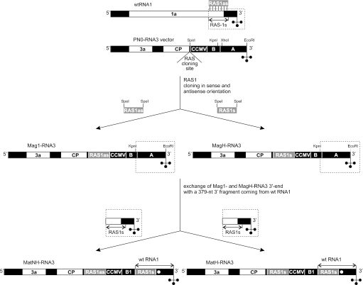
To address both issues, we decided to construct a so-called mixed system, homo- and heteromolecular at the same time. To this end two RNA3 molecules, prototypes of a new vector carrying two RASes, were prepared. To obtain them we used PN0-RNA3, described earlier, and a well-characterized recombinationally active sequence from BMV RNA1 (RAS1, see Figure 1). The 137 nt RAS1 corresponding to RNA1 between positions 2856 and 2992 was inserted into the PN0-RNA3 RAS cloning site, in antisense (RAS1as) and sense (RAS1s) orientations (Figure 2). As a result, we obtained Mag1- and MagH-RNA3 derivatives (30). Then the 356 nt portion of Mag1- and MagH-RNA3 3′ end was replaced with a 379 nt sequence representing the wtRNA1 3′ end (fragment encompassing the entire 3′-UTR and RAS1s sequence) (Figure 2). In addition, a marker mutation (called ΔXho) was introduced within the RNA1-derived fragment to make it distinguishable from an analogous region present in wtRNA1. To this end, the XhoI restriction site (2988–2994) was disrupted by a 4 nt insertion (GATC) between C-2991 and G-2992. Resultant RNA3 derivatives, called MatNH- and MatH-RNA3, have unchanged 5′-UTR, intergenic and coding regions and a highly modified 3′-UTR. The latter includes 3′-UTR coming from wtRNA1 and two RAS1 sequences (3′RAS1 and 5′RAS1) separated by a 338 nt spacer (sequence CCMV and B1). In MatNH-RNA3, 3′RAS1 is located in sense and 5′RAS1 in antisense orientation, while in MatH-RNA3 both RAS1 sequences are in sense orientation (Figure 2).
Figure 2.
Construction of MatNH- and MatH-RNA3 mutants. BMV genomic molecules are schematically presented as in Figure 1, white, black and gray boxes represent coding, noncoding and recombinationally active sequences, respectively, in sense (RAS1s) or antisense (RAS1as) orientation, dashed line squares encompass replaced parts of wtRNA1 and modified RNA3 molecules. Mag1- and MagH-RNA3 were created by inserting the RAS1 sequence from wtRNA1 into the RAS cloning site of PN0-RNA3 in antisense (Mag1-RNA3) or sense (MagH-RNA3) orientation. To construct MatNH-RNA3 and MatH-RNA3, the 356 nt very 3′ end of Mag1-RNA3 or MagH-RNA3 (between KpnI and EcoRI sites) was replaced with a 379 nt portion of the wtRNA1 3′ end (fragment containing the entire 3′-UTR and RAS1). Thus, both constructs contain two copies of RAS1 sequence—MatNH-RNA3 includes 5′RAS1as and 3′RAS1s, while MatH-RNA3 comprises 5′RAS1s and 3′RAS1s. Furthermore, a marker mutation ΔXho (marked as a white dot), removing the XhoI restriction site, was introduced into the 3′ end of MatNH- and MatH-RNA3, to make it distinguishable from an analogous region present in wtRNA1.
Having these two RNA3 derivatives, we were able to construct two variants of the mixed system: one for homologous (MatH-BMV mutant) and the other for non-homologous (MatNH-BMV mutant) recombination studies. The MatH-BMV genome is composed of wtRNA1, wtRNA2 and MatH-RNA3 and the MatNH-BMV genome of wtRNA1, wtRNA2 and MatNH-RNA3 (Table 1). In genomes of both BMV mutants three copies of RAS1 are present: two in the RNA3 derivative (RAS1s-RAS1s or RAS1as-RAS1s in MatH- and MatNH-RNA3, respectively) and one in wtRNA1 (RAS1s). Thus, in the mixed systems two identical RASes or RAS and its complementary counterpart were capable of supporting, respectively, homologous or non-homologous (heteroduplex-mediated) recombination between the same or between different BMV genomic RNAs. As a result, we could directly compare homo- and heteromolecular recombination systems in one in vivo experiment and examine whether our presumptions concerning the new recombination vector are correct.
Table 1.
Recombination activity of RAS1 in heteromolecular and mixed hetero- and homomolecular systems
| BMV mutant | Genomic RNA | Infectivitya | Number of analyzed lesions | Number of recombinants | Recombination frequencyb (%) |
|---|---|---|---|---|---|
| MH-BMV | wtRNA1 wtRNA2 MagH-RNA3 | 18 (±2) | 40 | 0 | 0 |
| M1-BMV | wtRNA1 wtRNA2 Mag1-RNA3 | 19 (±2) | 40 | 40 | 100 |
| MatH-BMV | wtRNA1 wtRNA2 MatH-RNA3 | 17 (±2) | 40 | 34 | 85 |
| MatNH-BMV | wtRNA1 wtRNA2 MatNH-RNA3 | 18 (±2) | 40 | 38 | 95 |
aInfectivity was defined as the average number of lesions per leaf.
bRecombination frequency was defined as the ratio between the number of lesions that developed recombinants and the total number of analyzed lesions.
Homologous recombination in the mixed homo–heteromolecular system
Earlier, Nagy and Bujarski (33) demonstrated that the 66 nt portion of RAS1 did not support homologous recombination in heteromolecular system. We repeated this experiment using MH-BMV mutants. Recombinants also did not form although the entire RAS1 sequence was present in wtRNA1 and MagH-RNA3 molecules (Table 1 and Figure 3). To test RAS1 activity in the mixed homologous recombination system, a previously used, well-established procedure was applied (30,33). C.quinoa plants (local lesion host for BMV) were inoculated with a mixture containing in vitro transcribed wtRNA1, wtRNA2 and MatH-RNA3. After 2 weeks, when infection symptoms were well developed, the number of lesions formed on every inoculated leaf was counted to determine the infectivity of the MatH-BMV mutant. Individual local lesions were excised and total RNA was extracted separately from each of them. Then, the 3′-portion of RNA3 progeny accumulating in examined lesions was selectively amplified by RT–PCR involving RNA3 specific primers A and B (for their location see Figure 1). Reaction products were separated in a 1.5% agarose gel and their length was determined. The formation of an ∼800 or 400–500 nt DNA fragment indicated that the lesion contained parental or recombinant RNA3, respectively. In this way, we were able to determine the number of lesions in which a viable RNA3 recombinant was generated. The presence of recombinants in analyzed lesions was confirmed by standard northern blot hybridization. DNA fragments obtained during selective RT–PCR amplification of RNA3 were cloned and sequenced. Finally, the results obtained with our new mixed homologous recombination system were compared with analogous data previously got using the heteromolecular system (33) (see Table 1 and Figure 3).
Figure 3.
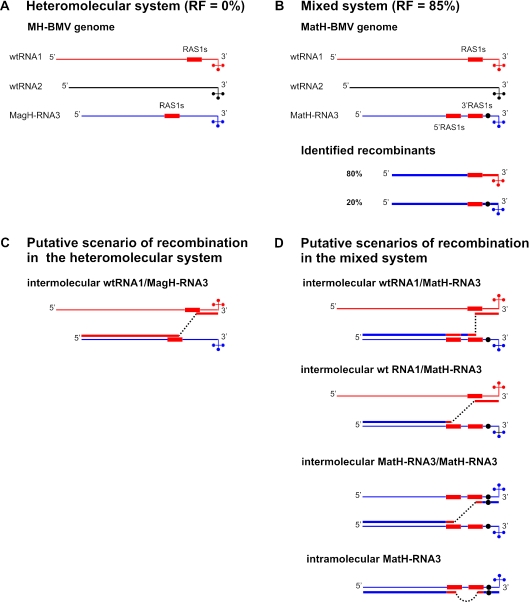
Homologous recombination in the heteromolecular and mixed homo-heteromolecular systems. Light and thicker lines represent viral genomic RNAs and nascent recombinant RNA, respectively. WtRNA1 is red (the recombinationally active sequence RAS1s which it contains is additionally boxed), wtRNA2 is black and modified RNA3 is blue. When inserted into RNA3, the wtRNA1-derived RAS1s sequence is also shown as a red box. The portion of the RNA3 recombinant synthesized on wtRNA1 is red and the fragment synthesized on RNA3 is blue. The black dot symbolizes the ΔXho mutation present in MatH-RNA3. RF, recombination frequency. (A) Heteromolecular system (MH-BMV). The presence of two RAS1s sequences in the MH-BMV mutant (one in wtRNA1 and one in MagH-RNA3) creates an opportunity for homologous recombination events between two different segments of the viral genome. However, during MH-BMV infection recombinants were not identified (RF = 0%). (B) Mixed system (MatH-BMV). Three identical RAS1s sequences are located in the MatH-BMV genome, two in MatH-RNA3 and one in wtRNA1. The recombination frequency observed during infection with MatH-BMV was 85%. Among the identified recombinants 80% did not have the ΔXho marker, and 20% had the ΔXho marker. (C) Putative scenario of RAS1-mediated homologous recombination in the heteromolecular system. BMV replicase starts nascent strand synthesis at the 3′ end of wtRNA1 is paused within the RAS1s sequence and then switches to the identical region located in MagH-RNA3. (D) Putative scenarios of RAS1-mediated homologous recombination in the mixed system. The presence of three identical RAS1s sequences in the MatH-BMV genome (3′RAS1s and 5′RAS1s sequences in MatH-RNA3 and RAS1s in wtRNA1) caused several scenarios of homologous recombination to be possible. BMV polymerase can initiate nascent strand synthesis at the 3′ end of wtRNA1 and then switch to 3′ or 5′RAS1s located in MatH-RNA3 (intermolecular wtRNA1/MatH-RNA3 crossover). Crossovers between 3′RAS1s and 5′RAS1s of two MatH-RNA3 molecules (intermolecular) and between two RAS1s sequences of the same MatH-RNA3 molecule (intramolecular) can also occur.
The data presented in Table 1 indicate that the exchange of MagH-RNA3 (carrying a single RAS1) into MatH-RNA3 (bearing two RAS1 sequences) did not affect the infectivity of the BMV mutants. The average numbers of lesions appearing on the leaves inoculated with MH- and MatH-BMV were similar: 18 and 17, respectively. Interestingly, although RAS1 did not support homologous crossovers in the heteromolecular system represented by MH-BMV, it was very active in the mixed system. About 85% of the local lesions developed during MatH-BMV infection accumulated the RNA3 recombinant instead of parental MatH-RNA3. In all of them, one RAS1 and a spacer were deleted. This indicates that crossovers occurred either within 3′ end 5′RAS1 present in MatH-RNA3 (inter- or intramolecular crossovers) or within RAS1 and 5′RAS1 located in wtRNA1 and MatH-RNA3, respectively. Recombinant junction sites were placed in identical regions; therefore, their location could not be precisely established.
The data presented till now also could not answer which molecules, exclusively MatH-RNA3 or wtRNA1 and MatH-RNA3, participated in recombination. In the heteromolecular system, only RAS1-mediated crossovers between wtRNA1 and MagH-RNA3 were permitted. The situation seems to be more complicated in the mixed homologous system where three copies of RAS1 are present, all in sense orientation: two of them in MatH-RNA3 (3′RAS1 and 5′RAS1) and one in wtRNA1. Consequently, RAS1-mediated homologous recombination may happen according to four different scenarios (Figure 3). It can engage MatH-RNA3 only and occur as intra- or intermolecular process or it can involve wtRNA1 and MatH-RNA3. In the latter case, recombination can be mediated by RAS1 present in wtRNA1 and either 5′- or 3′RAS1 located in MatH-RNA3.
To learn according to which scenario homologous recombination occurred, we checked whether mutation ΔXho introduced into MatH-RNA3 (just behind 3′RAS1) is still present in RNA3 recombinants. In this way, we were able to determine if their 3′-UTR was derived from MatH-RNA3 or wtRNA1 molecules. The undertaken analysis revealed that the mutation was present in ∼20% of recombinants. This result suggested that homologous crossovers preferentially occur between wtRNA1 and MatH-RNA3.
However, there are other explanations why ΔXho was absent in a large fraction of recombinants. It is possible that the mutation was removed either from MatH-RNA3, due to homologous recombination between its 3′RAS1 and wtRNA1, or from the RNA3 recombinant (carrying a single copy of RAS1) because it could also have recombined with wtRNA1. To examine the first possibility, progeny RNA3 extracted from the local lesions accumulating MatH-RNA3 (lesions in which recombinant was not generated) was analyzed. About 800 nt RT–PCR products obtained during selective amplification of RNA3's 3′-portion were cloned and sequenced. In all of 20 analyzed clones ΔXho was present. To test the second possibility, a full-length cDNA clone of RNA3 recombinant containing ΔXho (RNA3-ΔXhoR) was obtained. It was inserted into the pUC19 vector under the T7 polymerase promoter. The resultant plasmid named pRNA3-ΔXhoR was used after linearization to produce an infectious RNA3-ΔXhoR molecule by in vitro transcription. Then, RNA3-ΔXhoR was used together with wtRNA1 and wtRNA2 to inoculate C.quinoa plants. After 2 weeks, total RNA was extracted from individual lesions and a 3′-portion of the progeny RNA3 was amplified by RT–PCR. Obtained products were cloned and sequenced. As described previously, ΔXho was present in all of the analyzed 20 clones. These two experiments proved that homologous recombination between either 3′RAS1 of MatH-RNA3 or RAS1 present in RNA3 recombinant and wtRNA1 does not occur frequently enough to explain why most recombinants lack ΔXho. Altogether, these results supported our initial thesis that ΔXho was removed from 80% of homologous recombinants, since most of the crossovers occurred within 5′RAS1 from MatH-RNA3 and RAS1 from wtRNA1.
Non-homologous recombination in the mixed homo-heteromolecular system
Earlier we showed that RAS1 can effectively support non-homologous recombination if inserted into PN0-RNA3 in antisense orientation (30,33). The heteromolecular system used in our experiment was composed of wtRNA1, wtRNA2 and Mag1-RNA3 (M1-BMV mutant). Crossovers occurred within the local double-stranded region (local heteroduplex), which wtRNA1 and Mag1-RNA3 were capable of forming. In order to test RAS1 activity in the mixed non-homologous recombination system, C.quinoa plants were inoculated with the MatNH-BMV mutant (its genome is composed of wtRNA1, wtRNA2 and MatNH-RNA3). Two weeks later progeny RNA3 were analyzed as described above. The number of lesions developed on each leaf was counted and total RNA was extracted from individual local lesions. After RT–PCR amplification, the 3′-portion of BMV RNA3 accumulating in each lesion was analyzed in an agarose gel, cloned and sequenced. The presence of recombinants was confirmed by a standard Northern blot. The results obtained were compared with analogous data we had got using the heteromolecular system (30) (Table 1 and Figure 4).
Figure 4.
Non-homologous recombination in the heteromolecular and mixed homo-heteromolecular systems. As in Figure 3, light and thicker lines represent viral genomic RNAs and nascent recombinant RNA, respectively. WtRNA1 is red (the recombinationally active sequence RAS1s which it contains is additionally boxed), wtRNA2 is black and modified RNA3 is blue. When inserted into RNA3 the wtRNA1-derived RAS1s sequence is also shown as a red box, whereas the complementary RAS1as sequence is shown as a green box. The portion of the RNA3 recombinant synthesized on wtRNA1 is red, the portion synthesized on the RAS1as sequence is green and the fragment synthesized on RNA3 is blue. The black dot symbolizes the ΔXho mutation present in MatNH-RNA3. RF, recombination frequency. Dashed line squares encompass the region identical in both systems [the region where crossovers occur, shown in detail in (E)]. (A) Heteromolecular system (M1-BMV). A detailed description of the M1-BMV genome is presented in Figure 1. All nascent RNA3 molecules accumulating in M1-BMV infected plants were recombinants (RF = 100%). (B) Mixed system (MatNH-BMV). Three copies of RAS1 are located in the MatNH-BMV genome, two in MatNH-RNA3 (3′RAS1s and 5′RAS1as) and one in wtRNA1 (RAS1s). The recombination frequency observed during infection with MatNH-BMV was 95%. Of the identified recombinants, 10% were without the ΔXho marker, and 90% with the ΔXho marker. (C) Putative scenario of RAS1s/RAS1as-mediated non-homologous recombination in the heteromolecular system. Owing to the presence of RAS1s and RAS1as sequences in wtRNA1 and Mag1-RNA3, respectively, they are capable of forming a local double-stranded structure supporting non-homologous crossovers (for details see Figure 1). (D) Putative scenarios of RAS1s/RAS1as-mediated non-homologous recombination in the mixed system. The presence of 3′RAS1s and 5′RAS1as sequences in MatH-RNA3 and RAS1s in wtRNA1 creates several opportunities of heteroduplex formation: between wtRNA1 RAS1s and MatNH-RNA3 5′RAS1as (intermolecular), between two pairs of RAS1s/RAS1as sequences of two different MatNH-RNA3 molecules (intermolecular) and between 5′RAS1as and 3′RAS1s of the same MatNH-RNA3 molecule (intramolecular). Recombinants are generated if BMV replicase initiates nascent strand synthesis at the 3′ end of wtRNA1 or MatNH-RNA3 and then switches to MatNH-RNA3 within the local double-stranded region. (E) Recombinants identified during M1- and MatNH-BMV infection. Boxed fragments of recombining wtRNA1/Mag1-RNA3, wtRNA1/MatNH-RNA3 and MatNH-RNA3/MatNH-RNA3 molecules are practically identical in both systems (except for the ΔXho mutation present in MatNH-RNA3). The locations of the junction sites are marked with arrows and letters. The numbers indicate how many recombinants of the same type were isolated. Upper case letters refer to M1-BMV, lower case letters refer to MatNH-BMV.
As described previously, we observed that the exchange of Mag1-RNA3 (with a single RAS1as sequence) for MatNH-RNA3 (with two sequences: RAS1as and RAS1s) did not influence the infectivity of the BMV mutants. The average numbers of lesions developed on each leaf during infection with M1-BMV and MatNH-BMV were 19 and 18, respectively. There was also no difference between the recombination activity of M1-BMV and MatNH-BMV. RAS-1 (in fact RAS1s and RAS1as) supported non-homologous recombination equally in both systems. Recombination events occurred with a similar frequency (100 and 95% for M1- and MatNH-BMV, respectively) and recombinant junction sites were located within the same region of the heteroduplexes, which recombining molecules were capable of forming.
In the heteromolecular system, only one type of heteroduplex supporting non-homologous crossovers could possibly form: between wtRNA1 and Mag1-RNA3. In the mixed system, recombining molecules were capable of forming three types of heteroduplexes: (i) intermolecular, between 5′RAS1as from MatNH-RNA3 and RAS1s from wtRNA1, (ii) intermolecular, between 5′RAS1as and 3′RAS1s located in two MatHN-RNA3 molecules and (iii) intramolecular, between 5′RAS1as and 3′RAS1s located in the same MatNH-RNA3 molecule (Figure 4). To determine the molecules that participated in non-homologous recombination, the RT–PCR amplified 3′-portions of RNA3 were checked for ΔXho. It was present in 90% of recombinants. This result clearly showed that non-homologous crossovers almost always involve one (intramolecular recombination) or two (intermolecular recombination) MatNH-RNA3 molecules.
The universal BMV RNA3-based recombination vector
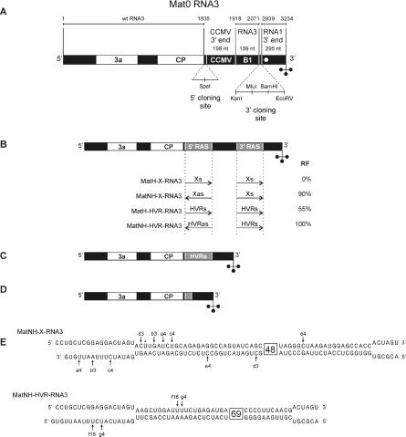
The results presented above indicated that the homomolecular system can provide new interesting data concerning the mechanism of RNA recombination, especially if it could be used for testing the recombination activity of RNA sequences derived from other RNA-based viruses. Consequently, we attempted to construct a universal BMV RNA3-based recombination vector called Mat0-RNA3 (for details see Materials and Methods and Figure 5A). In Mat0-RNA3, as in the former PN0-RNA3 vector, only 3′-UTR was modified. It is composed of the 295 nt very 3′ end of RNA1 followed by the 3′ RAS cloning site, a 338 nt spacer and the 5′ RAS cloning site.
Figure 5.
Mat0-RNA3 recombination vector. White, black and gray boxes represent coding, noncoding and recombinationally active sequences, respectively. (A) Schematic description of the Mat0-RNA3 vector. In Mat0-RNA3 only 3′-UTR was modified. It is composed of the 295 nt very 3′ end of wtRNA1 followed by the 3′RAS cloning site, a 338 nt spacer (containing a 139 nt sequence located in wtRNA3 between positions 1918 and 2071 and a 198 nt CCMV derived sequence) and the 5′RAS cloning site. (B) Preparation of Mat0-RNA3 derivatives carrying HCV-derived sequences. To test the recombination activity of HCV-derived sequences X and HVR the following Mat0-RNA3 derivatives were prepared: MatH-X-RNA3—with two copies of X in sense orientation (3′ and 5′Xs), MatNH-X-RNA3—with two copies of X located in different orientation (3′Xs and 5′Xas), MatH-HVR-RNA3—containing two copies of HVR in sense orientation (3′ and 5′HVRs) and MatNH-HVR-RNA3—with two copies of HVR, 3′-copy in sense and 5′ in antisense orientation (3′HVRs and 5′HVRas). MatH-X- and MatH-HVR-RNA3 were applied to test X's and HVR's ability to support homologous crossovers while MatNH-X- and MatNH-HVR-RNA3 were applied to examine X's and HVR's competence to induce non-homologous, heteroduplex-mediated recombination. RF, recombination frequency observed during infection involving each RNA3 derivative. (C). Homologous recombinants generated during infection involving MatH-HVR-BMV. RNA3 recombinants were only formed during MatH-HVR-BMV infection. In all of them, one recombinationally active sequence HVRs and a spacer were deleted and their 3′-UTR was composed of RNA1 derived sequence and HVRs followed by the CP coding region. Because crossovers occurred within identical regions, the location of recombinant junction sites could not be precisely established. All lesions developed on plants infected with MatH-X-BMV accumulated parental MatH-X-RNA3 molecules only. (D) Non-homologous recombinant which appeared during infection with MatNH-X- and MatNH-HVR-BMV mutants. During infection with MatNH-HVR- and MatNH-X-BMV, heteroduplex-mediated crossovers occurred with 100 and 90% frequency, respectively. Recombinant junction sites were located within the left portion of the local double-stranded region that could be formed either by Xs and Xas or by HVRs and HVRas [see (E)]. As a result, both sequences supporting non-homologous crossovers were almost entirely deleted, together with the whole spacer. (E) The distribution of non-homologous crossovers within the local double-stranded structure formed by MatNH-X-RNA3 and MatNH-HVR-RNA3 molecules. The locations of the junction sites are marked with arrows and letters. The numbers indicate how many times a given recombinant was isolated.
To determine the infectivity and stability of the new vector, C.quinoa plants were inoculated with a mixture containing wtRNA1, wtRNA2 and Mat0-RNA3 (Mat0-BMV mutant). After 2 weeks, the number of lesions developed on inoculated leaves was counted and then standard analysis of progeny RNA was carried out. Twenty separate lesions were excised, the total RNA was isolated and used for the selective RT–PCR amplification of the 3′-portion of progeny RNA3. The length of RT–PCR products was established by electrophoresis in a 1.5% agarose gel. In addition, reaction products were cloned and sequenced. This demonstrated that the Mat0-BMV mutant is infectious (usually 20 lesions were developed on each leaf, see Table 2) and Mat0-RNA3 is stable during the whole period of infection and thus it can be used as a recombination vector.
Table 2.
Recombination activity of HCV-derived sequences tested in the homomolecular systems
| BMV mutant | Genomic RNA | Infectivitya | Number of analyzed lesions | Number of recombinants | Recombination frequencyb (%) |
|---|---|---|---|---|---|
| Mat0-BMV | wtRNA1 wtRNA2 Mat0-RNA3 | 20 (±2) | 20 | 0 | 0 |
| MatH-HVR-BMV | wtRNA1 wtRNA2 MatH-HVR-RNA3 | 16 (±2) | 25 | 14 | 55 |
| MatNH-HVR-BMV | wtRNA1 wtRNA2 MatNH-HVR-RNA3 | 16 (±2) | 25 | 25 | 100 |
| MatH-X-BMV | wtRNA1 wtRNA2 MatH-RNA3 | 4 (±1) | 15 | 0 | 0 |
| MatNH-X-BMV | wtRNA1 wtRNA2 MatNH-RNA3 | 6 (±2) | 25 | 23 | 90 |
aInfectivity was defined as the average number of lesions per leaf.
bRecombination frequency was defined as the ratio between the number of lesions that developed recombinants and the total number of analyzed lesions.
Practical application of Mat0-RNA3 vector
In order to demonstrate that Mat0-RNA3 can be used as an effective tool in recombination studies, we applied it to examine the recombination activity of two specific sequences derived from the HCV genome. The first, 101 nt sequence is placed within the 5′-portion of the HCV genome (within the fragment encoding E2 protein) and is named HVR (40,42). The second, called the sequence X (X) constitutes a 98 nt 3′ end of HCV genomic RNA. It has been shown that X represents the most conservative fragment of HCV genome (43). Both sequences were obtained by a standard RT–PCR method involving viral RNA isolated from the blood of infected patients as a template (40,41). Amplified fragments were inserted into the 5′-cloning site of Mat0-RNA3 in two different orientations (sense and antisense), then only in sense orientation into the 3′-cloning site (for details see Materials and Methods). As a result, four different Mat0-RNA3 derivatives were generated: (i) MatH-HVR-RNA3, possessing two copies of HVR in sense orientation (3′ and 5′HVRs); (ii) MatH-X-RNA3, with two copies of X in sense orientation (3′ and 5′Xs); (iii) MatNH-HVR-RNA3, with two copies of HVR, the 3′-copy in sense and the 5′ in antisense orientation (3′HVRs and 5′HVRas); (iv) MatNH-X-RNA3, with two copies of X located in different orientation (3′Xs and 5′Xas) (Figure 5B). The former two were applied to test HVR's and X's ability to support homologous crossovers while the latter two to examine Xs/Xas' and HVRs/HVRas' capacity to induce non-homologous, heteroduplex-mediated recombination. Unlike previously tested mutants (MatNH- and MatH-BMV), in MatH-HVR-, MatNH-HVR-, MatH-X- and MatNH-X-BMV, the examined sequences were present only in the recombination vector. They were absent in the two other genomic RNAs, so that recombination crossovers could involve only RNA3 molecules.
To determine the recombination activity of HCV-derived sequences, C.quinoa plants were inoculated with four BMV mutants: MatH-HVR-BMV, MatNH-HVR-BMV, MatH-X-BMV and MatNH-X-BMV. Their genomes were composed of wtRNA1, wtRNA2 and one of the newly generated Mat0-RNA3 derivatives (either MatH-HVR-, MatNH-HVR-, MatH-X- or MatNH-X-RNA3) (Table 2). After 2 weeks, the standard procedure of BMV RNA3 progeny analysis was applied. The number of lesions developed during each infection was counted. The 3′-portion of progeny RNA3 was amplified by the RT–PCR method. The length of RT–PCR products was established (by electrophoresis in a 1.5% agarose gel), then they were cloned and sequenced.
We found that BMV mutants carrying HVRs/HVRs and HVRs/HVRas sequences are as infectious as Mat0-BMV; usually they developed 14–18 lesions on each inoculated leaf. The two others, MatH-X- and MatNH-X-BMV mutants, are visibly less infectious and developed 3–5 and 4–8 lesions/leaf, respectively. Despite differences in their infectivity, BMV mutants carrying 3′HVRs and 5′HVRas as well as 3′Xs and 5′Xas supported non-homologous, heteroduplex-mediated crossovers very efficiently. An RNA3 recombinant was generated in 100 and 90% of lesions developed during infection with MatNH-HVR- and MatNH-X-BMV, respectively. Recombinant junction sites were located within the left portion of the local double-stranded region that could potentially be formed either by HVRs and HVRas or by Xs and Xas. As a result, both sequences supporting non-homologous crossovers were almost entirely deleted, together with the whole spacer (Figure 5D and E).
Interestingly, homologous recombinants were generated only during infection involving MatH-HVR-BMV. Fifty-five percent of analyzed lesions contained the RNA3 recombinant. In all sequenced recombinants, one HVR and the spacer were deleted (Figure 5C). Their 3′-UTR was composed of a 295 nt RNA1-derived sequence and HVRs followed by the CP coding region. All lesions developed on plants infected with MatH-X-BMV accumulated parental MatH-X-RNA3 molecules only.
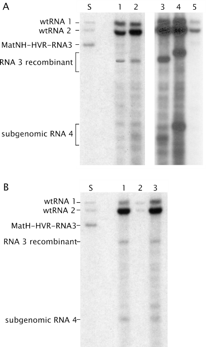
The presence of homologous and non-homologous recombinants in the examined lesions was always confirmed not only by RT–PCR but also by northern blot analysis (Figure 6). This revealed the same tendency as that observed earlier using the heteromolecular system (30,33). BMV accumulated to a very low level in lesions containing parental RNA3 (original molecules with duplicated sequences—Figure 6A, lane 5 and Figure 6B, lane 2). However, this changed in lesions where a recombinant was generated (Figure 6A, lanes 1–4 and Figure 6B, lanes 1 and 3).
Figure 6.
Northern blot analysis of the accumulation of BMV recombinants generated in the newly constructed homomolecular system. The total RNA was isolated from individual lesions, separated in a 1% agarose gel, blotted onto a nylon membrane and probed with a 32P-labeled RNA (200 nt probe complementary to the 3′-UTR shared by all BMV genomic RNAs). A mixture of the BMV RNA transcripts used for plant inoculation was applied as a molecular mass marker (lane S). (A) Non-homologous recombination. Representative data obtained for MatNH-HVR- and MatNH-X-BMV. All lesions developed on the MatNH-HVR-BMV infected plants contained one of two almost identical recombinants (lanes 1 and 2) (see Figure 5E). During infection with MatNH-X-BMV, several different recombinants were generated (lanes 3 and 4). In the 10% of lesions where a recombinant was not generated, the parental virus accumulated to a very low level (lane 5). (B) Homologous recombination. About 55% of lesions developed during MatH-HVR-BMV infection contain identical recombinants (lanes 1 and 3); the parental virus accumulated in the remaining ones (lane 2).
DISCUSSION
Recombination in the homo- and heteromolecular systems
Earlier it was shown that the BMV-based heteromolecular system can be used as an effective tool for investigating the mechanism of homologous and non-homologous recombination, although it is only suitable for testing the recombination activity of the sequences derived from the 3′-portion of BMV RNA1 or RNA2 (30,33). To overcome this problem, we attempted to create a new universal recombination in vivo system. The collected data suggested that a BMV RNA3-based homomolecular system would best fulfill our expectations. To confirm the correctness of the above presumption, to determine the efficacy of the homomolecular system and to compare it with the heteromolecular one, two mixed homo–heteromolecular systems were constructed—one to study homologous (MatH-BMV) and the other non-homologous (MatNH-BMV) recombination.
The mixed systems were prepared in such a way that two identical or two complementary sequences were capable of supporting homologous or non-homologous crossovers, respectively, either between molecules representing the same segment of the BMV genome (modified RNA3) or between molecules representing two different segments of the BMV genome (wtRNA1 and modified RNA3). Experiments involving MatH- and MatNH-BMV showed that recombination can occur both in homo- and heteromolecular systems and proved that the former should be at least as effective as the previously utilized heteromolecular one. Interestingly, the RAS1s sequence did not support homologous recombination during infection with MH-BMV (heteromolecular system) and it was quite active in the mixed system. This clearly demonstrates that not only primary and secondary structure but also the location of RAS within the viral genome affects its ability to mediate homologous crossovers. The undertaken experiments also revealed that homologous recombination occurs more often between two different RNAs (RNA1 and RNA3), while non-homologous recombination usually involves molecules representing the same segment of the BMV genome (RNA3). The obtained results constitute yet another piece of evidence that the mechanisms of homologous and non-homologous recombination are different. The same conclusion was reached by us earlier while studying the influence of specific mutations in BMV-encoded protein 2a (26,27). We identified among other the mutation in the 2a protein, which inhibits non-homologous crossovers without affecting the frequency of homologous ones.
At present, it is difficult to judge at which stage of the recombination process the observed differences occur. One can only suppose that the structural requirements of transfer of the replicase-nascent strand complex from the donor to the acceptor molecule must be different in homologous and non-homologous recombination. In the case of the former, a basic factor facilitating this process is complementarity between the acceptor and the nascent strand. Consequently, there is no necessity for the replication complex to be stable during homologous crossovers (22). Replicase can leave the donor template alone or together with a nascent strand. Then, the 3′ end of the newly synthesized RNA molecule can function as a guide; it can find a complementary sequence in the acceptor RNA, hybridize and serve as a primer allowing viral replicase to reinitiate RNA synthesis. In non-homologous heteroduplex-mediated recombination, a factor enhancing crossover seems to be the interaction between the donor and the acceptor (the formation of a local double-stranded region) (30,33). Considering that BMV genomic RNAs are copied within spherules (44), intramolecular hybridization between RAS1s and RAS1as, located in MatNH-RNA3, is much more likely. Thus, the results presented here indicate that the way the virus replicates can strongly affect the recombination process. However, further detailed studies are necessary in order to explain this phenomenon.
Recombination in the universal homomolecular system
Based on results obtained using the heteromolecular (30,33) and mixed systems, we constructed a new homomolecular one. Its most crucial element is the Mat0-RNA3 vector, into which both tested sequences can be introduced. In order to show the usefulness of this system, we employed it to test the recombination activity of two distinctly different sequences deriving from the genome of an RNA virus not related to BMV. Our choice was the 98 nt sequence X and an HVR both from HCV genome. There are many reasons as to why the two sequences can be deemed drastically different. The most important of them are (i) sequence X is placed in a non-coding region, while HVR in a coding one, (ii) sequence X is the least variable and HVR the most variable fragment of the HCV genome (42,43,45), (iii) unlike to HVR, sequence X possesses a very stable and well-defined secondary and tertiary structure (42,45).
We ascertained that the introduction of HVR into the Mat0-RNA3 vector (in sense/sense and antisense/sense orientation) does not influence BMV infectivity. The latter was, however, reduced if HVR was replaced with sequence X. We found that HVR supports homologous recombination and HVRs and its complementary counterpart HVRas mediate non-homologous crossovers. The frequency of homologous recombination amounted to 55% and of non-homologous to 100%. Sequence Xs did not support homologous crossovers but Xs and complementary sequence Xas were capable of inducing non-homologous ones (their frequency reaching 90%).
The obtained results testify that the local double-stranded structures induce non-homologous recombination crossovers very efficiently. This may reflect the capacity of RNA viruses to remove inverted repeats from their genomes. Viruses lacking such ability would be an easy target for double-stranded RNA-induced RNA silencing, which is known as the plant antiviral mechanism (46). Moreover, the data presented suggest that sequence X, which adopts a very compact and stable structure (45), is not able to mediate homologous recombination. It occurs efficiently within AU-rich HVR sequences whose structure is more labile and dynamic (42). Earlier research on homologous recombination in BMV led to similar conclusions. It was shown that homologous recombination occurs effectively in AU-rich regions (47) and is not observed within highly structured 3′- and 5′-UTR (48). These observations concur with the proposed mechanism of homologous RNA recombination (22). It assumes that AU-rich regions facilitate the detachment of the polymerase-nascent strand complex from donor RNA. On the other hand, it is thought that the stability of RNA structure makes the hybridization of the nascent strand and/or replicase to the acceptor difficult.
Currently, it is becoming increasingly clear that RNA recombination plays a very complex role in a virus' life cycle. Not only does it permit the exchange of genetic material between viruses (3,4,22), frequent homologous crossovers between molecules representing the same segment of the virus genome also stabilize genetic information (48). Moreover, here we showed that homologous and non-homologous recombination might control the organization of the virus genome by removing direct or inverted repeats, which affect the virus' ability to replicate or accumulate in the infected cells. Interestingly, we observed that complementary sequences are more effectively deleted than homologous ones. It seems that some of the latter can prevail in the viral genome probably due to their compact stable structure that prevents recombination events.
Altogether, the data presented here prove that the newly created BMV-based homomolecular recombination system can be used to examine in vivo recombination activity of various RNA sequences derived from the genomes of related or unrelated viruses. However, there are other factors which, in addition to RNA structure, can affect the course of the studied process. Specific properties of the viral replicase and the host proteins that are necessary for recombination events can be of equally great importance. Therefore, there is a need to create similar universal recombination systems in other viruses. We believe that these systems will be very helpful in finding some general rules in RNA recombination and will provide us with knowledge which is indispensable to understand how new RNA viruses or retroviruses are generated.
Acknowledgments
The authors would like to thank Wojciech Rypniewski for his advice and comments on the manuscript. This research was supported by the Polish Government through grants (6 PO5A 054 21 and 6 P04A 038 19) from the State Committee for Scientific Research (KBN). Funding to pay the Open Access publication charges for this article was also provided by KBN.
Conflict of interest statement. None declared.
REFERENCES
- 1.Hirst G.K. Genetic recombination in the Newcastle disease virus, poliovirus and influenza. Cold Spring Harbor Symp. Quant. Biol. 1962;27:303–309. doi: 10.1101/sqb.1962.027.001.028. [DOI] [PubMed] [Google Scholar]
- 2.Ledinko N. Genetic recombination in the poliovirus type 1 studies of crossovers between a normal horse serum-resistant mutant and several guanidine-resistant mutants of the same strain. Virology. 1963;20:107–119. doi: 10.1016/0042-6822(63)90145-4. [DOI] [PubMed] [Google Scholar]
- 3.Lai M.M.C. RNA recombination in animal and plant viruses. Microbiol. Rev. 1992;56:61–79. doi: 10.1128/mr.56.1.61-79.1992. [DOI] [PMC free article] [PubMed] [Google Scholar]
- 4.Alejska M., Kurzyniska-Kokorniak A., Broda M., Kierzek R., Figlerowicz M. How RNA viruses exchange their genetic material. Acta Bioch. Polon. 2001;48:391–407. [PubMed] [Google Scholar]
- 5.Suzuki Y., Gojobori T., Nakagomi O. Intergenic recombination in rotaviruses. FEBS Lett. 1998;427:183–187. doi: 10.1016/s0014-5793(98)00415-3. [DOI] [PubMed] [Google Scholar]
- 6.Plyusnin A., Kukkonen S.K.J., Plyusina A., Vapalahti O., Vaheri A. Transfection-mediated generation of functionally competent Tula hantavirus with recombinant S RNA segment. EMBO J. 2002;21:1497–1503. doi: 10.1093/emboj/21.6.1497. [DOI] [PMC free article] [PubMed] [Google Scholar]
- 7.Mindich L., Qiao X., Gottlieb P., Strassman J. Heterologous recombination in the dsRNA bacteriophage Φ6. J. Virol. 1992;66:2605–2610. doi: 10.1128/jvi.66.5.2605-2610.1992. [DOI] [PMC free article] [PubMed] [Google Scholar]
- 8.Onodera S., Qiao X., Gottlieb P., Strassman J., Frilander M., Mindich L. RNA structure and heterologous recombination in the dsRNA bacteriophage Φ6. J. Virol. 1993;67:4914–4922. doi: 10.1128/jvi.67.8.4914-4922.1993. [DOI] [PMC free article] [PubMed] [Google Scholar]
- 9.Coffin J.M. Structure, replication and recombination of retrovirus genome. Some unifying hypotheses. J. Gen. Virol. 1979;199:47–59. doi: 10.1099/0022-1317-42-1-1. [DOI] [PubMed] [Google Scholar]
- 10.Zhang J., Temin H.M. Rate and mechanism of nonhomologous recombination during single cycle of retroviral replication. Science. 1993;259:234–238. doi: 10.1126/science.8421784. [DOI] [PubMed] [Google Scholar]
- 11.Pathak V.K., Hu W.-S. ‘Might as well jump!’ Template switching by retroviral reverse transcriptase, defective genome formation and recombination. Semin. Virol. 1997;8:141–150. [Google Scholar]
- 12.Worobey M., Holmes E.C. Evolutionary aspects of recombination in RNA viruses. J. Gen. Virol. 1999;80:2535–43. doi: 10.1099/0022-1317-80-10-2535. [DOI] [PubMed] [Google Scholar]
- 13.Khatchikian D., Orlich M., Rott R. The increased pathogenicity after insertion of a 28s ribosomal RNA sequence into hemaglutinin gene of an influenza virus. Nature. 1989;340:156–157. doi: 10.1038/340156a0. [DOI] [PubMed] [Google Scholar]
- 14.Aaziz R., Tepfer M. Recombination in RNA viruses and in virus-resistant transgenic plants. J. Gen. Virol. 1999;80:1339–1346. doi: 10.1099/0022-1317-80-6-1339. [DOI] [PubMed] [Google Scholar]
- 15.Baroth M., Orlich M., Thiel H.J., Becher P. Insertion of cellular NEDD8 coding sequences in a pestivirus. Virology. 2000;278:456–466. doi: 10.1006/viro.2000.0644. [DOI] [PubMed] [Google Scholar]
- 16.Greene A.E., Alison R.F. Recombination between viral RNA and transgenic plants transcripts. Science. 1994;263:1423–1425. doi: 10.1126/science.8128222. [DOI] [PubMed] [Google Scholar]
- 17.Nagai M., Sakoda Y., Mori M., Hayashi M., Kida H., Akashi H. Insertion of a cellular sequence and RNA recombination in the structural protein coding region of cytopathogenic bovine viral diarrhea virus. J. Gen. Virol. 2003;84Pt2:447–452. doi: 10.1099/vir.0.18773-0. [DOI] [PubMed] [Google Scholar]
- 18.Figlerowicz M., Alejska M., Kurzyńska-Kokorniak A., Figlerowicz M. Genetic variability: the key problem in the prevention and therapy of RNA-based virus infections. Med. Res. Rev. 2003;23:488–518. doi: 10.1002/med.10045. [DOI] [PMC free article] [PubMed] [Google Scholar]
- 19.Jarvis T.C., Kirkegaard K. Poliovirus RNA recombination: Mechanistic studies in the absence of selection. EMBO J. 1992;11:3135–3145. doi: 10.1002/j.1460-2075.1992.tb05386.x. [DOI] [PMC free article] [PubMed] [Google Scholar]
- 20.Fu K., Baric R.S. Evidence for variable rates of recombination in the MHV genome. Virology. 1992;189:88–102. doi: 10.1016/0042-6822(92)90684-H. [DOI] [PMC free article] [PubMed] [Google Scholar]
- 21.Banner L.R., Keck J.G., Lai M.M.C. A clustering of RNA recombination sites adjacent to hyper-variable region of the peplomer gene of murine coronavirus. J. Virol. 1990;175:548–555. doi: 10.1016/0042-6822(90)90439-X. [DOI] [PMC free article] [PubMed] [Google Scholar]
- 22.Figlerowicz M., Bujarski J.J. RNA recombination in brome mosaic virus, a model plus stranded RNA virus. Acta Biochim. Polon. 1998;45:1–23. [PubMed] [Google Scholar]
- 23.Cascone P.J., Haydar T.F., Simon A.E. Sequences and structures required for recombination between virus-assisted RNAs. Science. 1993;260:801–805. doi: 10.1126/science.8484119. [DOI] [PubMed] [Google Scholar]
- 24.Zhang C., Cascone P.J., Simon A.E. Recombination between satellite and genomic RNAs of turnip crinkle virus. Virology. 1991;184:791–794. doi: 10.1016/0042-6822(91)90454-j. [DOI] [PubMed] [Google Scholar]
- 25.Panavas T., Nagy P.D. Yeast as a model host to study replication and recombination of defective interfering RNA of tomato bushy stunt virus. Virology. 2003;314:315–325. doi: 10.1016/s0042-6822(03)00436-7. [DOI] [PubMed] [Google Scholar]
- 26.Figlerowicz M., Nagy P.D., Bujarski J.J. A mutation in a putative RNA polymerase gene inhibits nonhomologous, but not homologous, genetic recombination in RNA virus. Proc. Natl Acad. Sci. USA. 1997;94:2073–2078. doi: 10.1073/pnas.94.5.2073. [DOI] [PMC free article] [PubMed] [Google Scholar]
- 27.Figlerowicz M., Nagy P.D., Bujarski J.J. Mutation in the N-terminus of the brome mosaic virus polymerase affect genetic RNA–RNA recombination. J. Virol. 1998;72:9192–9200. doi: 10.1128/jvi.72.11.9192-9200.1998. [DOI] [PMC free article] [PubMed] [Google Scholar]
- 28.Nagy P.D., Bujarski J.J. Efficient system of homologous RNA recombination in brome mosaic virus: sequence and structure requirements and accuracy of crossovers. J. Virol. 1995;69:131–140. doi: 10.1128/jvi.69.1.131-140.1995. [DOI] [PMC free article] [PubMed] [Google Scholar]
- 29.Nagy P.D., Zhang C., Simon A.E. Dissecting RNA recombination in vitro: role of RNA sequences and the viral replicase. EMBO J. 1998;17:2392–2403. doi: 10.1093/emboj/17.8.2392. [DOI] [PMC free article] [PubMed] [Google Scholar]
- 30.Figlerowicz M. Role of RNA structure in heteroduplex-mediated and site-specific nonhomologous recombination in brome mosaic virus. Nucleic Acids Res. 2000;28:1714–1723. doi: 10.1093/nar/28.8.1714. [DOI] [PMC free article] [PubMed] [Google Scholar]
- 31.Shapka N., Nagy P.D. The AU-rich RNA recombination hot spot sequence of brome mosaic virus is functional in tombusviruses: implications for the mechanism of RNA recombination. J. Virol. 2004;78:2288–2300. doi: 10.1128/JVI.78.5.2288-2300.2004. [DOI] [PMC free article] [PubMed] [Google Scholar]
- 32.Romanova L.I., Blinov V.M., Tolskaya E.A., Viktorova E.G., Kolesnikova M.S., Guseva E.A., Agol V.I. The primary structure of crossovers regions of intertypic poliovirus recombinants: a model recombination between RNA genomes. Virology. 1986;155:202–213. doi: 10.1016/0042-6822(86)90180-7. [DOI] [PubMed] [Google Scholar]
- 33.Nagy P.D., Bujarski J.J. Targeting the site of RNA–RNA recombination in brome mosaic virus with antisense sequences. Proc. Natl Acad. Sci. USA. 1993;90:6390–6394. doi: 10.1073/pnas.90.14.6390. [DOI] [PMC free article] [PubMed] [Google Scholar]
- 34.Figlerowicz M., Bibiłło A. RNA motifs mediating in vivo site-specific nonhomologous recombination in (+)RNA virus enforce in vitro nonhomologous crossovers with HIV-1 reverse transcriptase. RNA. 2000;6:339–351. doi: 10.1017/s1355838200991210. [DOI] [PMC free article] [PubMed] [Google Scholar]
- 35.Havelda Z., Dalmay T., Burgyan J. Secondary structure-dependent evolution of Cymbidium ringspot virus defective interfering RNA. J. Gen. Virol. 1997;78:1227–1234. doi: 10.1099/0022-1317-78-6-1227. [DOI] [PubMed] [Google Scholar]
- 36.Carpenter C.D., Oh J.-W., Zhang C., Simon A.E. Involvement of stem–loop structure in the location of junction sites in viral RNA recombination. J. Mol. Biol. 1995;245:608–622. doi: 10.1006/jmbi.1994.0050. [DOI] [PubMed] [Google Scholar]
- 37.Bruyere A., Wantroba M., Flasinski S., Dzianott A., Bujarski J.J. Frequent homologous recombination events between molecules of one RNA component in a multipartite RNA virus. J. Virol. 2000;74:4214–4219. doi: 10.1128/jvi.74.9.4214-4219.2000. [DOI] [PMC free article] [PubMed] [Google Scholar]
- 38.Ahlquist P. Bromovirus RNA replication and transcription. Curr. Opin. Genet. Dev. 1992;2:71–76. doi: 10.1016/s0959-437x(05)80325-9. [DOI] [PubMed] [Google Scholar]
- 39.Olsthoorn R.C.L., Bruyere A., Dzianott A., Bujarski J.J. RNA recombination in brome mosaic virus: effects of strand-specific stem–loop inserts. J. Virol. 2002;76:12654–12662. doi: 10.1128/JVI.76.24.12654-12662.2002. [DOI] [PMC free article] [PubMed] [Google Scholar]
- 40.Mizokami M., Ohno T. Methods in Molecular Medicine, Vol. 19: Hepatitis C Protocols. Humana Press; 1998. Determination of HCV quasispecies by cloning and sequencing; pp. 207–211. [DOI] [PubMed] [Google Scholar]
- 41.Aizaki H., Aoki Y., Harada T., Ishii K., Suzuki T., Nagamori S., Toda G., Matsura T., Miyamura T. Full-length complementary DNA of hepatitis C virus genome from an infectious blood sample. Hepatology. 1998;27:621–627. doi: 10.1002/hep.510270242. [DOI] [PubMed] [Google Scholar]
- 42.Tanaka M., Yokosuka O., Omata M. Structural and functional characterization of the hypervariable region in the HCV genome. Nippon Rinsho. 2004;62:43–47. [PubMed] [Google Scholar]
- 43.Kolykhalov A.A., Feinstone S.M., Rice C.M. Identification of a highly conserved sequence element at the 3′ terminus of hepatitis C virus genome RNA. J. Virol. 1996;70:3363–3371. doi: 10.1128/jvi.70.6.3363-3371.1996. [DOI] [PMC free article] [PubMed] [Google Scholar]
- 44.Schwartz M., Chen J., Janda M., Sullivan M., den Boon J., Ahlquist P. A positive-strand RNA virus replication complex parallels form and function of retrovirus capsids. Mol. Cell. 2002;9:505–514. doi: 10.1016/s1097-2765(02)00474-4. [DOI] [PubMed] [Google Scholar]
- 45.Blight K.J., Rice C.M. Secondary structure determination of the conserved 98-base sequence at the 3′ terminus of hepatitis C virus genome RNA. J. Virol. 1997;71:7345–7352. doi: 10.1128/jvi.71.10.7345-7352.1997. [DOI] [PMC free article] [PubMed] [Google Scholar]
- 46.Voinnet O. RNA silencing as a plant immune system against viruses. Trends Genet. 2001;17:449. doi: 10.1016/s0168-9525(01)02367-8. [DOI] [PubMed] [Google Scholar]
- 47.Nagy P.D., Bujarski J.J. Engineering of homologous recombination hotspots with AU-rich sequences in brome mosaic virus. J. Virol. 1997;71:3799–3810. doi: 10.1128/jvi.71.5.3799-3810.1997. [DOI] [PMC free article] [PubMed] [Google Scholar]
- 48.Urbanowicz A., Alejska M., Formanowicz P., Błażewicz J., Figlerowicz M., Bujarski J.J. Frequent homologous crossovers among molecules of brome mosaic bromovirus RNA1 or RNA2 segments in vivo. J. Virol. 2005;79:5732–5742. doi: 10.1128/JVI.79.9.5732-5742.2005. [DOI] [PMC free article] [PubMed] [Google Scholar]


