

Abstract
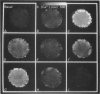
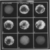
1. Changes in intracellular ionized calcium [Ca2+]i induced by human growth hormone releasing factor (hGRF) were analysed by quantitative fluorescent microscopy using a dual-wavelength, ratiometric video imaging system and low light level charge-coupled device (CCD) camera visualizing Fura-2 in dispersed male rat anterior pituitary cells. 2. In cells responding to hGRF, spontaneous basal oscillations in [Ca2+]i were frequently observed, and these were usually characterized by a gradient of [Ca2+]i localized in the subplasmalemmal region of the cell. 3. Of the cells which responded to hGRF, the peptide evoked a rise in [Ca2+]i, especially in the region of the subplasmalemma. Continuous application of 10 nM-hGRF produced several different temporal patterns of the [Ca2+]i response which were not attributable to spatial response profiles. A sustained rise in [Ca2+]i was the most common type of response to hGRF (44% of the cells examined). 4. One-third of the cells responding to 10 nM-hGRF showed spontaneous basal [Ca2+]i oscillations ranging from 100 to 500 nM. Mean values of basal and 10 nM-hGRF-induced [Ca2+]i of these cells were 81 +/- 11 nM (mean +/- S.E.M., n = 27) and 560 +/- 47 nM (n = 27) respectively. There was no significant correlation between basal [Ca2+]i and the hGRF-induced [Ca2+]i increase, nor was there any consistent correlation with regard to the spatial response profile. 5. Application of 2 mM-Co2+ abolished the hGRF-induced rise in [Ca2+]i. Quantitative analysis of this effect, performed by comparing the mean [Ca2+]i evoked during the application of hGRF with and without Co2+, respectively, also showed significant inhibition of the hGRF-induced rise in [Ca2+]i by the application of Co2+ (P less than 0.001). 6. The hGRF-induced rise in [Ca2+]i was completely suppressed by replacing extracellular Na+ with impermeant molecules such as mannitol. The onset and offset of suppression was as rapid as that induced by Co2+. Quantitative analysis showed significant inhibition of the hGRF-induced rise in [Ca2+]i by Na+ replacement (P less than 0.01). 7. Tetrodotoxin, a potent blocker of voltage-sensitive Na+ channels (5 and 20 microM), did not affect the hGRF-induced rise in [Ca2+]i. 8. Extracellular application of the membrane permeable dibutyryl cyclic AMP (DBcAMP) to elevate intracellular levels of cyclic AMP caused a large rise in [Ca2+]i, which was dependent on extracellular Na+ and was abolished by 2 mM-Co2+ applied in the bath.(ABSTRACT TRUNCATED AT 400 WORDS)
Full text
PDF


Images in this article
Selected References
These references are in PubMed. This may not be the complete list of references from this article.
- Akerman S. N., Zorec R., Cheek T. R., Moreton R. B., Berridge M. J., Mason W. T. Fura-2 imaging of thyrotropin-releasing hormone and dopamine effects on calcium homeostasis of bovine lactotrophs. Endocrinology. 1991 Jul;129(1):475–488. doi: 10.1210/endo-129-1-475. [DOI] [PubMed] [Google Scholar]
- Aronson P. S. Kinetic properties of the plasma membrane Na+-H+ exchanger. Annu Rev Physiol. 1985;47:545–560. doi: 10.1146/annurev.ph.47.030185.002553. [DOI] [PubMed] [Google Scholar]
- Ashcroft F. M., Harrison D. E., Ashcroft S. J. Glucose induces closure of single potassium channels in isolated rat pancreatic beta-cells. 1984 Nov 29-Dec 5Nature. 312(5993):446–448. doi: 10.1038/312446a0. [DOI] [PubMed] [Google Scholar]
- Baker P. F., Hodgkin A. L., Ridgway E. B. Depolarization and calcium entry in squid giant axons. J Physiol. 1971 Nov;218(3):709–755. doi: 10.1113/jphysiol.1971.sp009641. [DOI] [PMC free article] [PubMed] [Google Scholar]
- Bilezikjian L. M., Vale W. W. Stimulation of adenosine 3',5'-monophosphate production by growth hormone-releasing factor and its inhibition by somatostatin in anterior pituitary cells in vitro. Endocrinology. 1983 Nov;113(5):1726–1731. doi: 10.1210/endo-113-5-1726. [DOI] [PubMed] [Google Scholar]
- Cheek T. R., Jackson T. R., O'Sullivan A. J., Moreton R. B., Berridge M. J., Burgoyne R. D. Simultaneous measurements of cytosolic calcium and secretion in single bovine adrenal chromaffin cells by fluorescent imaging of fura-2 in cocultured cells. J Cell Biol. 1989 Sep;109(3):1219–1227. doi: 10.1083/jcb.109.3.1219. [DOI] [PMC free article] [PubMed] [Google Scholar]
- DiPolo R., Beaugé L. Ca2+ transport in nerve fibers. Biochim Biophys Acta. 1988 Oct 11;947(3):549–569. doi: 10.1016/0304-4157(88)90007-x. [DOI] [PubMed] [Google Scholar]
- Grynkiewicz G., Poenie M., Tsien R. Y. A new generation of Ca2+ indicators with greatly improved fluorescence properties. J Biol Chem. 1985 Mar 25;260(6):3440–3450. [PubMed] [Google Scholar]
- Israel J. M., Denef C., Vincent J. D. Electrophysiological properties of normal somatotrophs in culture. An intracellular study. Neuroendocrinology. 1983 Sep;37(3):193–199. doi: 10.1159/000123542. [DOI] [PubMed] [Google Scholar]
- Kato M., Hattori M. A., Suzuki M. Inhibition by extracellular Na+ replacement of GRF-induced GH secretion from rat pituitary cells. Am J Physiol. 1988 Apr;254(4 Pt 1):E476–E481. doi: 10.1152/ajpendo.1988.254.4.E476. [DOI] [PubMed] [Google Scholar]
- Kato M., Suzuki M. Effect of Li+ substitution for extracellular Na+ on GRF-induced GH secretion from rat pituitary cells. Am J Physiol. 1989 Apr;256(4 Pt 1):C712–C718. doi: 10.1152/ajpcell.1989.256.4.C712. [DOI] [PubMed] [Google Scholar]
- Kato M., Suzuki M. Growth hormone releasing factor depolarizes rat pituitary cells in Na+-dependent mechanism. Brain Res. 1989 Jan 2;476(1):145–148. doi: 10.1016/0006-8993(89)91547-3. [DOI] [PubMed] [Google Scholar]
- Kato M., Suzuki M. The time course and extracellular Ca2+ involvement of growth hormone (GH) releasing factor-induced GH secretion in perifused dispersed rat pituitary cells. Jpn J Physiol. 1986;36(6):1225–1239. doi: 10.2170/jjphysiol.36.1225. [DOI] [PubMed] [Google Scholar]
- Mason W. T., Rawlings S. R. Whole-cell recordings of ionic currents in bovine somatotrophs and their involvement in growth hormone secretion. J Physiol. 1988 Nov;405:577–593. doi: 10.1113/jphysiol.1988.sp017349. [DOI] [PMC free article] [PubMed] [Google Scholar]
- Mason W. T., Sikdar S. K., Zorec R., Akerman S., Rawlings S. R., Cheek T., Moreton R., Berridge M. Ion channels, intracellular calcium, and exocytosis: control of hormone secretion in cultured bovine pituitary lactotrophs. Soc Gen Physiol Ser. 1989;44:225–238. [PubMed] [Google Scholar]
- Nakamura T., Gold G. H. A cyclic nucleotide-gated conductance in olfactory receptor cilia. 1987 Jan 29-Feb 4Nature. 325(6103):442–444. doi: 10.1038/325442a0. [DOI] [PubMed] [Google Scholar]
- Neylon C. B., Hoyland J., Mason W. T., Irvine R. F. Spatial dynamics of intracellular calcium in agonist-stimulated vascular smooth muscle cells. Am J Physiol. 1990 Oct;259(4 Pt 1):C675–C686. doi: 10.1152/ajpcell.1990.259.4.C675. [DOI] [PubMed] [Google Scholar]
- Ozawa S., Sand O. Electrophysiology of excitable endocrine cells. Physiol Rev. 1986 Oct;66(4):887–952. doi: 10.1152/physrev.1986.66.4.887. [DOI] [PubMed] [Google Scholar]
- Schlegel W., Winiger B. P., Mollard P., Vacher P., Wuarin F., Zahnd G. R., Wollheim C. B., Dufy B. Oscillations of cytosolic Ca2+ in pituitary cells due to action potentials. Nature. 1987 Oct 22;329(6141):719–721. doi: 10.1038/329719a0. [DOI] [PubMed] [Google Scholar]
- Schuldiner S., Rozengurt E. Na+/H+ antiport in Swiss 3T3 cells: mitogenic stimulation leads to cytoplasmic alkalinization. Proc Natl Acad Sci U S A. 1982 Dec;79(24):7778–7782. doi: 10.1073/pnas.79.24.7778. [DOI] [PMC free article] [PubMed] [Google Scholar]
- Swandulla D., Lux H. D. Changes in ionic conductances induced by cAMP in Helix neurons. Brain Res. 1984 Jul 2;305(1):115–122. doi: 10.1016/0006-8993(84)91126-0. [DOI] [PubMed] [Google Scholar]
- Thorner M. O., Holl R. W., Leong D. A. The somatotrope: an endocrine cell with functional calcium transients. J Exp Biol. 1988 Sep;139:169–179. doi: 10.1242/jeb.139.1.169. [DOI] [PubMed] [Google Scholar]