


Introduction
Anomalous origin of the coronary artery from the pulmonary artery is a rare anomaly characterized by the left or right coronary artery arising from the pulmonary artery instead of the aortic sinuses. Its incidence in live‐born infants is 1 in 300 000, accounting for 0.25%–0.5% of all congenital heart diseases. 1 Among these, anomalous origin of the left coronary artery from the pulmonary artery (ALCAPA) is more common and is classified into infantile and adult forms. 2 The diagnosis of ALCAPA primarily relies on various imaging modalities such as echocardiography, CT angiography and coronary angiography. 3 , 4 Here, we have presented a unique case of ALCAPA in an adult patient who presented with PH as an initial symptom.
Case report
A 21‐year‐old male was referred to our pulmonary hypertension (PH) centre because of progressive dyspnoea on exertion, and PH detected on echocardiography. He reported a decrease in exercise tolerance 2 years after a cold, followed by multiple episodes of syncope during exertion. Two months prior, his symptoms had worsened after a pulmonary infection.
Upon arrival, the patient was afebrile with a heart rate of 78 b.p.m., blood pressure of 116/72 mmHg and oxygen saturation of 98%. Physical examination revealed a loud P2 and 3/6 systolic murmur heard at the apex. B‐type natriuretic peptide was 778.5 pg/mL. The electrocardiogram revealed sinus rhythm and chest radiography showed no obvious abnormalities. The patient denies any significant past medical history.
Echocardiography revealed an anomalous origin of the left coronary artery (Figure S1) and significant mitral regurgitation on colour Doppler imaging (Video S1). Mild tricuspid regurgitation was also observed at a velocity of 4.2 m/s, and the estimated pulmonary arterial systolic pressure was 80 mmHg. He underwent right heart catheterization, and the result was consistent with combined post‐ and pre‐capillary PH (Table 1).
Table 1.
Haemodynamic data from right heart catheterization
| Right atrial pressure, mmHg | 3 |
| Systolic pulmonary artery pressure, mmHg | 74 |
| Diastolic pulmonary artery pressure, mmHg | 31 |
| Mean pulmonary artery pressure, mmHg | 45 |
| Pulmonary capillary wedge pressure, mmHg | 28 |
| Cardiac index, L/min/m2 | 2.54 |
| Pulmonary vascular resistance, Wood Units | 4.3 |
| Qp/Qs | 1.07 |
Qp/Qs, flow ratio between the pulmonary and systemic circulation.
Coronary artery computed tomography (CT) angiography revealed an anomalous origin of the left main coronary artery from the right wall of the main pulmonary artery (Figure 1) with left atrial and ventricular enlargement. Coronary angiography revealed a rich collateral circulation between the right and left coronary arteries (Figure 2; Video S2). Contrast injection into the right coronary artery resulted in retrograde opacification of the left coronary artery (Video S3).
Figure 1.
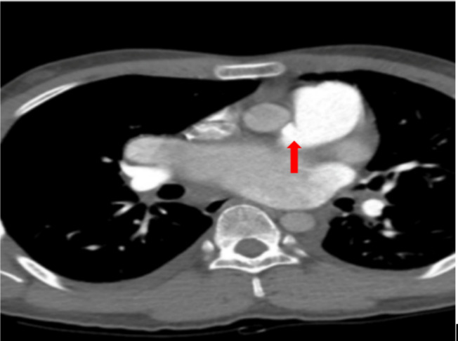
Coronary artery computed tomography angiography. The image shows an anomalous origin of the left main coronary artery from the right wall of the main pulmonary artery (highlighted by the red arrow).
Figure 2.
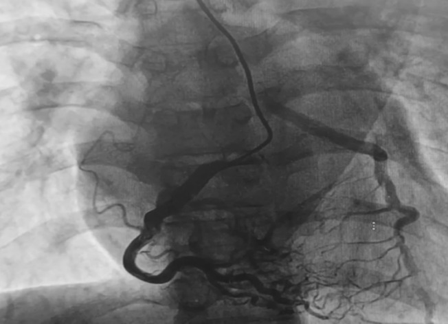
Coronary angiography. The image shows rich collateral circulation between the right and left coronary arteries.
Diagnosis and management
In patients presenting with unexplained dyspnoea and significantly elevated peak tricuspid regurgitation velocity, which raises suspicion of PH, the initial evaluation should focus on left heart disease (Group 2) and lung disease or hypoxia (Group 3), followed by the exclusion of pulmonary artery obstructions (Group 4), as these are the predominant causes of PH. Additionally, differential diagnosis with pulmonary arterial hypertension (PAH) is required. In this case, echocardiography indicated left heart disease as the likely cause. To systematically rule out other potential aetiologies, we collected a detailed medical history and conducted a series of tests. First, the patient had no history of smoking or chronic cough. Arterial blood gas analysis upon admission showed normal partial pressure of oxygen and oxygen saturation. Chest CT scans revealed no significant lung abnormalities. Additionally, polysomnography showed an AHI of 1 with no nocturnal hypoxemia, effectively ruling out hypoxia or lung disease (Group 3). Second, CT pulmonary angiography did not reveal any signs of pulmonary artery thrombosis, and ventilatory perfusion imaging showed no segmental or global perfusion defects. This effectively ruled out the possibility of Group 4 PH. Third, based on the patient's history and laboratory results, we excluded autoimmune diseases (immunological marker results are shown in Table S1 ), haematological disorders, HIV infection, portal hypertension, drug/toxin exposure and heritable PAH. Therefore, the patient does not appear to belong to Group 1 PH. Finally, right heart catheterization confirmed combined post‐ and pre‐capillary PH. Further evaluation with coronary artery CT and angiography confirmed ALCAPA. Therefore, the diagnosis was Group 2 PH secondary to ALCAPA.
After surgical consultation, the patient underwent coronary artery re‐implantation and mitral valve repair. Postoperatively, spironolactone, furosemide and potassium citrate were prescribed to improve heart failure symptoms. Four days postoperatively, a follow‐up ultrasound examination revealed a normal mitral valve function with normal leaflet motion. Doppler imaging revealed normal diastolic blood flow velocity across the mitral valve, with mild‐to‐moderate regurgitation during systole and mild regurgitation across the tricuspid valve. The diameter of the left atrium significantly decreased on postoperative ultrasound compared to preoperative measurements (pre: 52 mm vs. post: 30 mm). The estimated systolic pressure in the pulmonary artery was 54 mmHg. Three‐dimensional imaging revealed the corrected abnormal origin in the left coronary artery (Figure 3). The patient was discharged 1 week later and subsequently had regular telephone follow‐ups over 3 years, reporting significant improvement in exercise tolerance, no recurrent symptoms and an overall good condition. The timeline of significant events is summarized in Table S2.
Figure 3.

Three‐dimensional cardiac imaging. Imaging demonstrates successful correction of the origin of the left coronary artery.
Discussion
Anomalous origin of the coronary artery from the pulmonary artery is a rare coronary artery anomaly first reported in 1865 by Krause. 5 It manifests as the left or right coronary artery arising from the pulmonary artery instead of the corresponding left or right coronary sinus of the aorta. The aetiology may involve abnormal separation of the aorta and pulmonary artery during fetal development or the persistence of pulmonary buds that connect with the forming coronary artery. 6 ALCAPA is more common than anomalous right coronary artery from the pulmonary artery and typically classified into infantile and adult types.
During the fetal and neonatal periods, the pulmonary artery pressure equals systemic pressure, allowing the left coronary artery to be perfused by antegrade flow from the pulmonary artery. However, after birth, as pulmonary vascular resistance decreases and the ductus arteriosus closes, pulmonary artery pressure gradually falls, leading to a reversal of blood flow in the left coronary artery. In infantile‐type ALCAPA patients, poor development of collateral vessels between coronary arteries leads to myocardial ischaemia, myocardial infarction and congestive heart failure due to the progressive decline in pulmonary circulation pressure post‐birth and the resulting coronary steal phenomenon from the left coronary artery. Without timely treatment, the mortality rate for ALCAPA infants is as high as 90% within the first year of life. 7
Adult‐type ALCAPA is less common and characterized by the presence of extensive collateral vessels between the left and right coronary arteries. Retrograde perfusion of the left coronary artery with arterial blood from the right coronary artery allows these patients to survive into adulthood. In infantile‐type ALCAPA patients, mitral regurgitation is very common due to papillary muscle dysfunction and annular dilation caused by left ventricular wall ischaemia. 8 Similarly, in adult‐type ALCAPA patients, collateral blood flow may not fully meet the oxygen and energy demands of the left heart, particularly the subendocardial myocardium. This chronic ischaemia places adult patients at higher risk for mitral valve insufficiency, ischaemic cardiomyopathy, malignant arrhythmias and sudden cardiac death. In this case, mitral valve prolapse with severe regurgitation due to chronic myocardial ischaemia was the primary cause of the patient's pulmonary hypertension.
Adult‐type ALCAPA with PH has been reported in a few cases (Table 2). 8 , 9 , 10 , 11 , 12 , 13 , 14 , 15 These cases illustrate that the causes of PH in patients with ALCAPA can vary, including mitral regurgitation, mitral stenosis and myocardial ischaemia. In line with other reports, this case falls under Group 2 PH, which is PH due to left heart disease. 16 Specifically, right heart catheterization provided detailed haemodynamic data, revealing pulmonary vascular remodelling and elevated pulmonary vascular resistance, leading to the diagnosis of combined post‐ and pre‐capillary PH.
Table 2.
Literature review of cases of ALCAPA combined with PH
| Case [reference] | Age | Sex | Aetiology of PH | Degree of mitral regurgitation | Treatment | Prognosis |
|---|---|---|---|---|---|---|
| 19 | 47 | F | Unknown | Unknown | Bypass surgery | Little to no objective improvement at the 3‐ and 6‐month follow‐ups |
| 210 | 47 | F | Mitral regurgitation | Significant | Bypass surgery and mitral valve replacement | LVEDD decreased at the 3‐year follow‐up |
| 38 | 45 | F | Mitral regurgitation | Significant | Ligation of the anomalous origin of the LCA and and mitral valve replacement | Improved symptoms at 3‐month follow‐up |
| 411 | 20 | F | Rheumatic mitral stenosis | ‐ | Bypass surgery and open commissurotomy | Reduction in estimated mPAP postoperatively |
| 512 | 70 | F | Rheumatic mitral stenosis | ‐ | Medication only | Stable symptoms at 6‐month follow‐up |
| 613 | 26 | M | Ischaemic cardiomyopathy | Unknown | Bypass surgery | Improved symptoms and PH at 1‐year follow‐up |
| 714 | 36 | F | Myocardial infarction | Mild | Surgery | LVEF significantly improved at 6‐month follow‐up |
| 815 | 19 | F | Cardiomyopathy | Mild | Coronary artery reimplantation | Marked decrease in RAP and mPAP at 10‐month follow‐up |
ALCAPA, anomalous left coronary artery originating from the pulmonary artery; LVEDD, left ventricular end‐diastolic diameter; LVEF, left ventricular ejection fraction; mPAP, mean pulmonary artery pressure; PH, pulmonary hypertension; RAP, right atrial pressure.
Patients with left heart disease who develop PH have a worse prognosis; however, the use of PAH‐specific drug therapy in patients with PH‐LHD who exhibit elevated PVR remains a contentious issue. This is because such therapies may have variable and potentially detrimental effects in these patients. Moreover, the evidence supporting the use of PAH‐specific drugs in patients with group 2 PH is limited and conflicting. Considering these factors, we did not employ PAH‐specific drug therapy post‐surgery. Instead, we opted for treatment with spironolactone, furosemide and potassium citrate. Current guidelines recommend that PH‐LHD treatment should primarily focus on managing the underlying left heart disease, with targeted therapy not recommended. 16 Therefore, in this case, the initial consideration was the treatment of primary left heart disease, ALCAPA and mitral valve disease.
The preferred treatment for ALCAPA is surgical correction to reconstruct the coronary artery circulation. In most centres, the favoured approach is either coronary artery reimplantation or bypass surgery. 17 Although many patients with ALCAPAs have concomitant mitral valve regurgitation, whether to repair the mitral valve simultaneously during ALCAPA correction is controversial. Most centres suggest that routine repair of mitral valve regurgitation during ALCAPA correction surgery is not necessary for several reasons: (1) mitral valve regurgitation secondary to left ventricular dilation or papillary muscle dysfunction often improves with restoration of myocardial perfusion, and most patients do not require additional mitral valve repair surgery; (2) patients with ALCAPA are mostly infants and young children, and mitral valve repair in this population prolongs the surgical duration, increases complexity, and poses higher operative risks; and (3) many patients with ALCAPA have impaired left ventricular function, further increasing the surgical risks. 18 , 19
However, some centres are open to considering repair of severe mitral valve regurgitation to improve early postoperative cardiac output and to reduce the risk of mitral valve reintervention. 20 , 21 In this case, significant mitral valve regurgitation resulting from mitral valve prolapse caused severe postcapillary PH. Therefore, simultaneous ALCAPA correction surgery and mitral valve repair were performed after diagnosis. The postoperative recovery was uneventful, with normalization of the left atrial size, reduction of tricuspid regurgitation, decreased estimated pulmonary artery systolic pressure and restoration of mitral valve function to near‐normal levels observed during follow‐up. This highlights the importance of actively addressing mitral valve disease in such cases for patient improvement.
Conclusions
ALCAPA is a rare cause of PH. Imaging studies should be optimized to screen for potential causes of PH, and right heart catheterization is necessary for haemodynamic classification. Patients with ALCAPA should undergo early surgical intervention after the diagnosis is confirmed. For patients with concurrent mitral valve disease causing haemodynamic compromise, mitral valve repair surgery may be performed concomitantly with aortic reimplantation to maximize the improvement in symptoms and pulmonary haemodynamics.
Funding
This work was supported by Beijing Municipal Science and Technology Project (Z181100001718200) and the Capital's Funds for Health Improvement and Research (CFH) (2020‐2‐4033, 2020‐4‐4035).
Conflict of interest
None declared.
Supporting information
Figure S1. Echocardiography. The image shows an anomalous origin of the left coronary artery from pulmonary artery (highlighted by the red arrow).
Table S1. Summary of Immunological Marker Test Results.
Table S2. Summary of important timely arranged problems.
Video S1. Mitral Regurgitation on Color Doppler Imaging. Color Doppler imaging demonstrates significant mitral regurgitation.
Video S2. Collateral Circulation on Coronary Angiography. Coronary angiography reveals rich collateral circulation between the right and left coronary arteries.
Video S3. Retrograde Opacification of the Left Coronary Artery. Contrast injection into the right coronary artery results in retrograde opacification of the left coronary artery.
Duan, A. , Huang, Z. , Zhao, Z. , Zhao, Q. , Luo, Q. , and Liu, Z. (2025) Pulmonary hypertension associated with anomalous left coronary artery originating from the pulmonary artery. ESC Heart Failure, 12: 717–722. 10.1002/ehf2.15094.
Anqi Duan and Zhihua Huang contributed equally to this work and share first authorship.
[Correction added on 24 January 2025, after first online publication: The article category has been changed from ‘Clinical Correspondence’ to ‘Clinical Correspondences’ in this version.]
Contributor Information
Qin Luo, Email: luoqin2009@163.com.
Zhihong Liu, Email: zhihongliufuwai@163.com.
References
- 1. Oncel G, Oncel D. Anomalous origin of the left coronary artery from the pulmonary artery: diagnosis with CT angiography. J Clin Imaging Sci 2013. Jan;30:4. doi: 10.4103/2156-7514.106618 [DOI] [PMC free article] [PubMed] [Google Scholar]
- 2. Goo HW. Anomalous origin of the coronary artery from the pulmonary artery in children and adults: a pictorial review of cardiac imaging findings. Korean J Radiol 2021;22:1441‐1450. doi: 10.3348/kjr.2021.0034 [DOI] [PMC free article] [PubMed] [Google Scholar]
- 3. Peña E, Nguyen ET, Merchant N, Dennie C. ALCAPA syndrome: not just a pediatric disease. Radiographics 2009;29:553‐565. doi: 10.1148/rg.292085059 [DOI] [PubMed] [Google Scholar]
- 4. Gentile G, Caruso S. Adult‐type anomalous origin of the left coronary artery from the pulmonary artery. Radiology 2021;300:287. doi: 10.1148/radiol.2021204401 [DOI] [PubMed] [Google Scholar]
- 5. Krause W. Cber den Ursprungeinerakzessorischen A. coronariaaus der A. pulmonalis, Ztschr. Rat Med 1865;24:225‐227. [Google Scholar]
- 6. Blickenstaff EA, Smith SD, Cetta F, Connolly HM, Majdalany DS. Anomalous left coronary artery from the pulmonary artery: how to diagnose and treat. J Pers Med 2023. Oct 31;13:1561. doi: 10.3390/jpm13111561 [DOI] [PMC free article] [PubMed] [Google Scholar]
- 7. Kubota H, Endo H, Ishii H, Tsuchiya H, Inaba Y, Terakawa K, et al. Adult ALCAPA: from histological picture to clinical features. J Cardiothorac Surg 2020. Jan 13;15:14. doi: 10.1186/s13019-020-1048-y [DOI] [PMC free article] [PubMed] [Google Scholar]
- 8. Barbetakis N, Efstathiou A, Efstathiou N, Papagiannopoulou P, Soulountsi V, Fessatidis I. A long‐term survivor of Bland‐White‐Garland syndrome with systemic collateral supply: a case report and review of the literature. BMC Surg 2005. Dec;15:23. doi: 10.1186/1471-2482-5-23 [DOI] [PMC free article] [PubMed] [Google Scholar]
- 9. Christensen ED, Johansen JB, Thayssen P, Andersen PE, Alstrup P. Treatment of anomalous origin of the left coronary artery from the pulmonary artery in adulthood. Cardiology 1996. May‐Jun;87:260‐262. doi: 10.1159/000177098 [DOI] [PubMed] [Google Scholar]
- 10. Tiroke A, Herrmann G, Lins M, Mokhtari N, Reinecke A, Wieckhorst A, et al. Das Bland‐White‐Garland‐Syndrom im Erwachsenenalter [Bland‐White‐Garland syndrome in an adult]. Z Kardiol 2004. Jan;93:58‐62. German. doi: 10.1007/s00392-004-1039-3 [DOI] [PubMed] [Google Scholar]
- 11. Das M, Mahindrakar P, Das D, Behera SK, Chowdhury SR, Bandyopadhyay B. Anomalous left coronary artery from pulmonary artery with mitral stenosis. Ann Thorac Surg 2011. Aug;92:735‐737. doi: 10.1016/j.athoracsur.2011.02.022 [DOI] [PubMed] [Google Scholar]
- 12. Sinha SK, Khanra D, Jha MJ, Singh K, Razi M, Goel A, et al. Unusual survival of anomalous left coronary artery from the pulmonary artery with severe rheumatic mitral stenosis in septuagenarian women: foes becoming friends? J Clin Med Res 2016. Oct;8:760‐763. doi: 10.14740/jocmr2674w [DOI] [PMC free article] [PubMed] [Google Scholar]
- 13. Trivedi KR, Assaidi A, Macé L, Cuisset T, Fraisse A. Management of anomalous origin of the left coronary artery from the pulmonary artery in an adult with ischemic cardiomyopathy and pulmonary hypertension. Ann Thorac Surg 2014. Aug;98:e41‐e43. doi: 10.1016/j.athoracsur.2014.04.055 [DOI] [PubMed] [Google Scholar]
- 14. Gangadharan V, Sivagnanam K, Murtaza G, Ponders M, Teixeira O, Paul T. Anomalous origin of the left coronary artery from the pulmonary artery: an uncommon coronary anomaly with serious implications in adulthood. J Investig Med High Impact Case Rep 2017. Jan 1;5:2324709616684629. doi: 10.1177/2324709616684629 [DOI] [PMC free article] [PubMed] [Google Scholar]
- 15. Kilickiran Avci B, Ozden Tok O, Kalangos A, Alkan Bozkaya T, Ongen Z. A 19‐year‐old pregnant woman with pulmonary hypertension with progressive dyspnea. Chest 2021. Jun;159:e371‐e375. doi: 10.1016/j.chest.2020.12.048 [DOI] [PubMed] [Google Scholar]
- 16. Humbert M, Kovacs G, Hoeper MM, Badagliacca R, Berger RMF, Brida M, et al. 2022 ESC/ERS Guidelines for the diagnosis and treatment of pulmonary hypertension. Eur Heart J 2022. Oct 11;43:3618‐3731. doi: 10.1093/eurheartj/ehac237 [DOI] [PubMed] [Google Scholar]
- 17. Mongé MC, Eltayeb O, Costello JM, Sarwark AE, Carr MR, Backer CL. Aortic implantation of anomalous origin of the left coronary artery from the pulmonary artery: long‐term outcomes. Ann Thorac Surg 2015;100:154‐160. doi: 10.1016/j.athoracsur.2015.02.096 [DOI] [PubMed] [Google Scholar]
- 18. Weixler VHM, Zurakowski D, Baird CW, Guariento A, Piekarski B, Del Nido PJ, et al. Do patients with anomalous origin of the left coronary artery benefit from an early repair of the mitral valve? Eur J Cardiothorac Surg 2020. Jan 1;57:72‐77. doi: 10.1093/ejcts/ezz158 [DOI] [PubMed] [Google Scholar]
- 19. Cashen K, Kwiatkowski DM, Riley CM, Buckley J, Sassalos P, Gowda KN, et al. Anomalous origin of the left coronary artery from the pulmonary artery: a retrospective multicenter study. Pediatr Crit Care Med 2021;22:e626‐e635. doi: 10.1097/PCC.0000000000002820 [DOI] [PubMed] [Google Scholar]
- 20. Lv L, Lang X, Zhang S, Wang C, Jin Y, Zhi A, et al. Effectiveness and safety of mitral valve plasty in patients with an anomalous origin of the coronary artery from the pulmonary artery. J Cardiovasc Dev Dis 2023. Feb 9;10:75. doi: 10.3390/jcdd10020075 [DOI] [PMC free article] [PubMed] [Google Scholar]
- 21. Triglia LT, Guariento A, Zanotto L, Zanotto L, Cattapan C, Hu R, et al. Anomalous left coronary artery from pulmonary artery repair: outcomes from the European Congenital Heart Surgeons Association Database. J Card Surg 2021. Jun;36:1910‐1916. doi: 10.1111/jocs.15448 [DOI] [PubMed] [Google Scholar]
Associated Data
This section collects any data citations, data availability statements, or supplementary materials included in this article.
Supplementary Materials
Figure S1. Echocardiography. The image shows an anomalous origin of the left coronary artery from pulmonary artery (highlighted by the red arrow).
Table S1. Summary of Immunological Marker Test Results.
Table S2. Summary of important timely arranged problems.
Video S1. Mitral Regurgitation on Color Doppler Imaging. Color Doppler imaging demonstrates significant mitral regurgitation.
Video S2. Collateral Circulation on Coronary Angiography. Coronary angiography reveals rich collateral circulation between the right and left coronary arteries.
Video S3. Retrograde Opacification of the Left Coronary Artery. Contrast injection into the right coronary artery results in retrograde opacification of the left coronary artery.