Abstract
Background
"Alternatively-activated" macrophages are found in Th2-mediated inflammatory settings such as nematode infection and allergic pulmonary inflammation. Due in part to a lack of markers, these cells have not been well characterized in vivo and their function remains unknown.
Results
We have used murine macrophages elicited by nematode infection (NeMφ) as a source of in vivo derived alternatively activated macrophages. Using three distinct yet complementary molecular approaches we have established a gene expression profile of alternatively activated macrophages and identified macrophage genes that are regulated in vivo by IL-4. First, genes abundantly expressed were identified by an expressed sequence tag strategy. Second, an array of 1176 known mouse genes was screened for differential expression between NeMφ from wild type or IL-4 deficient mice. Third, a subtractive library was screened to identify novel IL-4 dependent macrophage genes. Differential expression was confirmed by real time RT-PCR analysis.
Conclusions
Our data demonstrate that alternatively activated macrophages generated in vivo have a gene expression profile distinct from any macrophage population described to date. Several of the genes we identified, including those most abundantly expressed, have not previously been associated with macrophages and thus this study provides unique new information regarding the phenotype of macrophages found in Th2-mediated, chronic inflammatory settings. Our data also provide additional in vivo evidence for parallels between the inflammatory processes involved in nematode infection and allergy.
Background
Macrophages play a crucial role in innate as well as adaptive immune responses to pathogens, and are thought to be critical mediators of many chronic inflammatory diseases [1-4]. During inflammation, the signals that monocytes encounter during migration to the inflammatory site direct their maturation into macrophages with distinct phenotypes. The best-studied macrophage phenotype is the classically-activated macrophage which develops in response to pro-inflammatory stimuli such as Th1 cytokines or bacterial products. Activation of macrophages by bacterial products such as LPS and CpG DNA often occurs as a result of engaging receptors of the Toll family [5], leading to the activation of microbicidal and pro-inflammatory pathways. The activation status of macrophages can determine whether infection is resolved successfully or progresses to a chronic state [6]. Accordingly, live intracellular pathogens such as Leishmania [7,8], Toxoplasma [9] and Mycobacteria [10] modulate macrophage phenotype as effective immune evasion strategies.
In contrast to intracellular pathogens, little is known about the behavior or function of macrophages after exposure to extracellular nematode parasites. Nematodes generally induce a Th2 cytokine response and as with allergic inflammation, macrophages and eosinophils are prominent components of the cellular infiltrate associated with infection. Macrophages that differentiate in the presence of Th2 cytokines have been called alternatively-activated macrophages [11] to distinguish them from classically-activated macrophages. Although IL-4 and IL-13 activated macrophages have been described in several in vitro systems [12-14], studies describing the recruitment or activation of these cells in vivo remain scarce. Furthermore, relative to pro-inflammatory Th1 pathways, the influence of Th2 activation signals or IL-4 on the phenotype and gene expression profiles of these macrophages is poorly understood.
We have previously described the induction of an alternatively activated macrophage population in mice implanted intraperitoneally with the filarial nematode Brugia malayi [15,16]. In these mice, which develop a profound Th2-type immune response, macrophages and eosinophils are recruited in high numbers to the site of parasite infection [17]. The composition of the peritoneal exudate is stable for many weeks with little overt pathology for the host or damage to the parasite. These nematode elicited macrophages (NeMφ) thus represent an in vivo model for macrophages found in chronic inflammatory settings with high levels of Th2 cell activation. NeMφ possess several distinctive characteristics, the most striking of which is the ability to profoundly suppress the proliferation of other cells with which they are co-cultured [15,16]. The suppressive phenotype of these macrophages is dependent on IL-4 since macrophages recruited in IL-4-deficient mice are not suppressive [15,18]. However, infected IL-4-deficient mice do not show either increased parasite burden or pathology [19], suggesting that suppressive macrophages in this setting are not essential for parasite survival. Interestingly, when these macrophages are used as antigen presenting cells to stimulate naïve T cells from TCR transgenic mice, they induce the differentiation of IL-4 producing Th2 cells [20].
In this study, we used a combination of EST analysis, expression array analysis and subtractive hybridization to establish a profile of IL-4 dependent gene expression in macrophages associated with nematode infection. Although a recent serial analysis of gene expression (SAGE) study provided extensive and valuable information regarding gene expression by in vitro derived human macrophages [21,22], little is known about gene expression in this functional subset of macrophages that have been activated in vivo under potent Th2 conditions. Our analysis validated that some genes known to be upregulated (e.g. arginase 1) or suppressed (e.g. MIP-1α, MIP-1β) in vitro by Th2 cytokines are indeed modulated in an IL-4 dependent manner in vivo. Importantly, a number of genes were identified that have not previously been associated with macrophage function. Of significant interest, FIZZ1/RELM-α [23,24], a resistin homologue recently discovered in a murine model of asthma [25], was identified as a major target of IL-4 action in macrophages. These analyses strengthen the link between allergic inflammation and helminth infection. In addition they suggest that reprogramming of gene expression by IL-4 and marked induction of novel secreted products such as FIZZ1/RELMα might define the progression to the chronic state characteristic of these conditions.
Results
EST analysis of suppressive NeMφ
We have previously shown that infection with B. malayi induces both suppressive macrophages and eosinophils in WT mice, and that the suppressive phenotype is intact in IL-5-/- mice [15], in the absence of co-recruitment of eosinophils. We therefore carried out an EST project by randomly sequencing clones from a cDNA library constructed from F4/80 purified peritoneal macrophages that were recruited by Brugia malayi into the peritoneal cavity of IL-5-/- mice. This analysis provided a profile of the most abundantly expressed genes in the suppressive NeMφ. From this macrophage library, a total of 651 clones were sequenced, processed and clustered as described in the methods. Of these, 244 clones could be grouped into 48 clusters containing two or more sequences. The remaining 407 clones were unique in our dataset. The full dataset is available at http://nema.cap.ed.ac.uk/seq_tables/macrophage/macro.html.
This strategy identified a number of very abundantly expressed genes (Table 1). Strikingly, more than 10% of the clones that were sequenced encoded a novel eosinophil chemotactic factor (Ym1/ECF-L) that shares close similarity with chitinases [17,26-28]. The second most abundantly expressed transcript was FIZZ1 [25], a newly identified gene not previously associated with macrophage function. The observation that hepatic arginase 1 was highly expressed was consistent with previous reports showing that this enzyme is closely associated with alternative macrophage activation [29]. Other genes that were abundantly expressed included serum amyloid A3 (SAA3, an acute phase protein [30]); Spα (a secreted scavenger receptor cysteine-rich protein [31]); C4 complement and Fc gamma receptor III.
Table 1.
Abundantly expressed genes in NeMφ
| No. of clones | % of dataset | GenBank Match (accession no.) |
|---|---|---|
| 69 | 10.60 | Ym1/ECF-L (BAA13458) |
| 13 | 1.99 | FIZZ1/RELM-α (NP_065255) |
| 9 | 1.38 | Cytoskeletal gamma-actin (NP_033739) |
| 8 | 1.23 | Arginase 1 (NP_031508) |
| 7 | 1.07 | Cytochrome oxidase subunit 1 (AB042432) |
| 7 | 1.07 | EF-1-ALPHA-1/EF-TU (P20001) |
| 7 | 1.07 | Serum amyloid A3 (NP_035445) |
| 4 | 0.61 | Sp-alpha (AF018269) |
| 3 | 0.46 | B(2)-microglobulin (M84367) |
| 3 | 0.46 | Fibronectin (P11276) |
| 3 | 0.46 | 40 kDprotein (1405340A) |
| 3 | 0.46 | ATP synthase F0 subunit 6 (NP_008113) |
| 3 | 0.46 | C4 complement (M11789) |
| 3 | 0.46 | acidic ribosomal phosphoprotein PO (NP_031501) |
| 3 | 0.46 | Fc gamma receptor III (AF197930) |
Arginase 1 is an IL-4 dependent gene in vivo
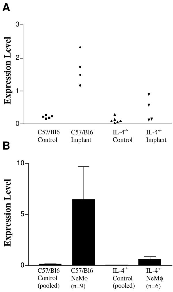
Arginase 1 is an inducible enzyme that metabolizes L-arginine, and has been shown to be upregulated in a Stat6 dependent manner by the Th2 cytokines IL-4 and IL-13 in vitro [29,32,33]. Arginase 1 competes with iNOS for L-arginine and thus can prevent nitric oxide release [33]. The abundant representation of arginase 1 in our EST dataset led us to test whether this gene was indeed upregulated in vivo as a result of exposure to a Th2 inducing nematode parasite. B. malayi were implanted into the peritoneal cavity of WT and IL-4-/- C57BL/6 mice and the expression of arginase 1 assessed in the recovered PEC. Consistent with the observation that the addition of IL-4 in vitro could induce arginase 1 [29,32,33], the lack of IL-4 in vivo prevented a significant upregulation of arginase1 over that found in resident peritoneal cells (Figure 1A). These experiments were repeated with F4/80+ macrophages purified with magnetic beads (Figure 1B). The results with purified macrophages were directly comparable to the results with total PEC verifying that macrophages were the source of upregulated arginase 1.
Figure 1.
Arginase 1 expression is increased in response to parasite implant in C57BL/6 but not IL-4 -/- mice. Real-time PCR analysis of arginase 1 expression by total cells (A) or F4/80 purified macrophages (B) obtained from peritoneal lavages of implanted or control mice. For the purified NeMφ, each cDNA sample was obtained from individual mice. For the purified control macrophages, the cDNA samples represent pooled macrophages from 5 mice. Expression levels were measured as a percentage of β-Actin expression. Error bars represent one SD from the mean of each analysis.
Although IL-13 has also been shown to induce arginase 1 upregulation [29,32] in vitro, the clear phenotypic difference between IL-4 deficient mice and WT mice suggests that IL-13 could not compensate in vivo for IL-4 in the induction of arginase 1 at least in C57BL/6 mice. Since the induction of arginase 1 is one of the clearest indicators of alternative activation [2,11], these results were consistent with our designation of NeMφ as 'alternatively activated'. Our findings were also consistent with a recent study demonstrating that type 2 cytokines regulate arginase production during helminth infection [34]
Identification of IL-4-regulated NeMφ genes using expression arrays
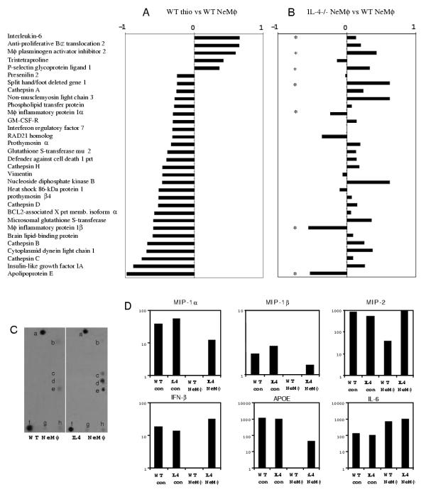
Although the EST strategy was very informative about the most abundantly expressed genes in NeMφ, we wished also to identify IL-4 dependent genes that are transcribed at lower levels. We therefore compared expression of 1176 known mouse genes using the Clontech Mouse 1.2 Atlas Array. The array contains a broad range of well-characterized genes involved in cellular pathways and functions, including cytokines, chemokines and associated receptors http://atlas.clontech.com. In order to distinguish between genes upregulated during the recruitment/maturation process and those whose expression was specific for parasite infection, we compared WT thioglycollate elicited macrophages with NeMφ (Figure 2A). We then compared gene expression in NeMφ derived from IL-4 deficient mice and WT mice (Figure 2B). From this approach we hoped to identify genes whose expression is altered by nematode infection in an IL-4 dependent manner.
Figure 2.
Mouse array analysis and real time PCR verfication. (A) Chart of the genes that showed the highest differential expression between NeMφ and thioglycollate-recruited macrophages. Values represent the difference between the gene expression in WT NeMφ and thioglycollate-recruited Mφ (arbitrary units). (B) Chart showing the difference between the gene expression in WT NeMφ and IL-4 -/- NeMφ. Genes selected for real-time PCR analysis are marked with an asterisk (*). (C) An example of a section of the Atlas array that contains MIP-1α (c), MIP-1β (d), MIP-2 (e). The left panel is probed with WT NeMφ and the right panel with IL-4-/- NeMφ. This section also contains the genes: prothymosin beta 4 (a), insulin-like growth factor IA (b), GADPH (f), myosin I alpha (g), and ornithine decarboxylase (h), which are all not differentially expressed. (D) Verification of Atlas array results by real-time PCR. cDNA samples from F4/80 purified macrophages from either resident peritoneal cells (con) or from parasite implanted mice (NeMφ) were diluted and normalized to contain equal levels of β-actin transcript (data not shown) before quantification of the different genes. Expression levels are shown in arbitrary units that are based on a comparison with β-actin expression (designated as 10,000 units). The results shown represent the mean values of 2 experiments with independent sources of experimental macrophage RNA.
Of the 1176 genes on the array, 141 genes were expressed by at least one of the macrophage populations. The expression pattern of the majority of genes found on the Mouse 1.2 Atlas Array was very similar between the 3 different macrophage populations. However, exceptions included genes that are upregulated by parasite infection but are apparently not IL-4 dependent such as IL-6 (Figure 2A &2B &2D). The most notable observation was that several pro-inflammatory cytokines and chemokines such as MIP-1α, MIP-1β and MIP-2 were more highly expressed in the IL-4-deficient NeMφ relative to WT NeMφ (Figure 2C &2D). As a confirmation of the results observed on the array, expression levels of several differentially expressed genes were further analyzed by real-time PCR. RNA was obtained from F4/80+ macrophages derived from WT and IL-4-/- mice and both resident macrophages and NeMφ were assessed (Figure 2D). These results confirmed that the capacity to produce several proinflammatory cytokines has been abolished in alternatively activated macrophages recruited under Th2 conditions. The suppression of proinflammatory cytokines is an IL-4 dependent process, since macrophages recruited in IL-4 deficient mice have not suppressed gene expression as effectively as the WT mice. Some genes, such as Mφ plasminogen activator inhibitor 2 and P-selectin glycoprotein ligand 1, which appeared by array analysis to be differentially regulated, failed to show differences on subsequent real-time PCR analysis.
Few genes were identified that were upregulated in the WT macrophages compared to the IL-4 deficient macrophages and were specific for parasite infection. This is probably due to a bias in our knowledge (and thus on the array) towards genes expressed in classically activated macrophages in comparison to alternative activation pathways and associated factors. Indeed, many of the abundantly expressed genes from our EST project were not represented on the Mouse 1.2 Atlas Array. Hence alternative strategies are necessary for the identification of novel genes that could be involved in alternative activation.
Comparative analysis of suppressive vs non-suppressive IL-4 deficient macrophages
In order to identify novel IL-4 dependent genes in the suppressive NeMφ, a subtractive hybridization library was constructed, using biotinylated RNA from IL-4-/- NeMφ to remove commonly expressed genes found in both WT and IL-4-/- populations. A total of 5760 primary recombinants were recovered. The library was gridded onto nylon membranes and was differentially screened for IL-4 dependent genes by probing separate membranes with cDNA from WT and IL-4-/- NeMφ. Of the 288 clones selected for further sequencing analysis, 66 yielded sequences greater than 150 bp. These were clustered and analyzed as described in the methods. Sequences with significant similarity to known genes are shown in Table 2.
Table 2.
Genes identified in subtractive NeMφ cDNA library
| No. of sequenced clones | % of total sequenced clones | GenBank Match (accession no.) |
|---|---|---|
| 25 | 37.88 | FIZZ1/RELM-α (NP_020509) |
| 10 | 15.15 | Serum amyloid A3 (NP_011315) |
| 2 | 3.03 | Similar to Rat RNA polymerase II transcription factor SIII or elongin B (L42855) |
| 2 | 3.03 | Similar to extracellular proteinase inhibitor (BC002038) |
| 2 | 3.03 | Metallopanstimulin 1 (NM_001030) |
| 1 | 1.51 | Inhibitor of DNA binding 2 (BC006921) |
| 1 | 1.51 | Adipocyte differentiation-related protein (M93275) |
| 1 | 1.51 | Methallothionein-1 gene transcription activator (AA146152) |
| 1 | 1.51 | Ring-box 1 or Rbx1 (NM_019712) |
| 1 | 1.51 | Cyclophilin (X52803) |
| 1 | 1.51 | Thrombospondin (M62470) |
| 1 | 1.51 | Ym1/ECF-L (D87757) |
| 1 | 1.51 | Cellular apoptosis susceptibility gene (AF301152) |
| 1 | 1.51 | Thymosin beta-4 (X16053.1) |
| 1 | 1.51 | H3 histone (BC002052) |
| 1 | 1.51 | CD9 antigen (NM_007657) |
| 1 | 1.51 | Ribosomal protein S29 (NM_009093.1) |
| 1 | 1.51 | 16S ribosomal RNA (AY011146) |
The most abundantly expressed clone (37.9%) in the subtractive library was the second most abundantly expressed clone (2%) in the suppressive NeMφ cDNA library. This gene is identical to recently described genes called FIZZ1 [25] and RELM-α [23]. Analysis of the mouse EST sequences in dbEST revealed that expression of FIZZ1/RELM-α was much more highly represented in our NeMφ EST dataset (13/651) than in any other tissue type.
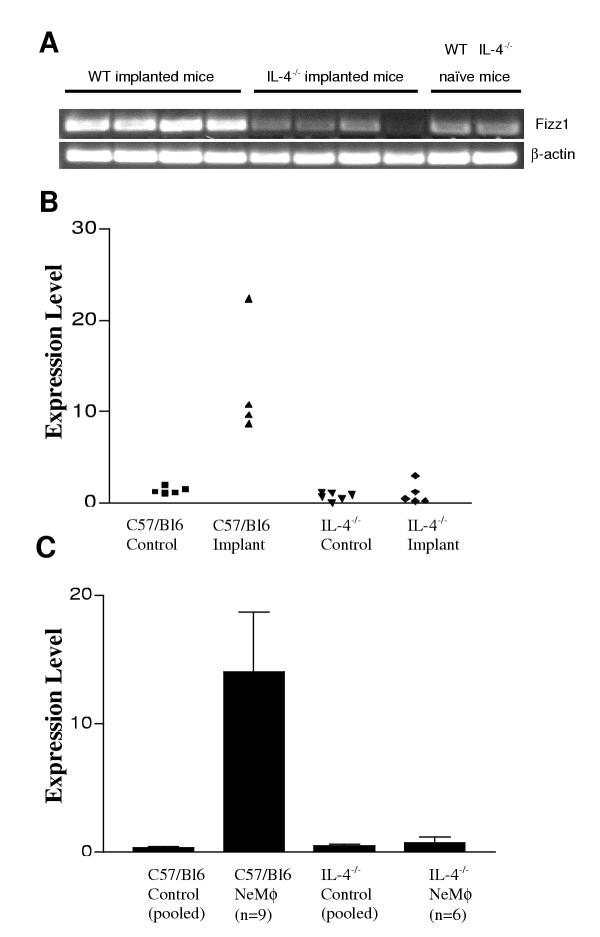
To confirm that FIZZ1/RELM-α was regulated by IL-4 in vivo, we compared the expression in PEC recovered from WT or IL-4-/- mice implanted with B. malayi using quantitative real-time PCR. Consistent with the results from the subtractive hybridization, the lack of IL-4 in vivo prevented a significant upregulation of FIZZ1/RELM-α (Figure 3A &3B). Expresssion was further analyzed using F4/80 purified macrophages. A 40-fold induction of gene expression at the level of mRNA, was found in WT NeMφ though expression remained basal in IL-4-/- NeMφ (Figure 3C).
Figure 3.
FIZZ1/RELMα expression is increased in response to parasite implant in C57BL/6 but not IL-4 -/- mice. Semiquantitative RT-PCR of FIZZ1/RELMα and β-actin was performed on total RNA samples from the peritoneal lavages of individual mice (A). Real Time PCR analysis of FIZZ1 expression by total cells (B) or F4/80 purified macrophages (C) obtained from peritoneal lavages of implanted or naïve control mice. For the purified NeMφ, each cDNA sample was obtained from individual mice. For the purified control macrophages, the cDNA samples represent pooled macrophages from groups of 2 or 3 mice for a total of 5 mice per control group. Expression levels were measured as a percentage of β-actin expression. Error bars represent one SD from the mean.
Apart from FIZZ1/RELM-α, the acute phase protein SAA3 was the most abundantly represented cluster in the subtractive library dataset (Table 2). We are currently investigating the implications of its production by alternatively activated macrophages. Also, six novel sequences were identified that had not been previously described in other EST projects as well as six genes sequences seen previously in other EST surveys but with no functional identification. We are currently in the process of characterizing these novel gene products.
Discussion
In this study, using NeMφ as a source of alternatively activated macrophages, we have identified novel "marker" genes that will be important in defining these cells in inflammatory responses in vivo. Our data also contributes to the evidence that genes identified in vitro (e.g. arginase) are expressed at elevated levels during Th2 mediated inflammation. Further, we have identified novel genes that could play important biological roles in the function of alternatively activated macrophages.
A key observation from our EST analysis was the high representation of relatively few transcripts. Our analysis would suggest that transcriptional activity of these macrophages is biased to relatively few genes that encode for secreted proteins not previously associated with macrophage function. Almost 10% of the transcripts in our alternatively activated macrophages encoded for a recently recognized eosinophil chemotactic factor termed Ym1/ECF-L. In subsequent analysis, we have found that Ym1/ECF-L expression can be induced in macrophages by the secretion of a nematode homologue of macrophage migration inhibitory factor (MIF) [17]. These data suggest that macrophages can act as intermediaries in the recruitment of eosinophils to the site of parasite residence. Futher, the extraordinarily high levels of Ym1 expression by NeMø suggest the possibility that this molecule, a chitin-binding protein [28], may also act as an effector molecule through direct interaction with chitin-containing pathogens.
One of the exciting findings to arise from this analysis was the discovery of a novel cysteine rich gene (FIZZ1/RELM-α) that was the second most abundantly expressed gene representing 2% of the NeMφ library. Two very recent unrelated publications have described this gene in entirely different contexts. Holcomb et al. demonstrated expression of this gene product in the bronchoalveolar lavage fluid of mice with OVA-induced pulmonary inflammation and termed it FIZZ1 (for found in inflammatory zone) [25]. In vitro studies showed that FIZZ1 could inhibit the nerve growth factor-mediated survival of rat neurons, and thus it may modulate neuron function in the lungs and act to control the local tissue response to allergic inflammation. Strikingly, Holcomb et al. did not find the expression of this protein in the alveolar macrophages of the inflamed lung.
FIZZ1 is a member of a family of tissue-specific genes with high similarity to resistin, termed 'resistin like molecules' (RELM) [23]. Resistin was first described as a protein produced by adipocytes that can antagonize insulin action and thus may be an important link between diabetes and obesity [24]. FIZZ1 is identical to RELM-α, a gene also expressed in white adipose tissue, as well as mammary tissue and tongue. Our results add a significant new dimension to the understanding of the biological role of FIZZ1/RELM-α by demonstrating that it is produced by activated macrophages and its upregulation is crucially dependent on IL-4.
It is interesting to note that the FIZZ/RELM gene family may consist of genes that are highly induced upon exposure of cells to exogenous stimuli. The majority of the mouse EST sequences submitted to dbEST thus far are from cDNA libraries made from normal tissues and therefore many interesting genes that are induced as a result of immune stimulation in vivo have not been identified. The expression of both Ym1 and FIZZ by alternatively activated macrophages in vivo is validated by recent unpublished data with phospholipase-C-deficient T. brucei. In these mouse studies, Ym1 and FIZZ1 were identified in macrophage populations isolated during the late stage of infection where Th2 responses dominate but not from classically activated macrophages found early in infection (G. Hassanzadeh, personal communication).
SAA3 was highly represented in both the NeMφ library and the subtractive library. Serum amyloid A proteins are type1 acute phase proteins that are rapidly synthesized following acute inflammation as a result of increased transcription. The regulation of the mouse SAA3 gene expression has been especially well studied [35-37]. Although it has been shown to be produced by macrophages [38] and is upregulated by such pro-inflammatory factors as LPS, IL-1 and IL-6 [35,37,39], SAA3 is also induced by glucocorticoids and can be inhibited by IFN-γ [40]. Interestingly, it has recently been shown that NFkB is involved in activation of SAA3 transcription [36]. Although this gene is upregulated under "classical" inflammatory conditions, we have found a strong representation of this gene in an IL-4 dependent manner under "alternative" activation conditions. This suggests that there is a category of genes that are upregulated as a result of macrophage activation under any inflammatory conditions, which are distinct from genes that are upregulated only in response to "classical" or "alternative" activation conditions. It further emphasizes that macrophage subpopulations may not fit neatly into these activation categories and are likely to represent a wide spectrum of phenotypes.
It is perhaps surprising that there was not a higher representation of chemokines and cytokines in the EST dataset. The only notable chemokine found was C10, which has also been previously described as an IL-4 dependent gene in macrophages [41]. The macrophages we observe may be representative of cells found in the later stages of chronic inflammation and the failure to observe significant cytokine or chemokine responses may be due to temporal effects; the expression of these effector molecules may have been and gone. The array analysis suggests that IL-4 specifically downregulates some pro-inflammatory chemokines such as MIP-1α, MIP-1β and MIP2. Downregulation of these chemokines has been previously observed [42,43] and this along with reduced iNOS expression has led to the assumption that alternatively activated macrophages are predominantly anti-inflammatory in function [11]. However, it is important to consider that these macrophages are found in highly inflammatory settings and may themselves be involved in promoting Th2-mediated pathology seen in both allergy and helminth infection. For example, increased expression of arginase 1 is associated with collagen deposition and other fibrogenic features [32]. This may be directly relevant to the pathology of filarial infection where elephantiasis is associated with infiltrates of macrophages and the deposition of thick collagen fibre bundles [44]. Consistent with this are the recent findings by Hesse et al. that arginase activated by type 2 cyokines plays a pivotal role in the granulomatous pathology in mice infected with Schistosoma mansoni[34].
Conclusions
The role of alternatively activated macrophages is still far from clear but these gene expression studies suggest that their designation as anti-inflammatory cells may not be a fully accurate reflection of their function. The high level expression of Ym1 suggests that alternatively activated macrophages may play a role in eosinophil chemotaxis and thus promote rather than prevent inflammation. Further, Ym1 is a molecule involved in crystal deposition [45], a phenomenon with no known function but which may have the potential to directly damage tissues [46]. The IL-4 dependent expression of FIZZ1/RELM-α suggests the possibility that these macrophages can regulate neuro-immunological cross-talk. These hypotheses remain to be tested and we are currently trying to establish the in vivo functional roles of Ym1/ECF-L and FIZZ1/RELM-α. Since these two genes have also recently been shown to be strongly induced in pulmonary inflammation, it suggests that "alternatively" activated macrophages are key effector cells in mediating many different features of Th2 associated inflammation.
Materials and methods
Preparation of in vivo derived alternatively activated macrophages
For all experiments, mice used were 6–8 week old male C57BL/6, either wild type (WT) or genetically deficient for IL-4 or IL-5. C57BL/6 IL-4 deficient (IL-4-/-) breeding pairs were purchased from B&K Universal Ltd. (North Humberside, UK) with permission of the Institute of Genetics, University of Cologne. C57BL/6 IL-5 deficient (IL-5-/-) mice were the kind gift of Prof. Manfred Kopf (Basel, Switzerland). B. malayi adult parasites were removed from the peritoneal cavity of infected jirds purchased from TRS Laboratories (Athens, GA) or maintained in house. Mice were surgically implanted intra-peritoneally (i.p.) with 6 live adult female B. malayi. Three weeks later the mice were euthanized by cardiac puncture and peritoneal exudate cells (PEC) were harvested by thorough washing of the peritoneal cavity with 15 ml of media (RPMI). Macrophages were purified by isolating adherent cells or by magnetic bead sorting as indicated in the text. When checked by flow cytometry, purification of macrophages by adherence typically yielded 80–85% F4/80+ cells, whereas magnetic bead sorting yielded 90–95% F4/80+ cells. Before magnetic bead cell purification, PEC were passed through a 70μm cell strainer and purified by centrifugation over Histopaque (Sigma) in order to remove any microfilariae. F4/80+ macrophages were purified with biotin conjugated F4/80 (rat IgG2b; Caltag) and Streptavidin Microbeads (Miltenyi Biotec), with either MS+ or VS+ columns in conjunction with MiniMacs or VarioMacs magnet (Miltenyi Biotec). Macrophage purity was confirmed by both flow cytometry and cytospin analysis.
cDNA library construction and EST analysis
Total RNA from magnetically purified macrophages, extracted using RNAStat60 (Biogenesis), was used to synthesize cDNA and unidirectionally cloned into the pCMV-Script plasmid vector, using the cDNA library construction kit (Stratagene, CA). Single clones from the unamplified library were picked at random and the cDNA inserts were amplified using vector primers T3 and T7. Aliquots of the amplified PCR products were checked by agarose gel and inserts greater than 200 bp were selected for sequencing. PCR products were prepared for sequencing using shrimp alkaline phosphatase and exonuclease I (Amersham Pharmacia Biotech Ltd, Sweden) [47]. Inserts were sequenced using the 5' vector primer SAC (GGGAACAA AAGCTGGAG) and ABI Big DYE terminators (Perkin Elmer Corporation, CT). Sequencing reactions were analysed using an ABI 377 automated sequencer.
Bioinformatics
Processing of ABI trace files was carried out using PHRED [48] (for basecalling) and cross_match [49] (for vector trimming). Only sequences 150 bp or longer were submitted to dbEST and used in subsequent analyses. Sequences >=150 bp in length were clustered on the basis of sequence similarity using an in-house program (Parkinson et al., unpublished). Clusters of sequences (which usually derive from one gene) were assembled into contigs using the program CAP3 [50].
cDNA expression array analysis
Transcripts from the RNA extracted from WT and IL-4-/- F4/80+ magnetically purified NeMφ were PCR amplified from cDNA tagged at the 5' and 3' ends during reverse transcription using the Generacer kit (Invitrogen). cDNA probes from thioglycollate elicited macrophages were generated as above, but macrophages were purified by adherence instead of using magnetic beads. The resulting cDNA probes were labelled with 33P and hybridized to separate Mouse 1.2 Atlas Array (Clontech) nylon membranes. The hybridization signal was quantified using ImageQuant Mac version 1.2. Comparison of signals from the positive control genes on the array indicated that the labeling intensities of the cDNA probes from the different macrophage populations were comparable. Expression values for individual genes were calculated in arbitrary units relative to the strongest positive control hybridization signal.
Subtractive library construction
Total RNA (1 μg) from F4/80 purified peritoneal macrophages from B. malayi implanted WT mice was converted into 1st strand cDNA using the Clontech SMART system. After synthesis of first strand cDNA from WT macrophages, 2 rounds of subtractive hybridization were undertaken with 20 μg RNA from peritoneal macrophages of IL-4-/- mice infected with B. malayi, using the Invitrogen 'Subtractor kit'. After 2 rounds of subtractive hybridization and phenol-choloroform extraction, the remaining single stranded cDNA was PCR amplified for 25 cycles. The PCR products were then size fractionated and fractions representing different size PCR products were cloned separately into the eukaryotic expression vector (pcDNA3.1/V5/His-TOPO) (Invitrogen). The ligated plasmids were transformed into XL-10 gold ultracompetent cells (Stratagene).
Subtractive library screen
5760 clones were gridded in duplicate onto a 22.2 × 22.2 cm nylon filter. cDNA from B. malayi recruited Mφ from WT and IL-4-/- mice were labeled with DIG-High Prime (Roche) and hybridized to separate nylon filters at 65°C. The filters were imaged using a Fuji LAS1000 and the resultant images were analysed using ArrayVision 4.3 (Imaging Research Inc.). Two hundred and eighty-eight clones showing the greatest differential expression in WT Mφ were selected for subsequent analysis and sequenced by MWG-Biotech. The sequences were subjected to the same bioinformatics analyses as the EST sequences (see above).
Quantitative RT-PCR analysis
Total RNA was extracted in RNAStat60 (Biogenesis) and single-stranded cDNA was synthesised using MMLV reverse transcriptase (Stratagene). Relative quantification of the expression of the genes of interest was measured by real-time PCR, using the LightCycler (Roche Molecular Biochemicals). PCR amplifications were performed in 10 μl, containing 1 μl cDNA, 4 mM MgCl2, 0.3 μM primers and the LightCycler-DNA SYBR Green I mix. The reaction was performed in the following conditions: 30 s denaturation at 95°C, 5 s annealing of primers at 55°C and 12 s elongation at 72°C, for 40–60 cycles. The fluorescent DNA binding dye SYBR Green was monitored after each cycle at 86°C. Five serial 1:5 dilutions of a positive control sample of cDNA were used as a standard curve in each reaction, and the expression levels of the genes of interest were measured as a percentage of β-Actin expression. Semi-quantitative PCR was carried out using Taq polymerase (Qiagen) for 30 cycles. PCR products were electrophoresed on 1% agarose gels and visualised by ethidium bromide staining. Primers for RT-PCR analysis were: β-Actin: TGGAATCCTGTGGCATCCATGAAAC and TAAAACG CAGCTCAGTAACAGTCCG; product size 300 bp. FIZZ1: GGTCCCAGTGCATATG GATGAGACCATAGA and CACCTCTTCACTCGAGGGACAGTTGGCAGC; product size 300 bp.
List of abbreviations
NeMφ: Nematode elicited macrophages, PEC: Peritoneal exudate cells; WT: wild type
Authors' contributions
Author 1 (PL) developed the approaches used in this study, constructed the libraries, coordinated the sequencing, performed the arrays, and drafted the manuscript. Author 2 (MGN) performed real time PCR analysis in this paper, assisted with the arrays and contributed to the analysis of the data and writing of the manuscript. Author 3 (JP) performed the bioinformatics. Author 4 (DG) helped with library construction and development of approaches. Author 5 (MLB) gave essential advice on development of the approaches and assisted with the bioinformatics. Author 6 (JEA) supervised the studies, participated in their design, contributed to the analysis and finalised the manuscript for publication.
Contributor Information
P'ng Loke, Email: ploke@uclink.berkeley.edu.
Meera G Nair, Email: m.g.nair@ed.ac.uk.
John Parkinson, Email: john.parkinson@ed.ac.uk.
David Guiliano, Email: David.Guiliano@ed.ac.uk.
Mark Blaxter, Email: mark.blaxter@ed.ac.uk.
Judith E Allen, Email: j.allen@ed.ac.uk.
Acknowledgements
We thank Ian Dransfield and Rick Maizels for helpful discussion and review of the manuscript. We thank Yvonne Harcus for invaluable technical assistance. This work was supported in part by a Wellcome Trust Prize Fellowship awarded to P Loke
References
- Glass CK, Witztum JL. Atherosclerosis. the road ahead. Cell. 2001;3:503–16. doi: 10.1016/s0092-8674(01)00238-0. [DOI] [PubMed] [Google Scholar]
- Allen JE, Loke P. Divergent roles for macrophages in lymphatic filariasis. Parasite Immunology. 2001;3:345–352. doi: 10.1046/j.1365-3024.2001.00394.x. [DOI] [PubMed] [Google Scholar]
- Kiefer R, Kieseier BC, Stoll G, Hartung HP. The role of macrophages in immune-mediated damage to the peripheral nervous system. Prog Neurobiol. 2001;3:109–27. doi: 10.1016/S0301-0082(00)00060-5. [DOI] [PubMed] [Google Scholar]
- Kinne RW, Brauer R, Stuhlmuller B, Palombo-Kinne E, Burmester GR. Macrophages in rheumatoid arthritis. Arthritis Res. 2000;3:189–202. doi: 10.1186/ar86. [DOI] [PMC free article] [PubMed] [Google Scholar]
- Aderem A, Ulevitch RJ. Toll-like receptors in the induction of the innate immune response. Nature. 2000;3:782–7. doi: 10.1038/35021228. [DOI] [PubMed] [Google Scholar]
- Reiner SL, Locksley RM. The regulation of immunity to Leishmania major. Annual Review of Immunology. 1995;3:151–177. doi: 10.1146/annurev.immunol.13.1.151. [DOI] [PubMed] [Google Scholar]
- Weinheber N, Wolfram M, Harbecke D, Aebischer T. Phagocytosis of Leishmania mexicana amastigotes by macrophages leads to a sustained suppression of IL-12 production. European Journal of Immunology. 1998;3:2467–77. doi: 10.1002/(SICI)1521-4141(199808)28:08<2467::AID-IMMU2467>3.0.CO;2-1. [DOI] [PubMed] [Google Scholar]
- Kane MM, Mosser DM. The role of IL-10 in promoting disease progression in leishmaniasis. J Immunology. 2001;3:1141–1147. doi: 10.4049/jimmunol.166.2.1141. [DOI] [PubMed] [Google Scholar]
- Bliss SK, Butcher BA, Denkers EY. Rapid recruitment of neutrophils containing prestored IL-12 during microbial infection. J Immunology. 2000;3:4515–21. doi: 10.4049/jimmunol.165.8.4515. [DOI] [PubMed] [Google Scholar]
- Balcewicz-Sablinska MK, Gan H, Remold HG. Interleukin 10 produced by macrophages inoculated with Mycobacterium avium attenuates mycobacteria-induced apoptosis by reduction of TNF-alpha activity. Journal of Infectious Diseases. 1999;3:1230–7. doi: 10.1086/315011. [DOI] [PubMed] [Google Scholar]
- Goerdt S, Orfanos CE. Other functions, other genes: alternative activation of antigen-presenting cells. Immunity. 1999;3:137–142. doi: 10.1016/s1074-7613(00)80014-x. [DOI] [PubMed] [Google Scholar]
- Doyle AG, Herbein G, Montaner LJ, Minty AJ, Caput D, Ferrara P, Gordon S. Interleukin-13 alters the activation state of murine macrophages in vitro: comparison with interleukin-4 and interferon-gamma. European Journal of Immunology. 1994;3:1441–1445. doi: 10.1002/eji.1830240630. [DOI] [PubMed] [Google Scholar]
- Stein M, Keshav S, Harris N, Gordon S. Interleukin 4 potently enhances murine macrophage mannose receptor activity: a marker of alternative immunologic macrophage activation. Journal of Experimental Medicine. 1992;3:287–292. doi: 10.1084/jem.176.1.287. [DOI] [PMC free article] [PubMed] [Google Scholar]
- Schebesch C, Kodelja V, Muller C, Hakij N, Bisson S, Orfanos CE, Goerdt S. Alternatively activated macrophages actively inhibit proliferation of peripheral blood lymphocytes and CD4+ T cells in vitro. Immunology. 1997;3:478–86. doi: 10.1046/j.1365-2567.1997.00371.x. [DOI] [PMC free article] [PubMed] [Google Scholar]
- Loke P, MacDonald AS, Robb AO, Maizels RM, Allen JE. Alternatively activated macrophages induced by nematode infection inhibit proliferation via cell to cell contact. European Journal of Immunology. 2000;3:2669–2678. doi: 10.1002/1521-4141(200009)30:9<2669::AID-IMMU2669>3.3.CO;2-T. [DOI] [PubMed] [Google Scholar]
- Allen JE, Lawrence RA, Maizels RM. APC from mice harboring the filarial nematode, Brugia malayi, prevent cellular proliferation but not cytokine production. International Immunology. 1996;3:143–151. doi: 10.1093/intimm/8.1.143. [DOI] [PubMed] [Google Scholar]
- Falcone FH, Loke P, Zang X, MacDonald AS, Maizels RM, Allen JE. A Brugia malayi homolog of macrophage mugration inhibitory factor reveals an important link between macrophages and eosinophil recruitment during nematode infection. J Immunol. 2001;3:5348–54. doi: 10.4049/jimmunol.167.9.5348. [DOI] [PubMed] [Google Scholar]
- MacDonald AS, Maizels RM, Lawrence RA, Dransfield I, Allen JE. Requirement for in vivo production of IL-4, but not IL-10, in the induction of proliferative suppression by filarial parasites. J Immunol. 1998;3:1304–1312. [PubMed] [Google Scholar]
- Lawrence RA, Allen JE, Gregory WF, Kopf M, Maizels RM. Infection of IL-4 deficient mice with the parasitic nematode Brugia malayi demonstrates that host resistance is not dependent on a T helper-2 dominated immune response. J Immunol. 1995;3:5995–6001. [PubMed] [Google Scholar]
- Loke P, MacDonald AS, Allen JE. Antigen presenting cells recruited by Brugia malayi induce Th2 differentiation of naive CD4+ T cells. European Journal of Immunology. 2000;3:1127–1135. doi: 10.1002/(SICI)1521-4141(200004)30:04<1127::AID-IMMU1127>3.0.CO;2-R. [DOI] [PubMed] [Google Scholar]
- Hashimoto S, Suzuki T, Dong HY, Yamazaki N, Matsushima K. Serial analysis of gene expression in human monocytes and macrophages. Blood. 1999;3:837–44. [PubMed] [Google Scholar]
- Suzuki T, Hashimoto S, Toyoda N, Nagai S, Yamazaki N, Dong HY, Sakai J, Yamashita T, Nukiwa T, Matsushima K. Comprehensive gene expression profile of LPS-stimulated human monocytes by SAGE. Blood. 2000;3:2584–91. [PubMed] [Google Scholar]
- Steppan CM, Brown EJ, Wright CM, Bhat S, Banerjee RR, Dai CY, Enders GH, Silberg DG, Wen X, Wu GD, Lazar MA. A family of tissue-specific resistin-like molecules. Proc Natl Acad Sci USA. 2001;3:502–6. doi: 10.1073/pnas.98.2.502. [DOI] [PMC free article] [PubMed] [Google Scholar]
- Steppan CM, Bailey ST, Bhat S, Brown EJ, Banerjee RR, Wright CM, Patel HR, Ahima RS, Lazar MA. The hormone resistin links obesity to diabetes. Nature. 2001;3:307–12. doi: 10.1016/S0014-5793(97)00535-8. [DOI] [PubMed] [Google Scholar]
- Holcomb IN, Kabakoff RC, Chan B, Baker TW, Gurney A, Henzel W, Nelson C, Lowman HB, Wright BD, Skelton NJ. et al. FIZZ1, a novel cysteine-rich secreted protein associated with pulmonary inflammation, defines a new gene family. EMBO J. 2000;3:4046–55. doi: 10.1093/emboj/19.15.4046. [DOI] [PMC free article] [PubMed] [Google Scholar]
- Guo L, Johnson RS, Schuh JC. Biochemical characterization of endogenously formed eosinophilic crystals in the lungs of mice. Journal of Biological Chemistry. 2000;3:8032–8037. doi: 10.1074/jbc.275.11.8032. [DOI] [PubMed] [Google Scholar]
- Jin HM, Copeland NG, Gilbert DJ, Jenkins NA, Kirkpatrick RB, Rosenberg M. Genetic characterization of the murine Ym1 gene and identification of a cluster of highly homologous genes. Genomics. 1998;3:316–22. doi: 10.1006/geno.1998.5593. [DOI] [PubMed] [Google Scholar]
- Owhashi M, Arita H, Hayai N. Identification of a novel eosinophil chemotactic cytokine (ECF-L) as a chitinase family protein. Journal of Biological Chemistry. 2000;3:1279–1286. doi: 10.1074/jbc.275.2.1279. [DOI] [PubMed] [Google Scholar]
- Munder M, Eichmann K, Modolell M. Alternative metabolic states in murine macrophages reflected by the nitric oxide synthase/arginase balance: competitive regulation by CD4+ T cells correlates with Th1/Th2 phenotype. J Immunol. 1998;3:5347–5354. [PubMed] [Google Scholar]
- Uhlar CM, Whitehead AS. Serum amyloid A, the major acute-phase reactant. European Journal of Biochemistry. 1999;3:501–523. doi: 10.1046/j.1432-1327.1999.00657.x. [DOI] [PubMed] [Google Scholar]
- Gebe JA, Llewellyn M, Hoggatt H, Aruffo A. Molecular cloning, genomic organization and cell-binding characteristics of mouse Spalpha. Immunology. 2000;3:78–86. doi: 10.1046/j.1365-2567.2000.00903.x. [DOI] [PMC free article] [PubMed] [Google Scholar]
- Munder M, Eichmann K, Moran JM, Centeno F, Soler G, Modolell M. Th1/Th2-regulated expression of arginase isoforms in murine macrophages and dendritic cells. J Immunol. 1999;3:3771–7. [PubMed] [Google Scholar]
- Rutschman R, Lang R, Hesse M, Ihle JN, Wynn TA, Murray PJ. Cutting edge: Stat6-Dependent Substrate Depletion Regulates Nitric Oxide Production. J Immunol. 2001;3:2173–2177. doi: 10.4049/jimmunol.166.4.2173. [DOI] [PubMed] [Google Scholar]
- Hesse M, Modolell M, La Flamme AC, Schito M, Fuentes JM, Cheever AW, Pearce EJ, Wynn TA. Differential regulation of nitric oxide synthase-2 and arginase-1 by type 1/type 2 cytokines in vivo: granulomatous pathology is shaped by the pattern of L-arginine metabolism. J Immunol. 2001;3:6533–6544. doi: 10.4049/jimmunol.167.11.6533. [DOI] [PubMed] [Google Scholar]
- Bing Z, Reddy SA, Ren Y, Qin J, Liao WS. Purification and characterization of the serum amyloid A3 enhancer factor. Journal of Biological Chemistry. 1999;3:24649–56. doi: 10.1074/jbc.274.35.24649. [DOI] [PubMed] [Google Scholar]
- Bing Z, Huang JH, Liao WS. NFkappa B interacts with serum amyloid A3 enhancer factor to synergistically activate mouse serum amyloid A3 gene transcription. Journal of Biological Chemistry. 2000;3:31616–23. doi: 10.1074/jbc.M005378200. [DOI] [PubMed] [Google Scholar]
- Huang JH, Liao WS. Induction of the mouse serum amyloid A3 gene by cytokines requires both C/EBP family proteins and a novel constitutive nuclear factor. Molecular and Cellular Biology. 1994;3:4475–84. doi: 10.1128/mcb.14.7.4475. [DOI] [PMC free article] [PubMed] [Google Scholar]
- Meek RL, Eriksen N, Benditt EP. Murine serum amyloid A3 is a high density apolipoprotein and is secreted by macrophages. Proc Natl Acad Sci USA. 1992;3:7949–52. doi: 10.1073/pnas.89.17.7949. [DOI] [PMC free article] [PubMed] [Google Scholar]
- Meheus LA, Fransen LM, Raymackers JG, Blockx HA, Van Beeumen JJ, Van Bun SM, Van de Voorde A. Identification by microsequencing of lipopolysaccharide-induced proteins secreted by mouse macrophages. J Immunol. 1993;3:1535–47. [PubMed] [Google Scholar]
- Ishida T, Matsuura K, Setoguchi M, Higuchi Y, Yamamoto S. Enhancement of murine serum amyloid A3 mRNA expression by glucocorticoids and its regulation by cytokines. Journal of Leukocyte Biology. 1994;3:797–806. doi: 10.1002/jlb.56.6.797. [DOI] [PubMed] [Google Scholar]
- Orlofsky A, Wu Y, Prystowsky MB. Divergent regulation of the murine CC chemokine C10 by Th1 and Th2 cytokines. Cytokine. 2000;3:220–8. doi: 10.1006/cyto.1999.0535. [DOI] [PubMed] [Google Scholar]
- Flesch IE, Barsig J, Kaufmann SH. Differential chemokine response of murine macrophages stimulated with cytokines and infected with Listeria monocytogenes. International Immunology. 1998;3:757–65. doi: 10.1093/intimm/10.6.757. [DOI] [PubMed] [Google Scholar]
- Spergel JM, Mizoguchi E, Oettgen H, Bhan AK, Geha RS. Roles of TH1 and TH2 cytokines in a murine model of allergic dermatitis. Journal of Clinical Investigation. 1999;3:1103–11. doi: 10.1172/JCI5669. [DOI] [PMC free article] [PubMed] [Google Scholar]
- Olszewski WL, Jamal S, Manokaran G, Lukomska B, Kubicka U. Skin changes in filarial and non-filarial lymphoedema of the lower extremities. Trop. 1993;3:40–4. [PubMed] [Google Scholar]
- Guo L, Johnson RS, Schuh JC. Biochemical characterization of endogenously formed eosinophilic crystals in the lungs of mice. J Biol Chem. 2000;3:8032–7. doi: 10.1074/jbc.275.11.8032. [DOI] [PubMed] [Google Scholar]
- Feldmesser M, Kress Y, Casadevall A. Intracellular crystal formation as a mechanism of cytotoxicity in murine pulmonary Cryptococcus neoformans infection. Infection and Immunity. 2001;3:2723–2727. doi: 10.1128/IAI.69.4.2723-2727.2001. [DOI] [PMC free article] [PubMed] [Google Scholar]
- Daub J, Loukas A, Pritchard DI, Blaxter M. A survey of genes expressed in adults of the human hookworm, Necator americanus. 1999;3:171–184. doi: 10.1017/s0031182099005375. [DOI] [PubMed] [Google Scholar]
- Ewing B, Hillier L, Wendl MC, Green P. Base-calling of automated sequencer traces using phred. I. Accuracy assessment. Genome Research. 1998;3:175–85. doi: 10.1101/gr.8.3.175. [DOI] [PubMed] [Google Scholar]
- Green P. Documentation for Phrap. Genome Center, University of Washington; 1996.
- Huang X, Madan A. CAP3: A DNA sequence assembly program. Genome Research. 1999;3:868–77. doi: 10.1101/gr.9.9.868. [DOI] [PMC free article] [PubMed] [Google Scholar]


