



Abstract
Purpose
This study aims to compare the effects of two generations of the Isobar dynamic fixation system—Isobar TTL (first-generation, less mobile) and Isobar EVO (second-generation, more mobile)—on the surgical and adjacent intervertebral disc segments, based on MRI findings.
Methods
A retrospective analysis was conducted on 29 patients treated with fenestrated decompression and Isobar EVO stabilization and 34 patients treated with fenestrated decompression and Isobar TTL stabilization. Clinical outcomes and radiographic parameters, including the disc height index (DHI) and range of motion (ROM) of the surgical and upper adjacent segments, lateral neural foraminal protrusion value (LNPV) of the upper adjacent segment, and Pfirrmann grading of disc degeneration, were evaluated and compared between the two groups.
Results
At the 48-month follow-up, no significant differences in Visual Analog Scale (VAS) scores or Oswestry Disability Index (ODI) were observed between the two groups (P > 0.01). The EVO group demonstrated significantly higher surgical segment DHI and ROM compared to the TTL group (P < 0.01). Meanwhile, the upper adjacent segment ROM was significantly lower in the EVO group compared to the TTL group at the final follow-up (P < 0.01). Additionally, the upper adjacent segment LNPV was larger in the EVO group than in the TTL group (P < 0.01). Pfirrmann grading revealed greater degeneration in surgical segments treated with TTL compared to EVO (P = 0.008).
Conclusion
Compared to the first-generation, less mobile Isobar TTL, the second-generation, more mobile Isobar EVO offers superior preservation of the surgical segment and slows the progression of degeneration in the upper adjacent segments. These findings highlight the benefits of greater mobility in posterior dynamic stabilization devices.
Keywords: Dynamic stabilization, Non-fusion, Lumbar degenerative diseases, Adjacent segment degeneration, Isobar system
Introduction
Spinal fusion has been recognized as the gold standard for the treatment of spinal degenerative disease, because it can relieve nerve root compression of the surgical segment through spinal canal decompression and reconstruct the stabilization and sequence of the surgical segment. Fusion, on the other hand, altered the normal biomechanical environment of lumbar functional units, resulting in the surgical segment’s loss of motion, which causes stress to build up in the adjacent segment and promotes degeneration of the adjacent segment [1–4]. There are several risks associated with an iliac bone graft, including nonunion, instrumentation failure, infection, and pain in the donor location. Additionally, intervertebral foraminoscopic discectomy and conservative treatment are the most frequently used procedures, but they have very high postoperative recurrence rates, especially for heavy workers and people with high activity levels, which can result in a greater financial burden and psychological stress. As a result, dynamic stabilizing devices have been developed to counteract some of these adverse effects.
Pedicle screw-based posterior dynamic stabilization can preserve some mobility while providing moderate surgical segment stabilization, reducing compensatory range of motion (ROM) and stress concentration in the adjacent segment to postpone and even prevent adjacent segment degeneration. In addition, surgical segmented lumbar discs can be placed in a biomechanically favorable environment to promote disc self-healing [5]. The Isobar dynamic stabilization system is one of the most commonly used posterior dynamic stabilization devices. It is designed to stabilize the surgical segment while preserving some ROM, preventing the adjacent segment from becoming hypermobile. The first-generation product of Isobar dynamic stabilization system, Isobar TTL, has ± 2.25° flexion-extension mobility and ± 0.4 mm longitudinal displacement. Many clinical studies conducted over the last decade have reported favorable outcomes in patients with lumbar degenerative disease who were treated with the Isobar TTL [6]. However, whether TTL can prevent adjacent segment intervertebral disc degeneration is debatable [7–9]. In this case, Isobar EVO, the second-generation product of the Isobar dynamic stabilization system, has been developed, with flexion-extension mobility increased to ± 4.5°, longitudinal displacement increased to ± 0.8 mm, and titanium rod bending increased moderately. To date, no studies have been published on the effect of EVO with increased mobility on adjacent segment and surgical segment intervertebral discs. The purpose of this retrospective study was to compare and analyze changes in some measurement indexes of MRI images of patients with lumbar degenerative diseases treated in our hospital with the TTL system and EVO system combined with decompression at various time points before and after the operation.
Materials and methods
Patient selection
This study reviewed 189 consecutive patients with single segment lumbar degeneration diseases who undergoing surgical treatment with Isobar TTL or EVO between March 2015 and July 2018. Based on the established inclusion and exclusion criteria, a total of 126 patients were excluded. Among them, 15 patients underwent L3/4 segment fixation, and 34 patients were unable to participate in the final follow-up (either due to being unreachable or declining to attend). Additionally, 2 patients passed away for unrelated reasons, and 20 patients had a history of surgery in other areas which could potentially influence the assessment of therapeutic outcomes. Over the course of the postoperative to follow-up period, 8 patients experienced fractures in the lumbar spine resulting from reasons unrelated to the surgical procedure and internal fixation. Notably, a total of 47 cases necessitated revision surgery and were successfully treated using the Isobar system. Finally, 63 patients were included in this study, 34 in the TTL group, and 29 in the EVO group. This study has been approved by the Ethical Committee of the Affiliated Hospital of Shaanxi University of Chinese Medicine. All patients participated freely and with written informed consent to the study.
Inclusion and exclusion of patients
The study was carried out in accordance with the moral guidelines that were established by the Declaration of Helsinki and its following amendments [10].
Inclusion criteria were as follows: (1) Patients with degenerative changes such as lumbar spinal stenosis (including cases with potential neurogenic claudication or loss of control over bowel and bladder function) and disc herniation (including herniation at the L4/5 or L5/S1 levels or severe herniation involving more than 50% of the spinal canal); (2) Failure to improve after at least 3 months of conservative treatment; (3) Availability of complete preoperative and final follow-up functional X-rays; (4) Underwent dynamic stabilization surgery (Isobar TTL or EVO, Scient’x-Alphatec, France); (5) Completion of at least 48 months of follow-up with detailed clinical and radiological evaluations; (6) Patients aged under 60 years at the time of surgery; (7) The upper adjacent segment Pfirrmann grade must be lower than Grade IV to minimize the confounding effects of severe pre-existing degeneration; (8) Patients with a body mass index (BMI) of less than 30 kg/m² to reduce the risk of complications related to obesity and excessive mechanical load; (9) Mild instability or early degenerative changes detected on imaging but without severe spondylolisthesis (> Grade II) or advanced facet arthropathy, ensuring the appropriateness of dynamic stabilization.
Exclusion criteria were as follows: (1) with severe lumbar instability (lumbar spondylolisthesis≥II degree), severe scoliosis or rotational deformity and other lumbar spine diseases; (2) a history of lumbar surgery; (3) with lumbar fractures, tumors, infections, ankylosing spondylitis, etc.; (4) with hip disease, cervical spondylotic myelopathy, or other illnesses that impacted the assessment of the therapeutic outcome; (5) severe osteoporosis (T value ≤ − 2.5 with single or multiple fragility fractures or T value ≤ − 3.0).
TTL was used in earlier cases due to its earlier release, while EVO was used in subsequent cases after its introduction. Both systems were applied to patients meeting the same inclusion and exclusion criteria to ensure comparability. The TTL system and the EVO system are Isobar dynamic fixation devices produced by the French ScienceX company. The difference between them is that, compared with the EVO system, the flexion-extension mobility of the TTL system increases from ± 2.25°to ± 4.5°, the longitudinal displacement increases from ± 0.2 mm to ± 0.8 mm, the bending degree of the titanium rod increases from 8° to 12°, and the contour of the titanium ring decreases by 25% [10]. (Fig. 1)
Fig. 1.

The TTL system is on the left, and the EVO system is on the right [10]
Surgical procedure
We have described this part in detail in our previous research [10].
Decompression Alone: Indicated for isolated nerve compression without radiographic or clinical evidence of instability or degenerative changes threatening segmental motion.
Decompression + Dynamic Stabilization: Employed for patients with mild instability, early-stage degenerative changes, or significant risk factors for adjacent segment degeneration, especially in younger and more active patients.
Decompression + Fusion: Reserved for patients with severe instability (e.g., spondylolisthesis > Grade II), advanced facet arthropathy, or deformities requiring rigid fixation.
Clinical and radiological evaluation
Outpatient re-examinations were performed on all patients. X-ray films of the lumbar spine in the anterolateral, hyperextension and hyperflexion positions, as well as a lumbar spine MR examination, were taken one month, one year, and every year after the operation.
Clinical outcomes were assessed through the Visual Analog Scale (VAS) for back and the Oswestry Disability Index (ODI). Each patient admitted to the hospital and each patient undergoing postoperative review has their VAS and ODI scores recorded and entered into their outpatient medical record.
Posteroanterior, lateral, and dynamic radiographs with flexion and extension views were obtained preoperatively and at the last follow-up. The digital PACS radiographic imaging review system was used to obtain MRI of the two groups preoperatively and at each follow-up visit. The radiographic outcomes included the stabilized segment’s disc height index (DHI) [11] and Pfirrmann grading [12], as well as the upper adjacent segment’s Pfirrmann grading. Because ASD frequently occurred superior to the surgical segment, we removed the nucleus pulposus of the surgical segment to varying degrees, and the volume of the nucleus pulposus of stabilized segments in different patients varied greatly, we only evaluated the lumbar nucleus pulposus volume (LNPV) of the upper adjacent segment [13] (Fig. 2). Each radiograph was measured and analysed independently by two experienced spine surgeons (who are not involved in the procedure and do not know the type of instrument used by the patient) and 2 Imaging physicians to minimize human error. A senior surgeon resolved disagreements in measurements.
Fig. 2.

A and B show LNPV measurements, and Both A and C are median sagittal plane of the MRI image. A. The h represents 1/2 of the height of the highest point in the middle of the nucleus pulposus in the median sagittal position of MRI. B. The a and b represent 1/2 of the anterior posterior diameter and width of the MRI cross-sectional ellipse, respectively. C. DHI was calculated using the MRI sagittal parameter de / cd
Statistical analysis
All statistical analyses were performed in GraphPad Prism 9.4 (GraphPad Software, San Diego, CA) and SPSS 19.0(SPSS Inc., Chicago, IL, USA). Continuous variables are expressed in the form of mean ± standard deviation. Two independent samples t-tests were used to compare differences between groups, and paired samples t-tests were used to compare differences between preoperative and final follow-up. To compare differences between groups, enumeration data and grade data (the Pfirrmann grading) were compared using the chi-square test. we performed Pearson correlation coefficient or Spearman’s rank correlation coefficient. Correlations > 0.7 were considered very strong; 0.5–0.7, strong; 0.3–0.5, moderate; and < 0.3, weak. A Kruskal-Wallis test and Dunn’s multiple comparison was used to correlate ROM of surgical segment with the Pfirrmann grades. When P < 0.01, the difference was considered to be statistically significant.
Results
Patients data
By using inclusion and exclusion criteria, 29 patients underwent posterior decompression and were fixed with an Isobar EVO device, and 34 patients were fixed with an Isobar TTL device. There were no statistically significant differences between the EVO group and the TTL group in age, sex, the distribution of surgical segments, follow-up time and type of disease (P > 0.01, Table 1).
Table 1.
Patient demographic and baseline data
| EVO group (n = 29) | TTL group (n = 34) | P value | |
|---|---|---|---|
| Age (years) | 51.38 ± 6.71 | 49.74 ± 5.31 | 0.282 |
| Gender (male/female) | 14/15 | 18/16 | 0.712† |
| Follow-up times (months) | 52.55 ± 3.4 | 51.35 ± 2.58 | 0.118 |
| BMI(kg/m2) | 21.9 ± 2.8 | 22.5 ± 3.4 | 0.348 |
| Operating level | 0.924† | ||
| L4/5 | 19 | 22 | |
| L5/S1 | 10 | 11 | |
| Diseases | 0.923† | ||
| Spinal stenosis | 14 | 16 | |
| Lumbar disc herniation | 15 | 18 | |
| Surgical duration (min) | 128.97 ± 8.59 | 127.83 ± 8.28 | 0.452 |
| Blood loss (mL) | 139.31 ± 19.99 | 133.97 ± 19.14 | 0.284 |
Values are presented as mean ± standard deviation
P values are based on the Independent two‑sample t‑test * or chi-square test†
BMI, body mass index
Clinical outcomes
The ODI and VAS scores of both groups were significantly improved at the final follow-up as compared to baseline values (P < 0.01, Table 2), but the difference at the final follow-up between the two groups was not significant (P > 0.01, Table 2).
Table 2.
Clinical outcomes
| VASback | ODI(%) | |||
|---|---|---|---|---|
| Pre-op | Final follow-up | Pre-op | Final follow-up | |
| TTL | 7.11 ± 1.16 | 1.1 ± 0.82* | 75.35 ± 7.18 | 10.62 ± 3.10* |
| EVO | 7.27 ± 1.03 | 0.82 ± 0.67* | 75.78 ± 8.31 | 10.67 ± 2.99* |
| P value | 0.645 | 0.141 | 0.058 | 0.860 |
Data are presented as mean ± standard deviation
#Significant differences between the TTL and EVO groups in each time point were tested by independent two-sample t-test, P < 0.01
* Significant difference between pre- and post-operative condition in each group using the paired t‑test, P < 0.01
ODI, Oswestry disability index; VAS, visual analogue scale
Radiographic outcomes
Disc height index
The results of the consistency test show that the kappa coefficient of DHI is 0.834, which is greater than 0.75, indicating high reliability. The postoperative DHI in the TTL group was lower than preoperative at all time points for both the surgical segment and the upper adjacent segment, but there were no statistically significant differences (P > 0.01). The postoperative DHI in the EVO group was higher than preoperative at final follow-up for the surgical segment (P < 0.01). And for the operative segment, The DHI of the EVO group was significantly higher than that of the TTL group at final follow-up for the surgical segment (P < 0.01). (Table 3)
Table 3.
Radiographic outcomes
| n | Surgical segment | P value | Upper adjacent segment | P value | |||
|---|---|---|---|---|---|---|---|
| Pre-op | Final follow-up | Pre-op | Final follow-up | ||||
| DHI | |||||||
| TTL | 34 | 0.33 ± 0.02 | 0.35 ± 0.02 | 0.004* | 0.55 ± 0.02 | 0.56 ± 0.02 | 0.453 |
| EVO | 29 | 0.35 ± 0.03 | 0.38 ± 0.01 | 0.003* | 0.56 ± 0.02 | 0.55 ± 0.03 | 0.612 |
| P value | 0.08 | 0.004* | 0.734 | 0.602 | |||
| ROM (°) | |||||||
| TTL | 34 | 9.74 ± 0.59 | 2.62 ± 0.36 | < 0.01 | 9.92 ± 0.82 | 12.14 ± 0.84 | < 0.01 |
| EVO | 29 | 9.63 ± 0.83 | 5.32 ± 0.64 | < 0.01 | 10.10 ± 0.93 | 10.97 ± 0.93 | < 0.01 |
| P value | 0.546 | < 0.01 | 0.429 | < 0.01 | |||
| LNPV (mm3) | |||||||
| TTL | 34 | N/A | N/A | 5.59 ± 0.34 | 5.03 ± 0.32 | < 0.01 | |
| EVO | 29 | N/A | N/A | 5.61 ± 0.35 | 5.29 ± 0.22 | < 0.01 | |
| P value | 0.841 | < 0.01 | |||||
Data are presented as mean ± standard deviation
#Significant differences between the TTL and EVO groups in each time point were tested by independent two-sample t-test., P < 0.01
* Significant difference between pre- and post-operative condition in each group using the paired t‑test, P < 0.01
DHI, disc height index; ROM, range of motion; LNPV, lumbar nucleus pulposus volume
Rang of motion
The results of the consistency test show that the kappa coefficient of ROM values of the surgical segment and the upper adjacent segment is 0.873 and 0.845, which is greater than 0.75, indicating high reliability. There were no significant differences in the mean ROM values of the surgical segment and the upper adjacent segment between the two groups preoperatively. However, the mean ROM of the surgical segment at the final follow-up were significantly lower than that at pre-operation between the two groups, and the upper adjacent segment at the final follow-up were significantly higher than that at pre-operation between the two groups (P < 0.01; Figs. 3 and 4). And the ROM at surgical segments of the EVO group were significantly higher than that of the TTL group (5.32°±0.64° vs. 2.62°±0.36°, P < 0.01) at the final follow-up, the ROM at upper adjacent segments of the EVO group were significantly lower than that of the TTL group (10.97°±0.93° vs. 12.14°±0.84°, P < 0.01; Table 3; Figs. 3 and 4). The Typical cases of Isobar EVO and Isobar TTL were showed in Figs. 5 and 6.
Fig. 3.
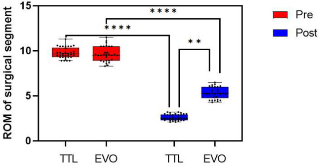
Rom of surgical segment between the TTL and EVO groups
Fig. 4.
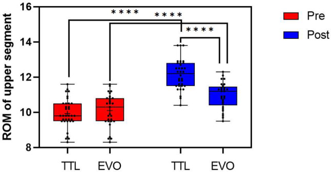
Rom of upper segment between the TTL and EVO groups
Fig. 5.

A-Eanda-e. A 47-year-old male patient underwent Isobar TTL stabilization due to spinal stenosis in L4/5. He underwent fenestration decompression at L4-5, and instrumentation at L4–5 using the Isobar TTL. A-E The preoperative radiographs. A-B. Preoperative frontal and lateral x-ray of lumbar spine. C. The preoperative lumbar sagittal T2WI with fat suppression. The Pfirrman grading of L4/5 was grade III, and that of L3/4 was grade II. D. The preoperative lumbar axial T2WI of L3/4 nucleus pulpous, and the LNPV of L3/4 nucleus pulpous was 4.86 mm3. F-G. The pre-operative ROM of the surgical segment was 8.3° based on X-ray. a-e. The radiographs obtained 48 months after the operation. a-b. Postoperative frontal and lateral x-ray of lumbar spine. c. The lumbar sagittal T2WI. The Pfirrman grading of L4/5 was grade IV, and that of L3/4 was grade III. d--e. The lumbar axial T2WI of L3/4 nucleus pulpous, and the LNPV of L3/4 nucleus pulpous was 4.32 mm3. f-g. The post-operative ROM of the surgical segment was 2.4° based on X-ray
Fig. 6.

A-E and a-e. A 43-year-old male patient underwent Isobar EVO stabilization due to spinal stenosis in L4/5. He underwent fenestration decompression at L4-5, and instrumentation at L4–5 using the Isobar EVO. A-B. Preoperative frontal and lateral x-ray of lumbar spine. C-E The preoperative MRI scans. C. The preoperative lumbar sagittal T2WI with fat suppression. The Pfirrman grading of L4/5 was grade IV, and that of L3/4 was grade II. D. The preoperative lumbar axial T2WI of L3/4 nucleus pulpous, and the LNPV of L3/4 nucleus pulpous was 4.93 mm3. F-G. The pre-operative ROM of the surgical segment was 8.7° based on X-ray. a-e. The radiographs obtained 50 months after the operation. a-b. Postoperative frontal and lateral x-ray of lumbar spine. c. The lumbar sagittal T2WI. The Pfirrman grading of L4/5 was grade III, and that of L3/4 was grade II. d-e. The lumbar axial T2WI of L3/4 nucleus pulpous, and the LNPV of L3/4 nucleus pulpous was 4.71 mm3. f-g. The post-operative ROM of the surgical segment was 4.6° based on X-ray
Lumbar nucleus pulposus volume
The results of the LNPV consistency test in the upper adjacent vertebrae revealed that the kappa coefficient of LNPV was 0.812, which was greater than 0.75, indicating high reliability. At the final follow-up, LNPV in both EVO group and TTL group alter significantly from before surgery (P < 0.01). At the final follow-up, the LNPV of the EVO group was higher than that of the TTL group (5.29 ± 0.22mm3 vs. 5.03 ± 0.32 mm3, < 0.01). (Table 3; Fig. 7) The typical cases in EVO group and TTL group are shown in Figs. 5 and 6.
Fig. 7.
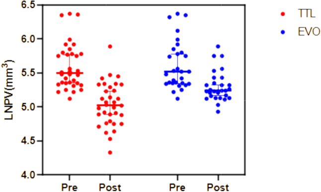
Preoperative and postoperative LNPV between the TTL and EVO groups
Correlation between ROM and LNPV
We used the surgical and upper adjacent segmental ROM to correlate the LNVP of upper adjacent segment. The LNPV increased with increasing surgical segment mobility, which were moderate positive correlation (r = 0.4212, p = 0.0006). The LNPV decreased with increasing upper adjacent segment mobility, which were moderate negative correlation (r=-0.3760, p = 0.0024). (Fig. 8)
Fig. 8.

The correlation between ROM and LNP
The pfirrmann grading
The Pfirrmann grading consistency test results show that the kappa coefficient is 0.783, which is greater than 0.75, indicating high reliability. Next, we used the Pfirrmann MRI grading system to correlate the degree of disc degeneration to the ROM of surgical segment. There was no difference in Pfirrmann classification between the two groups preoperatively (p = 0.658), grade 1 discs (n = 13), grade 2 discs (n = 21) in the TTL group and grade 1 discs (n = 15), grade 2 discs (n = 14) in the EVO group. Postoperative Pfirrmann grading showed differences between the two groups (p < 0.01), grade 1 discs(n = 0), grade 2 discs(n = 19), grade 3 discs(n = 15) in the TTL group and grade 1 discs(n = 11), grade 2 discs(n = 15), grade 3 discs(n = 3) in the EVO group. We found that ROM of TTL surgical segment had significantly upregulated Pfirrmann grade as compared ROM of EVO surgical segment (p = 0.008) (Fig. 9).
Fig. 9.
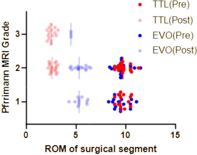
The correlation between ROM of surgical segment and Pfrrimann of upper segment disc
Discussion
Under normal physiological conditions, the intervertebral disc generates hydrostatic pressure via the hydrophilicity of proteoglycan in the nucleus pulposus in order to maintain biomechanical elasticity and to withstand isotropic stress [14, 15]. A decrease in proteoglycan due to a variety of factors lowers hydrostatic pressure, reducing volume and weakening the elasticity of the intervertebral disc [16]. When the nucleus pulposus loses its normal elasticity, it is unable to evenly distribute pressure between the vertebral bodies, resulting in gradual spinal segment instability and, eventually, degenerative changes such as narrowing of the intervertebral space, hyperplasia of the ligamentum flavum, joint degeneration, and osteophyte formation [17]. The most sensitive method for detecting lumbar nucleus pulposus degeneration is magnetic resonance imaging (MRI), which can detect water content and morphological changes in the early stages of intervertebral disc degeneration [18]. As a result, magnetic resonance imaging (MRI) was used to assess the degree of intervertebral disc degeneration in the stabilized segment and upper adjacent segments. The annulus fibrosus tissue has a low signal on any MRI sequence because the hydrogen atom in the annulus fibrosus tissue is fixed on the dense reticular structure formed by the polypeptide and does not participate in MRI imaging, whereas the nucleus pulposus tissue has a high signal in T2W1. As a result, the LNPV of the target segment of the intervertebral disc can be accurately measured in the T2W1 phase, and the degeneration or repair of the intervertebral disc nucleus pulposus can be studied more intuitively by comparing changes in LNPV [13, 19]. According to some studies, moderate intervertebral mobility and axial stress can boost the expression of extracellular matrix genes in the disc and increase nutrient supply by altering fluid circulation in the disc [20–22]. Putzier et al. compared the clinical outcomes of the dynamic stabilization system and nucleus pulposus resection in a retrospective study. The VAS and ODI of back pain in the two groups were significantly improved after 34 months, and there was no intervertebral disc degeneration in the surgical segment in the dynamic stabilization system group. The authors believe that the dynamic system provides favorable conditions for intervertebral disc repair and regeneration [23].
The Isobar TTL system has been widely used in clinical practice since the 1990s. The load transfer center of the isobar TTL system’s surgical segment is close to the anterior and middle columns of the spine, which is similar to the physiological state, according to in vitro biomechanical research. The internal fixation device bears significantly less compressive stress than the traditional rigid fixation device, while the intervertebral disc at the surgical segment can still bear a certain stress load and create conditions for intervertebral disc self-repair. However, because the Isobar TTL system retains limited mobility of the surgical segment, it is unclear whether it can prevent adjacent vertebral degeneration and promote disc repair. Tian et al. [8] discovered that the surgical segment mobility in the final follow-up was lower than pre-operation for 20 lumbar degeneration patients who underwent single segment Isobar TTL system, but there was no significant difference in surgical segment intervertebral space height and UCLA grade compared to pre-operation, indicating that the TTL system can effectively prevent surgical segment intervertebral disc degeneration. Through the apparent diffusion coefficient (ADC) and Pfirrmann grading of MRI diffusion weighted imaging, Gao et al. [24] investigated the degeneration of adjacent intervertebral discs in the TTL system and rigid internal fixation system supported interbody fusion. The ADC value of adjacent intervertebral discs in the TTL system group was higher and the Pfirrmann grading was lower 24 months after surgery, indicating that the TTL system can effectively prevent degeneration of adjacent intervertebral discs and may promote self-repair. However, because of the TTL system’s low mobility, it is questioned whether it can prevent disc degeneration of adjacent segments and whether it can be called a dynamic fixation system [9, 25]. Furthermore, the reduced dynamic system mobility increases the likelihood of surgical segment fusion. Yu et al. [26] hypothesized that fretting the surgical segment and increasing the anterior column load would improve bone reconstruction and growth. During the postoperative follow-up of 30 cases of Isobar TTL dynamic internal fixation system (without bone graft), Zhang et al. [27] discovered that 20 patients had surgical segment fusion, and the possible fusion rate was 66.7%, indicating that the surgical segment had a high tendency of interbody fusion. Xing et al. [28] discovered that joint bone fusion appeared in the CT images of 13 patients, which explains why the TTL system was initially used for bone graft fusion rather than non-fusion. As a result, increasing the mobility of the dynamic device may help to increase surgical segment fusion rates. This also provides an idea for us to explore whether the EVO system with higher mobility can increase more segmental fusion rates in the future.
In addition, the Isobar system can also be applied to non-fusion internal fixation. However, there are few reports on the application of the higher mobility Isobar EVO system to non-fusion, except our team. Yu et al. [29] investigated the use of EVO system non-fusion dynamic fixation to treat adjacent vertebral disease after lumbar fusion. The average DHI of postoperative surgical segments was significantly higher than that before operation, while the average DHI of upper adjacent segments was not statistically significant compared to that before operation, and adjacent vertebral disease did not occur again, indicating that EVO system non-fusion dynamic fixation may be an effective way to treat adjacent segment degeneration. In this study, our aim is to offer a thorough and insightful exploration of our rationale behind choosing dynamic stabilization and to illuminate the potential clinical advantages it holds within the context of herniated nucleus pulposus (HNP) and lumbar spinal stenosis (L-stenosis) cases. One of the core tenets driving our decision is the emphasis on preserving segmental motion. Dynamic stabilization presents a unique approach that seeks to strike a delicate balance between providing stability and allowing controlled movement. In cases of HNP and L-stenosis, where neural compression and instability often coexist, this approach holds the promise of addressing both factors simultaneously. Moreover, the potential effects of dynamic stabilization on adjacent segments are of paramount importance. By allowing a degree of motion at the treated level, we anticipate a potential reduction in stress transferred to neighboring segments. This consideration is especially significant in cases of HNP and L-stenosis, where preserving the health and functionality of adjacent discs is crucial to long-term outcomes.
Based on our results, Isobar system with varying levels of mobility was used to conduct a clinical cohort study. This system provides spinal stability while retaining some surgical segment mobility, and the load transfer center of the surgical segment is close to the anterior-middle column of the spine, resulting in less compressive stress than traditional rigid fixation devices. Given the minor mobility and contour differences between the EVO and TTL systems, we believe that investigating the relationship between mobility and other radiological outcomes using the EVO and TTL systems is more scientific. There was a significant difference in the DHI and LNPV of the intervertebral disc at the surgical segment and the upper adjacent segment in the TTL group at the last follow-up and before surgery. This demonstrates that, while the TTL system cannot slow down the surgical segment or the upper adjacent degenerative disc in the long term, the findings of Yang et al. [13], who examined the changes in DHI and LNPV before and after TTL system and PLIF, are inconsistent. We believe this may be due to the short follow-up period of the Yang’s study. In the EVO group, there were significant differences in DHI and LNPV between the final follow-up and preoperative (P<0.01), but DHI and LNPV in the EVO group were significantly higher than those in the TTL group at the same time point after operation (P<0.01). MRI results show that the EVO system can slow down the intervertebral disc of the surgical segment and adjacent segments when compared to the TTL dynamic internal fixation system. The reason could be that EVO dynamic system can retain ± 4.5 ° mobility of the surgical segment, allowing the surgical segment to continue bearing the stress of the spine, reduce stress concentration and abnormal mobility of the adjacent segment, and reduce the impact of abnormal stress on the adjacent segment’s intervertebral disc, thereby protecting the nucleus pulposus and gradually increasing its mobility. The TTL system has a maximum range of motion of only ± 2.25 °. When the intervertebral range of motion is less than 4°, it is classified as strong fusion or possible fusion, according to Suk’s fusion standard [30]. As a result, non-fusion surgery with the TTL system carries the risk of surgical segmental fusion. Although no surgical segment fusion was observed in this study, this could be attributed to the short follow-up period.
We then performed a correlation analysis between mobility and LNPV, and the results showed that as fixed segment mobility increased, the LNPV of the upper adjacent segment disc decreased less (r = 0.4212, p = 0.0006). Although the LNPV of the upper adjacent segment decreased significantly in two groups compared to the preoperative period, the LNPV decreased less in the EVO group, implying that increasing dynamic fixed segment mobility may slow the rate of disc degeneration. Furthermore, we found that ROM of TTL surgical segment had significantly upregulated Pfirrmann grade as compared ROM of EVO surgical segment (p = 0.008) Therefore, we believe that EVO dynamic fixation may slow down the degeneration of intervertebral discs in the upper adjacent segment. The following are possible explanations, according to this paper. Firstly, the EVO system preserves a greater range of motion in the fixed segments compared to the TTL system (± 4.5° vs. ±2.25°). This also implies a lesser impact on the mobility of the adjacent segments, with the EVO group showing a closer approximation to the physiological ROM. The influence of motion on intervertebral discs may be elucidated through the following aspects.
Lumbar and segmental mobility plays a significant role in distributing mechanical forces across the spinal column. Reduced mobility or excessive motion can alter load distribution on intervertebral discs, leading to abnormal stress concentration on specific areas of the discs. Over time, these altered loading patterns can contribute to accelerated disc degeneration [31, 32]. Intervertebral discs are avascular structures that rely on diffusion for nutrient and waste exchange. Spinal movement aids in creating pressure gradients within the discs, promoting the movement of fluids and nutrients in and out of the disc. Reduced mobility can hinder this process, potentially leading to inadequate nutrient supply and accelerated degeneration. Spinal motion also helps with the imbibition of fluid into the discs. During motion, fluid is squeezed out of the discs, and upon relaxation, it is drawn back in. This pumping action assists in maintaining disc hydration and nutrition [33]. Reduced mobility can disrupt this imbibition process, contributing to disc desiccation and degeneration. Cellular activity within intervertebral discs is influenced by mechanical loading. Cyclic loading, as occurs during motion, helps maintain a balance between anabolic and catabolic processes within disc cells [34, 35]. Reduced mobility can disrupt this balance, potentially favoring catabolic processes, leading to cell apoptosis and tissue breakdown. Furthermore, the upper and lower articular processes, as well as the joint capsules of the diseased and neighboring segments, were preserved as much as possible throughout the procedure to preserve the motor function of the articular process joints and reduce the stress on surrounding segments. It is worth mentioning that the Isobar system may be less cost-effective in elderly patients because its primary role is to maintain lumbar and segmental mobility, and that it is more appropriate for young and middle-aged patients with higher quality of life requirements. Due to the decreased mobility in elderly patients, there is less emphasis on preserving lumbar spine and segmental mobility. Moreover, most of elderly patients experience severe degeneration of adjacent segments, resulting in non-fusion dynamic internal fixation showing less pronounced effects compared to fusion surgery. On the other hand, in younger and middle-aged patients who meet the criteria, adjacent segments display minimal degeneration and a higher demand for daily activities. The utilization of the Isobar system for internal fixation allows for a certain degree of segmental mobility retention. This approach proves superior in terms of both addressing adjacent segment degeneration and clinical efficacy for the lumbar, surpassing the outcomes of fusion.
Conclusion
Both the EVO and TTL systems have the potential to improve clinical outcomes in lumbar degenerative disease. In the treatment of lumbar degenerative diseases, the EVO system in conjunction with decompression can provide a more satisfactory medium-term radiological effect. Furthermore, a certain range of increased mobility of the dynamic stabilization device can effectively reduce compensatory mobility of the upper adjacent segment, thereby slowing disc degeneration in the upper adjacent segment.
Limitation
To begin with, because this clinical study was only conducted in one center, patient selection and data evaluation are both subjective. As a result, more multi-center studies and cases are required to improve the data’s persuasiveness. Furthermore, the lack of a traditional fusion or rigid fixation control group in this clinical study may limit the comparability of the results. To increase the study’s reliability, the mid-term follow-up data from the control group must be included in the follow-up study for comparative analysis. Next, this study did not investigate the relationship between imaging parameters and clinical outcomes. And the patients were only followed for a short period of time, and the cases in this study will need to be followed up in order to determine the long-term effect of the Isobar dynamic system on the intervertebral disc. Finally, participants were not electromyographically screened for any neurodynamic pathology prior to the procedure. It is true that this is a limitation of the article that calls into question some of the outcomes because we only compared the participants’ ODI and VAS scores before and after surgery. Additionally, this study did not include a comparison with conventional rigid fixation or cases where no fixation was performed. Therefore, the advantages of dynamic stabilization over traditional fixation or decompression alone in patients without documented instability remain unclear. Future studies should explore these comparisons to better define the indications for dynamic fixation.
Acknowledgements
This work was jointly supported by Honghui-hospital, Xi’an Jiaotong University, Xi’an, and Yangling Demonstration Zone Hospital.
Author contributions
GJB and SYL: substantial contributions to the study’s conception, data analysis, and manuscript writing. GJB and CH: analysis and interpretation of data. GJB and FQ: correction of the grammar of the article. YLS, YKT and SXJ: substantial contribution in design and conception of the study. FQ and SYL provided significant revisions, overall guidance, and supervision of the manuscript, earning the role of corresponding author. All authors read and approved of the final manuscript.
Funding
Not applicable.
Data availability
No datasets were generated or analysed during the current study.
Declarations
Ethics approval and consent to participate
We declare that the study has been performed in accordance with the Declaration of Helsinki and has been approved by the institutional review board of Affiliated Hospital of Shaanxi University of Chinese Medicine. All patients participated freely and with written informed consent to the study.
Consent for publication
The authors declare that there are no conflicts of interest regarding the publication of this paper.
Competing interests
The authors declare no competing interests.
Footnotes
Publisher’s note
Springer Nature remains neutral with regard to jurisdictional claims in published maps and institutional affiliations.
References
- 1.Michael AP, Weber MW, Delfino KR, et al. Adjacent-segment disease following two-level axial lumbar interbody fusion[J]. J Neurosurg Spine. 2019;31(2):1–8. 10.3171/2019.2.SPINE18929. [DOI] [PubMed] [Google Scholar]
- 2.Hao DJ, Zhang JN. Letter: prognostic factors for adjacent segment disease after L4-L5 lumbar fusion[J]. Neurosurgery. 2021;89(3):E167. 10.1093/neuros/nyab189. [DOI] [PubMed] [Google Scholar]
- 3.Nessim A, Cho W, Yang XA, et al. Infra-adjacent segment disease after lumbar fusion: an analysis of pelvic parameters[J]. Spine (Phila Pa 1976). 2021;46(16):E888–92. 10.1097/BRS.0000000000003998. [DOI] [PubMed] [Google Scholar]
- 4.Li CH, Zhao H, Yu X. er al(2019). The research status and progress of impact of pre-existing adjacent segment pathology on adjacent segment degeneration after lumbar spinal fusion[J]. Orthopaedic Biomechanics Materials and Clinical Study,16(3):67–70. 10.3969/j.issn.1672-5972.2019.03.015
- 5.Cho BY, Murovic J, Park KW, et al. Lumbar disc rehydration postimplantation,of a posterior dynamic stabilization system[J]. J Neurosurg Spine. 2010;13(5):576–80. 10.3171/2010.5.SPINE08418. [DOI] [PubMed] [Google Scholar]
- 6.Guan J, Liu T, Li W, et al. Effects of posterior lumbar nonfusion surgery with isobar devices versus posterior lumbar interbody fusion surgery on clinical and radiologicalfeatures in patients with lumbar degenerative diseases: a meta-analysis. J OrthopSurgRes. 2022;17(1):116. 10.1186/s13018-022-03015-6. [DOI] [PMC free article] [PubMed] [Google Scholar]
- 7.Yao XQ, Jiang H, Cheng YQ, et al. Over two-year results of Isobar TTL fixation system for the treatment of lumbar degenerative disease[J]. Orthop J China. 2017;25(03):199–203. 10.3977/j.issn.1005-8478.2017.03.02. [Google Scholar]
- 8.Tian YX, Bao ZH, Zou J, et al. Short-term effectiveness of single-level lsobarTTL dynamic internal fixation in the treatment of lumbar degenerative diseases[J]. Chin J Tissue Eng Res. 2018;22(07):1020–5. 10.3969/j.issn.2095-4344.0052. [Google Scholar]
- 9.Fu L, France A, Xie Y, et al. Functional and radiological outcomes of semi-rigid dynamic lumbar stabilization adjacent to single-level fusion after 2 years[J]. Arch Orthop Trauma Surg. 2014;134(5):605–10. 10.1007/s00402-014-1961-4. [DOI] [PubMed] [Google Scholar]
- 10.Guan JB, Zhao DY, Liu T et al. (2023). Correlation between surgical segment mobility and paravertebral muscle fatty infiltration of upper adjacent segment in single-segment LDD patients: retrospective study at a minimum of 2 years’ follow-up. BMC Musculoskelet Disord. 2023;24(1):28. 10.1186/s12891-023-06137-y [DOI] [PMC free article] [PubMed]
- 11.Kim KT, Park SW, Kim YB. Disc height and segmental motion as risk factors for recurrent lumbar disc herniation[J]. Spine (Phila Pa 1976). 2009;34(24):2674–8. 10.1097/BRS.0b013e3181b4aaac. [DOI] [PubMed] [Google Scholar]
- 12.Pfirrmann CW, Metzdorf A, Zanetti M, et al. Magnetic resonance classification of lumbar intervertebral disc degeneration[J]. Spine (Phila Pa 1976. 2001;26(17):1873–8. 10.1097/00007632-200109010-00011. [DOI] [PubMed] [Google Scholar]
- 13.Yang S, Liu Y, Bao Z, et al. Comparison of adjacent segment DegenerationAfter nonrigid fixation system and posterior lumbar Interbody Fusion for single-level lumbar disc herniation: a new method of MRI analysis of lumbar nucleus pulposus VOlume[J]. J Invest Surg. 2018;31(4):307–12. 10.1080/08941939.2017.1325542. [DOI] [PubMed] [Google Scholar]
- 14.Huang RC, Wright TM, Panjabi MM, et al. Biomechanics of nonfusion implants[J]. Orthop Clin North Am. 2005;36(3):271–80. 10.1016/j.ocl.2005.02.010. [DOI] [PubMed] [Google Scholar]
- 15.Binch A, Fitzgerald JC, Growney EA, et al. Cell-based strategies for IVD repair: clinical progress and translational obstacles[J]. Nat Rev Rheumatol. 2021;17(3):158–75. 10.1038/s41584-020-00568-w. [DOI] [PubMed] [Google Scholar]
- 16.Wang Y, Bai B, Hu Y, et al. Hydrostatic pressure modulates intervertebral disc cell survival and extracellular matrix homeostasis via regulating Hippo-YAP/TAZ pathway[J]. Stem Cells Int. 2021;2021:1–17. 10.1155/2021/5626487. [DOI] [PMC free article] [PubMed] [Google Scholar]
- 17.Kos N, Gradisnik L, Velnar T. A brief review of the degenerative intervertebral disc disease[J]. Med Arch. 2019;73(6):421–4. 10.5455/medarh.2019.73.421-424. [DOI] [PMC free article] [PubMed] [Google Scholar]
- 18.Tertti M, Paajanen H, Laato M, et al. Disc degeneration in magnetic resonance imaging. A comparative biochemical, histologic, and radiologic study in cadaver spines[J]. Spine (Phila Pa 1976). 1991;16(6):629–34. 10.1097/00007632-199106000-00006. [DOI] [PubMed] [Google Scholar]
- 19.Huang WQ, Shao H, Li B, et al. Influence of Isobar TTL dynamic internal fixation system on adjacent segment degeneration by MRI measurement of lumbar nucleus pulposus volume[J]. Chin J Reparative Reconstr Surg. 2016;30(07):855–60. 10.7507/1002-1892.20160174. [DOI] [PubMed] [Google Scholar]
- 20.Guehring T, Omlor GW, Lorenz H, et al. Disc distraction shows evidence of regenerative potential in degenerated intervertebral discs as evaluated by protein expression, magnetic resonance imaging, and messenger ribonucleic acid expression analysis[J]. Spine (Phila Pa 1976). 2006;31(15):1658–65. 10.1097/01.brs.0000224558.81765.56. [DOI] [PubMed] [Google Scholar]
- 21.Urban JP, Smith S, Fairbank JC. Nutrition of the intervertebral disc[J]. Spine (Phila Pa 1976). 2004;29(23):2700–9. 10.5455/medarh.2019.73.421-424. [DOI] [PubMed] [Google Scholar]
- 22.Putzier M, Schneider SV, Funk JF, et al. The surgical treatment of the lumbar disc prolapses: nucleotomy with additional transpedicular dynamic stabilization versus nucleotomy alone[J]. Spine (Phila Pa 1976). 2005;30(5):E109–14. 10.1097/01.brs.0000154630.79887.ef. [DOI] [PubMed] [Google Scholar]
- 23.Schnake KJ, Putzier M, Haas NP, et al. Mechanical concepts for disc regeneration[J]. Eur Spine J. 2006;15(3):S354–60. 10.1007/s00586-006-0176-y. [DOI] [PMC free article] [PubMed] [Google Scholar]
- 24.Gao J, Zhao W, Zhang X, et al. MRI analysis of the ISOBAR TTL internal fixation system for the dynamic fixation of intervertebral discs: a comparison with rigid internal fixation[J]. J Orthop Surg Res. 2014;9(1):43. 10.1186/1749-799X-9-43. [DOI] [PMC free article] [PubMed] [Google Scholar]
- 25.Li Z, Li F, Yu S, et al. Two-year follow-up results of the Isobar TTL Semi-rigid Rod System for the treatment of lumbar degenerative disease[J]. J Clin Neurosci. 2013;20(3):394–9. 10.1016/j.jocn.2012.02.043. [DOI] [PubMed] [Google Scholar]
- 26.Yu AK, Siegfried CM, Chew B, et al. Biomechanics of posterior dynamic fusion systems in the lumbar spine: implications for stabilization with improved arthrodesis[J]. Clin Spine Surg. 2016;29(7):E325–30. 10.1097/BSD.0b013e31827588b1. [DOI] [PubMed] [Google Scholar]
- 27.Zhang XZ, Wang QF, Zhao ZY, et al. Therapeutic effect of Isobar TTL fixation system on lumbar degenerative disease and discussion on postoperative fusion rate[J]. China J Orthop Traumatol. 2019;32(10):914–8. 10.3969/j.issn.1003-0034.2019.10.008. [DOI] [PubMed] [Google Scholar]
- 28.Xing R, Dou Q, Li X, et al. Posterior dynamic stabilization with direct pars repair via Wiltse approach for the treatment of lumbar spondylolysis: the application of a novel surgery[J]. Spine (Phila Pa 1976). 2016;41(8):E494–502. 10.1097/BRS.0000000000001295. [DOI] [PubMed] [Google Scholar]
- 29.Song JW, Yang YD, Yu X, et al. Mid-term effect of Isobar EVO non-fusiondynamic fixation in the treatment of adjacent segment disease after lumbar fusion[J]. Chin J Tissue Eng Res. 2022;26(06):908–13. [Google Scholar]
- 30.Suk SI, Lee CK, Kim WJ, et al. Adding posterior lumbar interbody fusion topedicle screw fixation and posterolateral fusion after decompression in spondylolytic spondylolisthesis[J]. Spine (Phila Pa 1976). 1997;22(2):220–0. 10.1097/00007632-199701150-00016. [DOI] [PubMed] [Google Scholar]
- 31.Lao L, Daubs MD, Scott TP, et al. Effect of disc degeneration on lumbar segmental mobility analyzed by kinetic magnetic resonance imaging. Spine (Phila Pa 1976). 2015;40(5):316–22. 10.1097/BRS.0000000000000738. [DOI] [PubMed] [Google Scholar]
- 32.Kong MH, Hymanson HJ, Song KY, et al. Kinetic magnetic resonance imaging analysis of abnormal segmental motion of the functional spine unit. J Neurosurg Spine. 2009;10(4):357–65. 10.3171/2008.12.SPINE08321. [DOI] [PubMed] [Google Scholar]
- 33.Bowles RD, Setton LA. Biomaterials for intervertebral disc regeneration and repair. Biomaterials. 2017;129:54–67. 10.1016/j.biomaterials.2017.03.013. [DOI] [PMC free article] [PubMed] [Google Scholar]
- 34.Zhao CQ, Wang LM, Jiang LS, et al. The cell biology of intervertebral disc aging and degeneration. Ageing Res Rev. 2007;6(3):247–61. 10.1016/j.arr.2007.08.001. [DOI] [PubMed] [Google Scholar]
- 35.Guan J, Liu T, Yu X et al. (2023). Biomechanical and clinical research of Isobar semi-rigid stabilization devices for lumbar degenerative diseases: a systematic review. Biomed Eng Online. 2023;22(1):95. 10.1186/s12938-023-01156-1 [DOI] [PMC free article] [PubMed]
Associated Data
This section collects any data citations, data availability statements, or supplementary materials included in this article.
Data Availability Statement
No datasets were generated or analysed during the current study.