Abstract
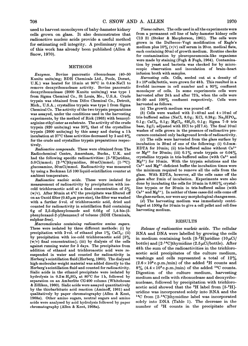
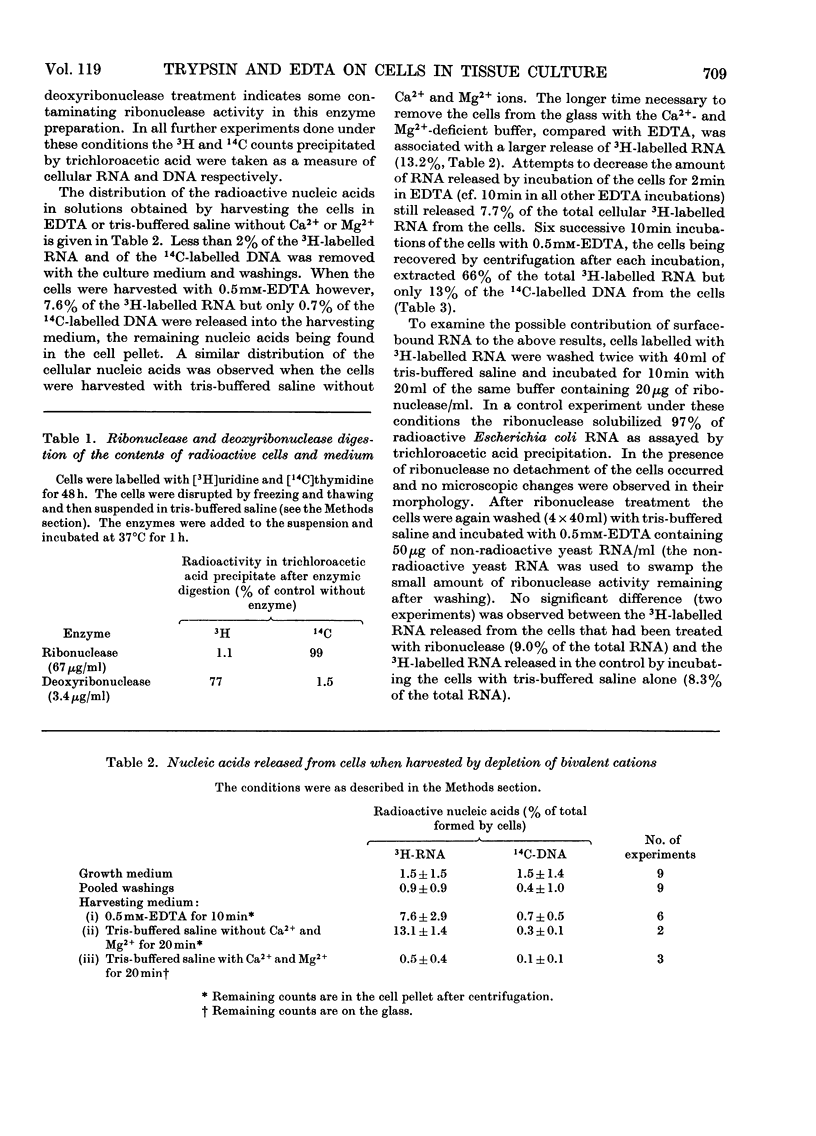
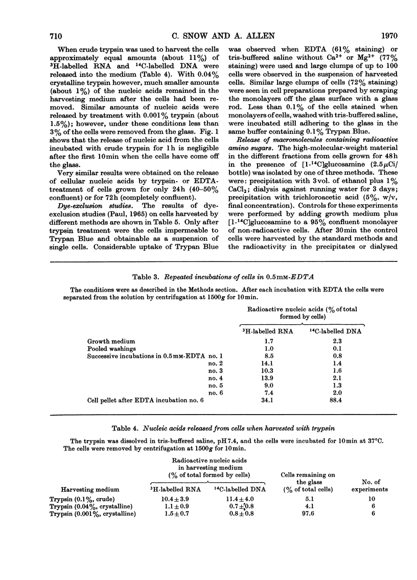
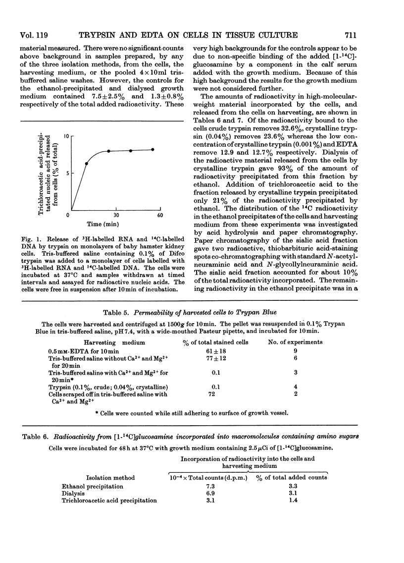
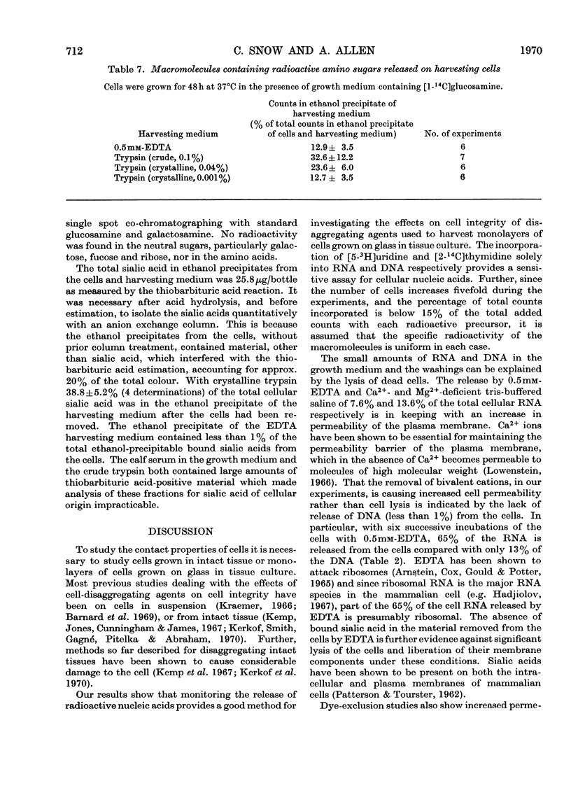
Monolayers of baby-hamster kidney cells were grown on glass in tissue culture and harvested with trypsin or EDTA in order to investigate the cell surface macromolecules removed by these cell-disaggregating agents. The release of nucleic acids from the cells during the harvesting procedure was monitored by labelling the cellular RNA with [5-3H]uridine and the cellular DNA with [2-14C]thymidine. Treatment of the cells with EDTA was found to cause an increase in the permeability of the plasma membrane with 7.6% of the cellular RNA, but less than 1% of the cellular DNA, being released. Moreover, 61% of the cells harvested with EDTA were permeable to Trypan Blue. With crude trypsin, lysis of the cell occurred with the release of similar amounts of RNA and DNA amounting to about 11% of the total cellular nucleic acid. In contrast, crystalline trypsin released only 1% of the cellular nucleic acids. Since virtually all the cells (99%) after harvesting in crystalline trypsin were impermeable to Trypan Blue, this method was suitable for obtaining cell surface macromolecules without contamination by intracellular damage. [1-14C]Glucosamine was incorporated by the cells only into bound hexosamines and sialic acids. [By monitoring the release of radioactivity in high-molecular-weight material in such experiments a measure of the release of macromolecules containing amino sugars was obtained.] Of the total macromolecules containing amino sugars in the cells 33%, 24% and 13% were released when the cells were harvested with crude trypsin, crystalline trypsin or EDTA respectively. Crystalline trypsin also released 39% of the total sialic acid of the cell, whereas less than 1% of the cellular sialic acid was present in the EDTA-treated fraction. It is concluded that the macromolecules containing amino sugars released with crude trypsin and EDTA are likely to be heavily contaminated with intracellular material. However, the macromolecules released by crystalline trypsin appear to come from the cell surface.
Full text
PDF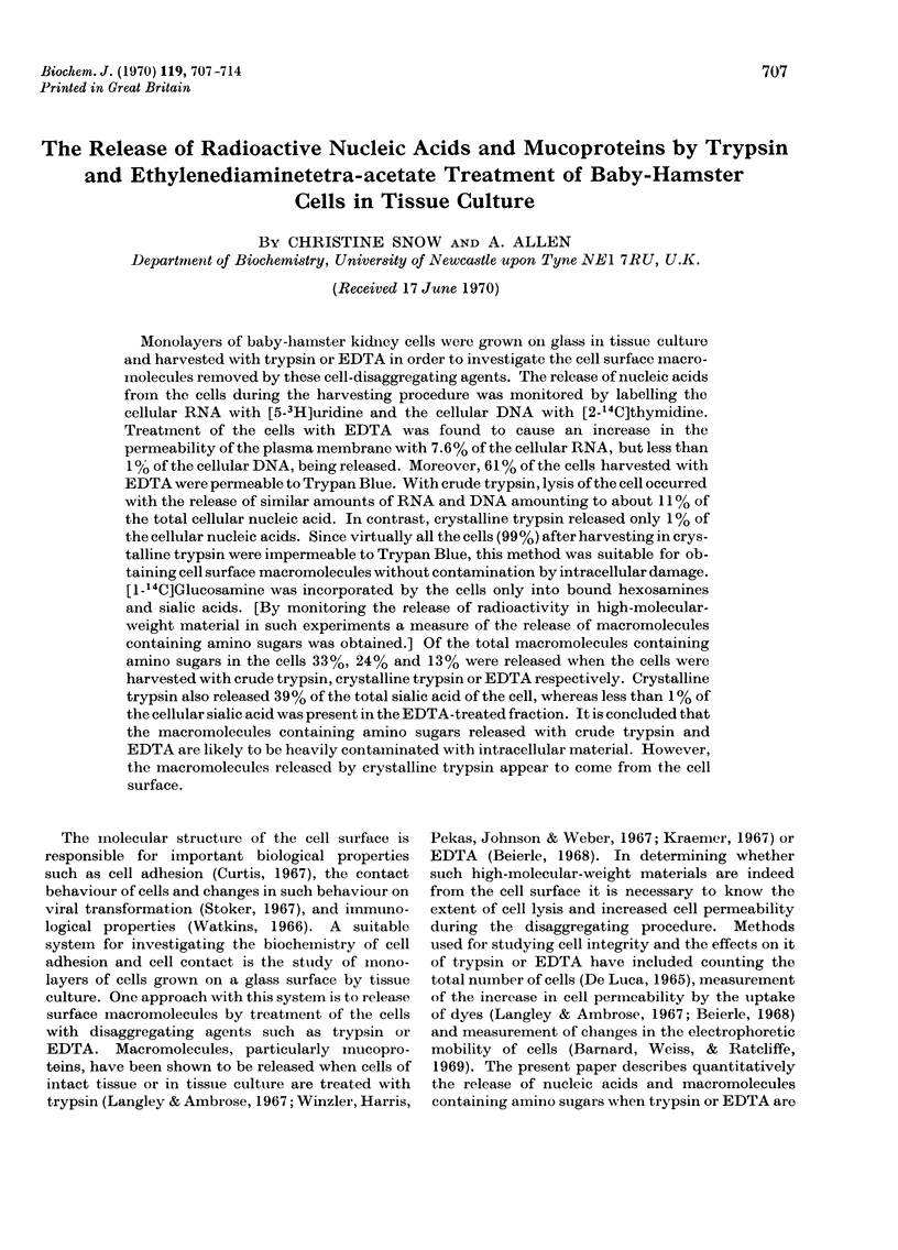
Selected References
These references are in PubMed. This may not be the complete list of references from this article.
- AMINOFF D. Methods for the quantitative estimation of N-acetylneuraminic acid and their application to hydrolysates of sialomucoids. Biochem J. 1961 Nov;81:384–392. doi: 10.1042/bj0810384. [DOI] [PMC free article] [PubMed] [Google Scholar]
- Allen A., Kent P. W. Biosynthesis of intestinal mucins. Effect of puromycin on mucoprotein biosynthesis by sheep colonic mucosal tissue. Biochem J. 1968 Jan;106(1):301–309. doi: 10.1042/bj1060301. [DOI] [PMC free article] [PubMed] [Google Scholar]
- Allen A., Kent P. W. Studies on the enzymic N-acylation of amino sugars in the sheep colonic mucosa. Biochem J. 1968 Apr;107(4):589–598. doi: 10.1042/bj1070589. [DOI] [PMC free article] [PubMed] [Google Scholar]
- Allen A., Snow C. The effect of trypsin or ethylenediaminetetraacetate on the surface of cells in tissue culture. Biochem J. 1970 Apr;117(2):32P–32P. doi: 10.1042/bj1170032pa. [DOI] [PMC free article] [PubMed] [Google Scholar]
- Arnstein H. R., Cox R. A., Gould H., Potter H. A comparison of methods for the isolation and fractionation of reticulocyte ribosomes. Biochem J. 1965 Aug;96(2):500–506. doi: 10.1042/bj0960500. [DOI] [PMC free article] [PubMed] [Google Scholar]
- Barnard P. J., Weiss L., Ratcliffe T. Changes in the surface properties of embryonic chick neural retina cells after dissociation. Exp Cell Res. 1969 Mar;54(3):293–301. doi: 10.1016/0014-4827(69)90205-5. [DOI] [PubMed] [Google Scholar]
- Beierle J. W. Cell proliferation: enhancement by extracts from cell surfaces of polyoma-virus-transformed cells. Science. 1968 Aug 23;161(3843):798–799. doi: 10.1126/science.161.3843.798. [DOI] [PubMed] [Google Scholar]
- FOGH J., FOGH H. A METHOD FOR DIRECT DEMONSTRATION OF PLEUROPNEUMONIA-LIKE ORGANISMS IN CULTURED CELLS. Proc Soc Exp Biol Med. 1964 Dec;117:899–901. doi: 10.3181/00379727-117-29731. [DOI] [PubMed] [Google Scholar]
- Hakomori S. I., Teather C., Andrews H. Organizational difference of cell surface "hematoside" in normal and virally transformed cells. Biochem Biophys Res Commun. 1968 Nov 25;33(4):563–568. doi: 10.1016/0006-291x(68)90332-x. [DOI] [PubMed] [Google Scholar]
- Kemp R. B., Jones B. M., Cunningham I., James M. C. QUuantitative investigation on the effect of puromycin on the aggregation of trypsin- and versene-dissociated chick fibroblast cells. J Cell Sci. 1967 Sep;2(3):323–340. doi: 10.1242/jcs.2.3.323. [DOI] [PubMed] [Google Scholar]
- Kerkof P. R., Smith S., Gagné H. T., Pitelka D. R., Abraham S. Nonviability of sodium tetraphenylboron-dissociated cells. Exp Cell Res. 1969 Dec;58(2):445–448. doi: 10.1016/0014-4827(69)90530-8. [DOI] [PubMed] [Google Scholar]
- Kraemer P. M. Production of heparin related glycosaminoglycans by an extablished mammalian cell line. J Cell Physiol. 1968 Apr;71(2):109–120. doi: 10.1002/jcp.1040710202. [DOI] [PubMed] [Google Scholar]
- Kraemer P. M. Regeneration of sialic acid on the surface of Chinese hamster cells in culture. II. Incorporation of radioactivity from glucosamine-1-14C. J Cell Physiol. 1967 Apr;69(2):199–207. doi: 10.1002/jcp.1040690210. [DOI] [PubMed] [Google Scholar]
- Langley O. K., Ambrose E. J. The linkage of sialic acid in the Ehrlich ascites-carcinoma cell surface membrane. Biochem J. 1967 Jan;102(1):367–372. doi: 10.1042/bj1020367. [DOI] [PMC free article] [PubMed] [Google Scholar]
- Loewenstein W. R. Permeability of membrane junctions. Ann N Y Acad Sci. 1966 Jul 14;137(2):441–472. doi: 10.1111/j.1749-6632.1966.tb50175.x. [DOI] [PubMed] [Google Scholar]
- PATTERSON M. K., Jr, TOUSTER O. Intracellular distribution of sialic acid and its relationship to membranes. Biochim Biophys Acta. 1962 Jan 29;56:626–628. doi: 10.1016/0006-3002(62)90623-6. [DOI] [PubMed] [Google Scholar]
- Richmond J. E., Glaeser R. M., Todd P. Protein synthesis and aggregation of embryonic cells. Exp Cell Res. 1968 Sep;52(1):43–58. doi: 10.1016/0014-4827(68)90546-6. [DOI] [PubMed] [Google Scholar]
- Stoker M. Contact and short-range interactions affecting growth of animal cells in culture. Curr Top Dev Biol. 1967;2:107–128. doi: 10.1016/s0070-2153(08)60285-9. [DOI] [PubMed] [Google Scholar]
- WHITEHOUSE M. W., ZILLIKEN F. Isolation and determination of neuraminic (sialic) acids. Methods Biochem Anal. 1960;8:199–220. doi: 10.1002/9780470110249.ch5. [DOI] [PubMed] [Google Scholar]
- Watkins W. M. Blood-group substances. Science. 1966 Apr 8;152(3719):172–181. doi: 10.1126/science.152.3719.172. [DOI] [PubMed] [Google Scholar]
- Weiss L. The cell periphery. Int Rev Cytol. 1969;26:63–105. doi: 10.1016/s0074-7696(08)61634-4. [DOI] [PubMed] [Google Scholar]
- Winzler R. J., Harris E. D., Pekas D. J., Johnson C. A., Weber P. Studies on glycopeptides released by trypsin from intact human erythrocytes. Biochemistry. 1967 Jul;6(7):2195–2202. doi: 10.1021/bi00859a042. [DOI] [PubMed] [Google Scholar]


