



ABSTRACT
Bacteria encounter numerous stressors in their constantly changing environments and have evolved many methods to deal with stressors quickly and effectively. One well-known and broadly conserved stress response in bacteria is the stringent response, mediated by the alarmone (p)ppGpp. (p)ppGpp is produced in response to amino acid starvation and other nutrient limitations and stresses and regulates both the activity of proteins and expression of genes. Escherichia coli also makes inorganic polyphosphate (polyP), an ancient molecule evolutionary conserved across most bacteria and other cells, in response to a variety of stress conditions, including amino acid starvation. PolyP can act as an energy and phosphate storage pool, metal chelator, regulatory signal, and chaperone, among other functions. Here we report that E. coli lacking both (p)ppGpp and polyP have a complex phenotype indicating previously unknown overlapping roles for (p)ppGpp and polyP in regulating cell division, cell morphology, and metabolism. Disruption of either (p)ppGpp or polyP synthesis led to the formation of filamentous cells, but simultaneous disruption of both pathways resulted in cells with heterogenous cell morphologies, including highly branched cells, severely mislocalized Z-rings, and cells containing substantial void spaces. These mutants also failed to grow when nutrients were limited, even when amino acids were added. These results provide new insights into the relationship between polyP synthesis and the stringent response in bacteria and point toward their having a joint role in controlling metabolism, cell division, and cell growth.
IMPORTANCE
Cell division is a fundamental biological process, and the mechanisms that control it in Escherichia coli have been the subject of intense research scrutiny for many decades. Similarly, both the (p)ppGpp-dependent stringent response and inorganic polyphosphate (polyP) synthesis are well-studied, evolutionarily ancient, and widely conserved pathways in diverse bacteria. Our results indicate that these systems, normally studied as stress-response mechanisms, play a coordinated and novel role in regulating cell division, morphology, and metabolism even under non-stress conditions.
KEYWORDS: (p)ppGpp, polyphosphate, cell division, cell morphology, stringent response, stress response
INTRODUCTION
Bacteria have evolved stress response systems to ensure their survival in ever-changing environments and against host defenses. Two widely conserved stress response strategies are the general stress response in bacteria known as the stringent response, and the nearly universally conserved polyphosphate (polyP) pathway (1–4). However, the extent and nature of the interactions between these stress response pathways is poorly understood, as are their roles in bacterial physiology under non-stress conditions.
The stringent response is mediated by the small molecule guanosine 5′-diphosphate 3′-diphosphate (ppGpp) and guanosine 5′-triphosphate 3′-diphosphate (pppGpp), known collectively as (p)ppGpp (5–7). In E. coli, (p)ppGpp is synthesized by the proteins RelA and SpoT (8–12), part of the RSH (RelA-SpoT homolog) family, with SpoT being able to both synthesize and degrade (p)ppGpp (8, 9, 13–17). SpoT is an essential gene in relA+ strains, as (p)ppGpp levels quickly rise to toxic levels in the absence of (p)ppGpp hydrolysis (18–22). (p)ppGpp can bind to and regulate the activity of proteins, including binding RNA polymerase and DksA to regulate genome-wide gene expression (23–25). (p)ppGpp downregulates DNA and RNA synthesis, protein production, and cell growth and modifies gene expression of up to one-third of the genome in E. coli in response to a wide variety of stressors (5, 6, 14, 26–30). In recent years, (p)ppGpp has emerged as a master regulator of bacterial cellular biology and is now known to affect nearly every aspect from growth rate, sporulation, motility, competence, biofilm formation, toxin production, and virulence of pathogenic bacteria (6, 27, 31–36). Relevant to the results we present here, the stringent response has also been linked to the regulation of cell division, albeit by currently unknown mechanisms (37–39).
Inorganic polyphosphate (polyP) is an evolutionarily ancient biopolymer and is found in nearly all bacterial and many eukaryotic cells (4, 40–44). In bacteria, polyP is involved in regulating gene expression, chelation of metals, acting as a protein-stabilizing chaperone, and can affect biofilm formation, stress sensing, quorum sensing, and motility (3, 4, 45–52). PolyP chains can be from dozens to thousands of phosphates long and are produced by polyphosphate kinase (PPK) and degraded by the enzyme exopolyphosphatase (PPX) (53–55). Pathogenic bacteria lose their virulence when polyP production is abolished, suggesting that polyP is vital for the ability of bacteria to cause harm, and motivating ongoing searches for PPK inhibitors as antivirulence drugs (4, 51, 56–59).
PolyP and the stringent response have long been suspected to be linked together, affecting virulence and the ability of bacteria to survive. Both relA spoT mutants lacking (p)ppGpp and ppk mutants lacking polyP have multiple amino acid auxotrophies and growth defects on minimal media, for example, suggesting parallel or linked roles in surviving nutritional stresses (48, 60, 61). (p)ppGpp is known to play a role in preventing the degradation of polyP in bacteria by inhibiting PPX (43, 44, 62, 63), but evidence also suggests other potential links between these two fundamental systems (43, 64). Recent work from our laboratory (42) showed that (p)ppGpp is not itself required for polyP synthesis, but did find a link between the production of polyP in E. coli and the transcription factor DksA, which acts in coordination with (p)ppGpp to regulate gene expression within the cell (63, 65–67). Mutations of RNA polymerase that mimic the effects of (p)ppGpp binding to that enzyme on transcription (a.k.a. stringent alleles) (68–72) also reduce polyP synthesis by an unknown mechanism (43).
While the effects of (p)ppGpp and polyP during stress have been the focus of many investigations, their effect on cell physiology during normal growth conditions remains poorly understood, and the interaction between these two fundamental systems is not clear (6, 8, 13, 36, 43, 73). In this work, we identify striking and unexpected combinatorial phenotypes in E. coli mutants lacking both (p)ppGpp and polyP that suggest that these two pathways coordinately regulate fundamental mechanisms of cell division and morphology, even in the absence of nutritional stress. This provides new insights into the roles of general stress response pathways under non-stress conditions and raises important questions about the mechanisms bacteria use to maintain their integrity during growth, highlighting gaps in our knowledge in an area that has been the subject of many decades of research scrutiny.
RESULTS
Triple mutants lacking ppk, relA, and spoT have a growth defect on a minimal medium that cannot be rescued with casamino acids
While both ppk and relA spoT mutants have well-described amino acid requirements in minimal media (44, 60, 74–76), both grow robustly on rich LB medium (Fig. 1). We were surprised, therefore, to find that a triple ppk relA spoT mutant, entirely lacking the ability to synthesize both polyP and (p)ppGpp (74, 77), has a substantial growth defect on LB (Fig. 1). Even more surprisingly, while we could easily rescue the growth defects of ppk, relA, ppk relA, and relA spoT mutants on minimal media by adding 0.05% (wt/vol) casamino acids (an undefined mixture of amino acids generated by acid hydrolysis of casein), casamino acids were unable to restore growth of the ppk relA spoT triple mutant on minimal medium. This was true both on phosphate-buffered M9 minimal medium (69.5 mM Pi) (78) and on MOPS minimal medium (1.23 mM Pi) (79). Growth of the mutant strains was not affected by plating on hypo-osmotic LBK (LB medium with no added NaCl) or hyper-osmotic LBK +500 mM sucrose (Fig. S1A). This indicated to us that there was an amino acid- and osmolarity-independent defect in the ppk relA spoT strain that was not present in either parent strain, suggesting a previously unsuspected redundant metabolic role for polyP and (p)ppGpp in E. coli.
Fig 1.

Triple mutants lacking ppk, relA, and spoT have a growth defect on a minimal medium that cannot be rescued with casamino acids. E. coli strains MG1655 (wild type), MJG0224 (MG1655 ∆ppk-749), MJG0226 (MG1655 ∆relA782), MJG1116 (MG1655 ∆ppk-749 ∆relA782), MJG1136 (MG1655 ∆relA782 spoT207::cat+), and MJG1137 (MG1655 ∆ppk-749 ∆relA782 spoT207::cat+) were grown overnight in LB broth, then rinsed and normalized to an A600 = 1 in PBS. Aliquots (5 µL) of serially diluted suspensions were spotted on LB, M9 glucose, M9 glucose containing 0.05% (wt/vol) casamino acids (c.a.a.), MOPS glucose, or MOPS glucose containing 0.05% (wt/vol) casamino acids (c.a.a.) plates and incubated overnight at 37°C (representative image from at least three independent experiments).
E. coli lacking (p)ppGpp have low levels of the general stress response sigma factor RpoS (80, 81), and polyP has also been reported to increase RpoS expression (82), so we tested whether the phenotype of a ppk relA spoT mutant was also present in a ppk rpoS mutation, but it was not (Fig. S1B) indicating that the role of (p)ppGpp in a ppk mutant is independent of RpoS-dependent transcriptional regulation (81). We also quantified guanosine nucleotide pools in E. coli in minimal media and found that (p)ppGpp accumulation was significantly higher in ∆ppk cells than in ppk+ strains (Fig. S2A and B), supporting the idea that these two molecules can in some way compensate for each other’s absence.
The growth defect of a ppk relA spoT mutant on a minimal medium with casamino acids can be rescued by the expression of either PPK or synthetase-active SpoT
Growth of the ppk relA spoT mutant on a minimal medium containing casamino acids was restored by ectopic expression of either PPK or SpoT (Fig. 2A). Complementation by SpoT was dependent on (p)ppGpp synthesis, since expression of a SpoTD73N mutant allele lacking (p)ppGpp hydrolase activity (83) restored growth, but the expression of SpoTD259N and SpoTD73N, D259N alleles, which lack (p)ppGpp synthetase activity (83), did not. Expression of wild-type SpoT enhanced the growth of a relA spoT mutant on a minimal medium containing casamino acids (Fig. 2B), but expression of SpoT alleles lacking (p)ppGpp synthetase activity did not, suggesting that (p)ppGpp synthesis underlies this effect. The relA spoT mutant could not be transformed with a plasmid encoding hydrolase-defective SpoTD73N (data not shown). This was unsurprising since accumulation of (p)ppGpp in E. coli cells lacking (p)ppGpp hydrolase activity is well known to prevent growth (74–76). The more surprising result from this experiment is that the ppk relA spoT mutant did tolerate SpoTD73N expression (Fig. 2A), suggesting that polyP is somehow involved in (p)ppGpp-dependent growth inhibition.
Fig 2.

The growth defect of a ppk relA spoT mutant on a minimal medium with casamino acids can be rescued by expression of either PPK or synthetase-active SpoT. E. coli strains (A) MJG1137 (MG1655 ∆ppk-749 ∆relA782 spoT207::cat+) or (B) MJG1136 (MG1655 ∆relA782 spoT207::cat+) containing the indicated plasmids (pppk+ =pPPK8, pspoT+ =pSPOT1, pspoTD73N = pSPOT2, pspoTD259N = pSPOT3, pspoTD73N, D259N = pSPOT4) were grown overnight in LB broth containing ampicillin, then rinsed and normalized to an A600 = 1 in PBS. Aliquots (5 µL) of serially diluted suspensions were spotted on LB, M9 glucose, or M9 glucose containing 0.05% (wt/vol) casamino acids (c.a.a.) plates containing ampicillin and incubated overnight at 37°C (representative image from at least three independent experiments).
To further probe the effect of polyP on the phenotype of these strains, we added exogenous polyP to the growth media, which had no effect on the growth of any tested strain (Fig. S3A), indicating that polyP must be produced intracellularly to impact these phenotypes. We also expressed the hyperactive PPKE245K variant (84, 85) from a plasmid to enhance polyP levels above natural levels (Fig. S3B). This rescued the growth defects of ppk, ppk relA, and, surprisingly, relA spoT mutants on MOPS glucose without amino acids as well as partially restoring growth to the ppk relA spoT mutant on MOPS glucose with 0.05% casamino acids (Fig. S3C), similar to the effect of the wild-type ppk plasmid in Fig. 2A.
Both tryptone and yeast extract contain components that rescue the growth defect of a ppk relA spoT mutant on a minimal medium with casamino acids
LB medium consists of 1% (wt/vol) of tryptone (a tryptic digest of casein), 0.5% (wt/vol) of yeast extract (prepared from the soluble fraction of boiled Saccharomyces cerevisiae cells), and 0.5% (wt/vol) of NaCl (78). Both tryptone and yeast extract at these concentrations restored growth of the ppk relA spoT triple mutant on a minimal medium containing casamino acids (Fig. 3A). The same effect could be seen in liquid media and with strains containing a complete deletion of spoT rather than the spoT207::cat+ insertion mutation (Fig. S4). Those experiments also showed that growth of all of the tested mutant strains in minimal media supplemented with a defined mixture of purified amino acids (yeast synthetic dropout mix supplement; Sigma Aldrich cat. #Y1501) was comparable to that in media supplemented with casamino acids, supporting our conclusions from Fig. 3A. However, we found that variability was high in liquid media, especially later in the growth curves (possibly due to unpredictable accumulation of suppressor mutations), and opted to continue using solid media to evaluate the growth phenotype of the ppk relA spoT mutant.
Fig 3.

Both tryptone and yeast extract contain components that rescue the growth defect of a ppk relA spoT mutant on a minimal medium with casamino acids. E. coli strains MG1655 (wild type), MJG0224 (MG1655 ∆ppk-749), MJG1136 (MG1655 ∆relA782 spoT207::cat+), and MJG1137 (MG1655 ∆ppk-749 ∆relA782 spoT207::cat+) were grown overnight in LB broth, then rinsed and normalized to an A600 = 1 in PBS. Aliquots (5 µL) of serially diluted suspensions were spotted on LB, M9 glucose, or M9 glucose containing the indicated percentages (wt/vol) of casamino acids (c.a.a.) and (A) tryptone or yeast extract (YE) or (B) yeast extract fractions containing compounds either greater than 3,500 Da or less than 3,000 Da and incubated overnight at 37°C (representative image from at least three independent experiments).
We used filtration and dialysis to divide yeast extract into fractions with molecular weights greater than 3,500 Da or less than 3,000 Da and found that the component(s) responsible for restoring growth of the ppk relA spoT mutant on minimal medium containing casamino acids were present in the small molecule (e.g., less than 3,000 Da) fraction (Fig. 3B). Because LB medium is regularly sterilized by autoclaving, the compound(s) in question must also be heat-stable, and we are currently working to identify the relevant molecule(s), which we expect to give insights into the metabolic pathway(s) responsible for the inability of the ppk relA spoT mutant to grow on minimal media.
Morphological defects of ppk relA spoT cells 1: filamentation
Since the ppk relA spoT mutant did not grow as well on LB plates as the other mutant strains, we wanted to see whether there were visible morphological defects present in this strain or its parent strains. We performed confocal time-lapse microscopy and noted that cells lacking either polyP or (p)ppGpp grown in LB are longer than wild-type cells, indicating defects in cell division (Fig. 4). Our results showed that a ppk mutant lacking polyP was slightly longer than the wild type MG1655 while growing in exponential phase on LB (Fig. 4). Strains deficient in (p)ppGpp were also filamentous, with a relA mutant being longer on average than a double relA spoT mutant (Fig. 4). The ppk relA mutant lacking polyP and only containing SpoT for production of (p)ppGpp were about the same length as the relA mutant or the relA spoT mutant (Fig. 4). These results are in general agreement with previous work in E. coli showing increased cell length in relA spoT mutants (86) and in Pseudomonas aeruginosa showing that strains lacking polyP become filamentous in stationary phase (87). The triple mutant ppk relA spoT contained filamentous cells as well (Fig. 4); however, these cells were much more heterogenous than any of the other mutants, appearing less stable with odd morphologies that we will explore in depth in the following sections. When grown on MOPS minimal media agarose pads, the mutant cells were no longer filamentous and had lengths more similar to the wild type (Fig. 4C) (compare, for example, the wild-type and ppk relA strains growing on MOPS in Videos SV1 and SV2). Nevertheless, even under these conditions, relA, relA spoT, and ppk relA spoT mutant cells were significantly longer than the wild type. The ppk relA spoT triple mutant immediately stopped growing on the MOPS agarose pad and slowly started to shrink over several hours (Video SV3). Over 3 hours on MOPS minimal media agarose pads, ppk relA spoT mutants shrank an average of 15.3% in total length (Video SV3; Table S1).
Fig 4.

Microscopy of polyP and (p)ppGpp mutants shows filamentous cell growth. (A) Confocal microscopy images of MG1655 (MJG0001), ∆ppk (MJG0224), ∆relA (MJG0226), ∆ppk ∆relA (MJG1116), ∆relA ∆spoT (MJG1287), and ∆ppk ∆relA ∆spoT (MJG1282). Images were captured on LB agarose pads incubated at 37°C and imaged every 5 minutes while growing. (B) Length of cells growing at 37°C on an LB agarose pad and (C) length of cells growing at 37°C on a MOPS minimal media agarose pad. Length of cells was determined and manually calculated using FIJI using the line tool and measuring length with built-in measurements to FIJI after setting the appropriate scale. n = number of cells. Statistical significance was calculated in Prism GraphPad by one-way ANOVA, *P-value <0.05, **P-value <0.005, ***P-value <0.0005, ****P-value <0.0001.
Morphological defects of ppk relA spoT cells 2: displaced FtsZ ring formation and deviant cell division
To determine where cell division was taking place in these filamentous cells, we used an FtsZ-GFP reporter to observe where the Z-ring was localizing within cells during cell division (88). During time-lapse microscopy, we observed the FtsZ reporter forming a Z-ring at the midpoint of wild-type cells as expected (Fig. S5; Video SV4) (89–91). The double ppk relA mutant and the triple mutant ppk relA spoT however showed that, while some FtsZ rings formed normally, there were many cells with profoundly disrupted Z-ring localization phenotypes. We observed cells containing multiple Z-rings within a single cell (Fig. 5A and B; Fig. S6), including instances with two Z-rings forming in the middle of a cell with both Z-rings constricting and resulting in the release of a non-growing mini-cell (Fig. 5A; Fig. S6).
Fig 5.

Strains lacking polyP and (p)ppGpp have disrupted cell division. (A) Confocal fluorescence time-lapse microscopy of the mutant ppk relA spoT FtsZ-GFP (MJG2405) on an LB agarose pad at 37°C. The triple mutant forms two Z-rings in the middle of the cell, releasing a mini-cell (red arrows). There are also two Z-rings that form at either pole of a single cell, both functional and releasing a mini cell (black arrows). Single channel images of TD and GFP alone can be viewed in Fig. S6. Examples of mini-cell formation and release can also be seen by transmission and cryo-electron microscopy (Fig. S9 and S11). (B) Confocal fluorescence time-lapse microscopy of the mutant ppk relA FtsZ-GFP (MJG2403) on an LB agarose pad at 37°C. This image shows a mutant forming three Z-rings at one pole, and at least three at the opposite pole as well, with no Z-rings forming in the middle of the cell, for a total of six Z-rings in a single cell. This cell continued to grow without lysing (Video SV5). (C) Quantification of FtsZ-ring location within the cell, and its relationship to cell length in E. coli strains MG1655 FtsZ-GFP (MJG2041), ppk relA FtsZ-GFP (MJG2403), and ppk relA spoT FtsZ-GFP (MJG2405). Cell length is shown on the y-axis, with the corresponding location of the FtsZ-ring on the x-axis as a fraction of the total cell length. Positions of FtsZ rings were obtained by measuring, in pixels, the distance between the center of the FtsZ band and the cell pole. Poles were arbitrarily designated either 0 or 1, with the midpoint of the total cell length being 0.5.
Not only were multiple Z-rings forming at the midpoint of cells, but we also observed Z-rings forming at the poles of cells dividing and releasing mini-cells (Fig. 5A and B; Video SV5). FtsZ rings appeared to be able to form nearly anywhere along the length of the cell in the ppk relA double and ppk relA spoT triple mutant (Fig. 5C). In the ppk relA strain roughly 4.7% of cells observed had disrupted FtsZ ring placement, while the ppk relA spoT mutant had about 0.8% of cells with disrupted FtsZ ring placement (Table S1). We also observed one instance of an FtsZ ring forming at the side wall of a branching ppk relA cell, pinching off and releasing a mini-cell from the side wall of a branching cell (Fig. S7). Most cells did appear to divide normally when cell division occurred, with filamentous cells failing to localize a Z-ring until division occurred. The ppk and relA spoT mutants did not have these errors with FtsZ formation and formed Z-rings normally in LB at 37°C (Fig. S5).
When the ppk relA spoT mutant was compared to the wild-type by transmission electron microscopy (TEM) and cryo-electron microscopy (CEM), we confirmed these observations (Fig. S8 to S11) and noticed other cell division defects in the triple mutant. We observed cells appearing to divide, but without forming a proper septum fully separating the dividing cells (Fig. 6). We observed a cell where cell division had mostly occurred and the cytoplasm was divided, with the two cells still connected by a membrane bridge (Fig. 6A and B). We also observed cells where cell division was actively occurring with a neck-like bridge between both cells still connecting the cytoplasm from one cell to another (Fig. 6C and D). None of these phenomena were observed in wild-type cells and could conceivably reflect a variety of defects in cell division in the ppk relA spoT mutant, either up- or downstream of the involvement of FtsZ itself.
Fig 6.

TEM of ppk relA spoT cells failing to divide properly. (A) TEM of ppk relA spoT (MJG1282) failing to divide properly. The cytoplasm of this cell appears to have condensed within the cell and away from the divisisome site but is still connected by what appears to be a small bridge of membrane that has yet to separate and could be the result of disrupted FtsZ-ring formation. (B) This image is the square section from (A) at a higher magnification. (C) TEM of ppk relA spoT (MJG1282) not completely forming a divisisome and staying connected through a bridge while still sharing cytoplasmic contents between the undivided cells. (D) This image is the square section from (C) at a higher magnification.
Morphological defects of ppk relA spoT cells 3: branched cells
While observing the ppk relA double mutant and the ppk relA spoT triple mutants, we discovered that these mutants were able to form very unusual branched cells, along with other odd and unexpected cell morphologies. Only cells lacking both polyP and one or both (p)ppGpp synthetases were able to form branched cells, including instances of cells with more than three distinct poles (Fig. 7A through C; Fig. S7). In the ppk relA double mutant, we were able to observe a branched cell developing over time with FtsZ still capable of forming Z-rings and causing cell division (Fig. 7A). The frequency of cell branching was relatively low but contrasted starkly with the fact that it never occurred in wild-type, ppk, relA, or relA spoT strains. The double mutant ppk relA developed branched cells slightly less frequently (~1.9% of cells) than the triple mutant ppk relA spoT (~2.2% of cells) (Table S1), but there was less uniformity in branched cells of the triple mutant. The triple mutant developed into more heterogeneous branching cells with more noticeable defects in cell wall morphology including the formation of spheroplasts (Fig. 7C; Videos SV6 to SV8). The development of branched cells did not depend on the presence of the FtsZ-GFP reporter, as we observed the same morphologies in strains lacking the reporter (Fig. 7C). We also observed branching cells by TEM (Fig. 7D). These results may suggest a combined or redundant role for polyP and (p)ppGpp in regulating cell wall synthesis and/or integration of newly synthesized peptidoglycan, defects in which have been reported to result in branching in E. coli cells (92, 93).
Fig 7.

Cells lacking polyP and (p)ppGpp can develop branching cell morphologies. (A) Confocal fluorescence time-lapse microscopy of the mutant ppk relA FtsZ-GFP (MJG2403) on an LB agarose pad at 37°C showing branching cells. Branched cells are still capable of dividing and growing. (B) Still image of confocal fluorescent microscopy of ppk relA FtsZ-GFP mutant (MJG2403) mutant on LB agarose pad at 37°C showcasing branched cells. (C) Still image of confocal microscopy of ∆ppk ∆relA ∆spoT (MJG1282) mutant on LB agarose pad at 37°C showcasing branched cells and other very odd cell morphologies. (D) Transmission electron microscopy image of ppk relA spoT (MJG1282) grown in LB at 37°C until log phase prior to imaging. This image shows a cell developing what appears to be a branching pole developed from the sidewall of the cell.
Morphological defects of ppk relA spoT cells 4: cell envelope defects and cytoplasmic condensation
Another unexpected phenotype we noted while observing the ppk relA spoT triple mutant was the presence of cells developing what appear as “holes” or void spaces within the bacterial cell, appearance of which preceded the leakage of cytoplasmic contents out of the cell, as represented by the FtsZ-GFP fusion protein (Fig. 8A). To investigate this phenotype more closely, we performed cryo-electron microscopy of the triple mutant, looking for evidence of disrupted cellular membranes. We imaged what appears to be an invagination of the cell wall, with cytoplasmic contents being released as blebs from the cell surrounded by a cell wall and membranes (Fig. 8B). The site where cytoplasmic contents appear to be “leaking” from may be a site of cell division, as we saw similar invaginated structures form in dividing cells (Fig. S10 and S11). We also observed disruptions in the outer membrane of the triple mutant (Fig. 8B), with what appears to be cytoplasmic contents condensing oddly within the cell (Fig. 8B; Fig S12 and S13).
Fig 8.
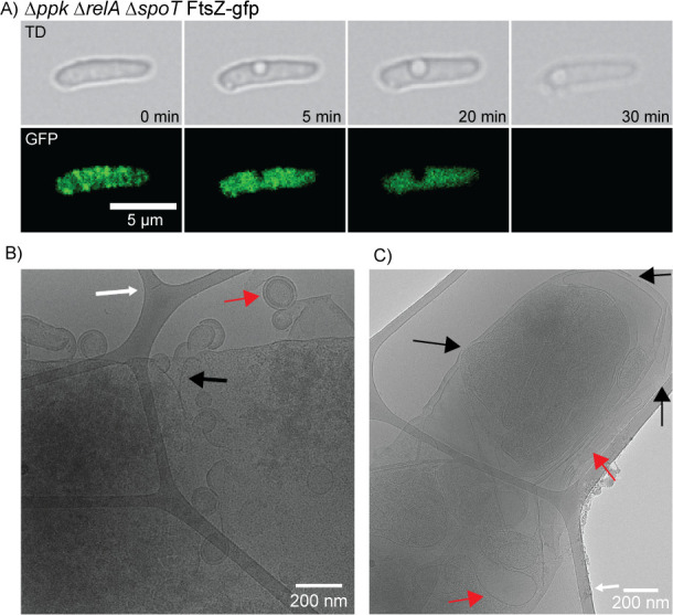
ppk relA spoT triple mutant cells can develop perforated membranes, leaking cytoplasmic contents out of the cell. (A) Time-lapse fluorescent microscopy of the ppk relA spoT mutant with the FtsZ-GFP reporter (MJG2405) showing empty space within the cell. FtsZ appears to fail to localize within the cell prior to losing its cytoplasmic contents just before cell death occurs. (B) Cryo-electron microscopy image of ppk relA spoT mutant (MJG2405) showing what appears to be a leaking cell wall with cytoplasmic contents blebbing off. White arrows point to the carbon lattice that cells were suspended on for imaging in CEM. There appears to be an invagination of the cell wall where cytoplasmic contents seem to be being released (black arrow). The cytoplasmic contents appear to be surrounded by a fully intact cell wall including an outer membrane, peptidoglycan, and inner membrane (red arrow). (C) CEM image of ppk relA spoT mutant (MJG2405) showing what appears to be holes in the cellular membrane (black arrows) where cytoplasmic contents and fluid may be lost. There also appears to be cytoplasm condensation, likely from fluid loss, with the cytoplasm condensing with the cell wall and membrane folding in on itself (red arrow).
Using TEM, we observed ppk relA spoT mutant cells that appeared to have their cytoplasm and inner membrane shrunk away from the cell wall, creating large periplasmic spaces in ~1.1% of cells observed (Table S1), a phenomenon known as plasmolysis (Fig. 8 and 9; Fig. S14 and S15). Plasmolysis has been known to be caused by a hyperosmotic shock in bacteria, although in that case the cytoplasmic shrinkage is relatively uniform among cells in a population (94, 95) and as shown in Fig. S1A, changes in medium osmolarity do not impact the growth of the ppk relA spoT mutant. These results suggest there might be a mechanism by which polyP and (p)ppGpp co-regulate turgor pressure or potentially for dysregulation of efflux pumps causing the loss of cytoplasmic contents and subsequent efflux of water. This dehydration, however, could also be due to loss of water through a leaking membrane (Fig. 8) or other unknown mechanisms. In another paper reporting similar-appearing void spaces in bacterial cytoplasms, the authors believed they were seeing cytoplasmic condensation caused by disruptions in the cell envelope due to treatment with sublethal concentrations of antibiotics (96), in which case our observations might imply a role for polyP and (p)ppGpp in synthesis and/or incorporation of the newly synthesized peptidoglycan into the cell wall. Regardless of the underlying mechanism(s), these results indicate that in a ppk relA spoT mutant, some cells are unable to properly maintain their growing cell wall or membranes.
Fig 9.
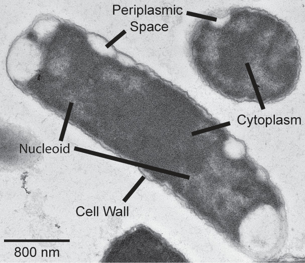
Transmission electron micrograph of ppk relA spoT mutant showing plasmolysis in both cross-sectional and trans-sectional viewpoint. TEM of ppk relA spoT (MJG1282) shows the inner membrane appearing to shrink and pull away from the cell wall (plasmolysis), leaving large periplasmic spaces within the cell. We can see here in both the longitudinal and horizontal axis of the cell wall surrounding the cell with large open spaces around the perimeter of the cell where there appears to be no cytoplasm creating these periplasmic spaces and resulting in plasmolysis. The cell appears to still be actively dividing as we can see in the longitudinal axis it appears that the nucleoid of the cell is being restricted to the poles of the cell away from the midline as we would expect in cells about to divide.
Stringent alleles of RNA polymerase restore some, but not all phenotypes of a ppk relA spoT mutant
The amino acid auxotrophy of relA spoT mutants can be rescued by mutations in RNA polymerase called stringent alleles, which mimic the regulatory effect of (p)ppGpp binding to RNA polymerase on transcription, including the activation of expression of amino acid synthesis operons (69, 70, 75, 76). Many of these mutations in RNA polymerase also confer rifampicin resistance (97). The stringent alleles rpoB3443 and rpoB3449 (70) were, as expected, able to restore the growth of a relA spoT mutant in the absence of casamino acids, while the rifampicin-resistant but non-stringent allele rpoB148 (97) did not (Fig. 10A). Notably, rpoB3443 and rpoB3449 restored growth of the ppk relA spoT triple mutant in the presence of casamino acids, but not in their absence. Microscopic observation of a ppk relA spoT rpoB3443 mutant grown on LB, however, revealed a dramatic restoration of wild-type cell morphology (Fig. 10B; Video SV9). The growth rates of the ppk relA spoT and ppk relA spoT rpoB3443 mutants in LB, while considerably slower than the wild type, were not different from each other (Fig. S16), so this morphological rescue was not due to a change in growth rate.
Fig 10.

Stringent alleles of RNA polymerase restore growth of ppk relA spoT mutants on minimal medium with casamino acids and rescue morphological defects on rich medium. (A) E. coli strains MG1655 (wild-type), MJG1136 (MG1655 ∆relA782 spoT207::cat+), MJG1137 (MG1655 ∆ppk-749 ∆relA782 spoT207::cat+), MJG1237 (MG1655 ∆relA782 spoT207::cat+ rpoB3449), MJG1241 (MG1655 ∆ppk-749 ∆relA782 spoT207::cat+ rpoB3449), MJG1579 (MG1655 ∆relA782 spoT207::cat+ rpoB3443), MJG1580 (MG1655 ∆relA782 spoT207::cat+ rpoB148), MJG1581 (MG1655 ∆ppk-749 ∆relA782 spoT207::cat+ rpoB3443), and MJG1582 (MG1655 ∆ppk-749 ∆relA782 spoT207::cat+ rpoB148) were grown overnight in LB broth, then rinsed and normalized to an A600 = 1 in PBS. Aliquots (5 µL) of serially diluted suspensions were spotted on LB, M9 glucose, or M9 glucose containing 0.05% (w/v) casamino acids (c.a.a.) plates and incubated overnight at 37°C (representative image from at least three independent experiments). (B) Confocal microscopy of ppk relA spoT rpoB3443 (MJG1581) grown on a LB agarose pad at 37°C.
These results taken together suggest that the nutritional (Fig. 1 to 3 and 10A) and morphological (Fig. 4 to 9 and 10B) phenotypes of the ppk relA spoT mutant can be genetically separated and that the morphological phenotypes of this strain in particular appear to be linked to transcriptional regulation by (p)ppGpp. By contrast, the combinatorial growth defect of the triple mutant on a minimal medium may be dependent on (p)ppGpp’s impacts on a protein or proteins other than RNA polymerase (66, 75, 76). These results also reinforce our conclusion that whatever compound(s) are present in LB that allow growth of the triple mutant (Fig. 3) are not likely to be amino acids.
DISCUSSION
Connections between (p)ppGpp and polyP in E. coli have been suspected for decades (44, 62), but the nature and consequences of those connections have remained obscure (42, 43, 47, 98). We have now identified a striking combinatorial phenotype that clearly demonstrates that these two conserved “stress response” molecules play important linked roles in controlling fundamental metabolic processes under non-stress growth conditions. The fact that these phenotypes appear only when both (p)ppGpp and polyP are eliminated suggests that either one alone is sufficient to maintain more or less normal cells, and that therefore some critical pathway or pathways must be regulated by both molecules. The challenge that remains is to dissect the mechanism(s) by which (p)ppGpp and polyP coordinate these processes.
Both (p)ppGpp and polyP can act at multiple regulatory levels in the bacterial cell. The regulatory consequences of (p)ppGpp synthesis are better studied and include dramatic changes in the genome-wide transcriptome of E. coli due to (p)ppGpp binding to RNA polymerase and the transcription factor DksA (2, 63). (p)ppGpp can also direct regulation of a variety of other enzymes (1, 26, 66), including notably the exopolyphosphatase PPX, which is inhibited by (p)ppGpp (44, 55, 62). We do not know at this time whether PPX impacts the phenotypes reported here. More than 700 genes are transcriptionally regulated by (p)ppGpp, approximately 400 of which are inhibited and 300 are stimulated (including, to a modest extent, ppk)(99), but the exact list depends on growth conditions and on how (p)ppGpp synthesis is induced (99, 100). Although stringent alleles of RNA polymerase have been known for many decades and are thought to mimic the effects of (p)ppGpp binding (69, 70, 75, 76), to our knowledge no genome-wide characterization of their impact on transcription has been performed. More than 50 other proteins that bind (p)ppGpp in E. coli have been identified (66), and the impact of that binding has been characterized for only a subset of those proteins (1, 26, 66). No experiments have been reported to date examining the polyP protein interactome in E. coli. It also remains possible that some effects of PPK might be either indirect, for example by impacts on nucleotide pools (Fig. S2)(41) or entirely independent of polyP synthesis, although there are no consistently identified PPK protein-binding partners in the literature for protein-protein interactions in E. coli MG1655 (101–104).
PolyP can also act at multiple levels, although its impact is considerably less well characterized. PolyP interacts directly with the Lon protease, for example, to modulate its activity and substrate specificity (105–108), which could affect a very broad range of potential protein targets in the cell both directly and indirectly. In combination with Hfq, polyP is also able to silence transcription of some genes (109), and a ∆ppk mutant has substantial changes in its transcriptome and proteome (110), although how much of this is due to direct regulatory impacts as opposed to indirect responses to a lack of polyP remains unclear. Whether other E. coli proteins might be impacted by polyP binding is not well known, since to our knowledge no systematic study identifying polyP-binding proteins in bacteria has been performed (111, 112). The fact that we can genetically distinguish between the metabolic and morphological phenotypes of ppk relA spoT mutants (Fig. 10) strongly suggests that at least two different pathways are co-regulated by (p)ppGpp and polyP, which further complicates the problem.
Based on the existing literature, we can identify a few potential overlaps between the (p)ppGpp and polyP regulons in E. coli which might be relevant to the phenotypes we observe in ppk relA spoT mutants. As one example, (p)ppGpp binds to the DapB protein (66) and upregulates transcription of many of the dap genes involved in the synthesis of the peptidoglycan precursor diaminopimelate (99, 113), while in a ∆ppk mutant, Varas et al. (110) reported an increase in DapA protein levels and a decrease in dapF transcription. In another example, FtsY, an essential component of the signal recognition particle that delivers integral membrane proteins to the inner membrane (114), is allosterically inhibited by (p)ppGpp binding (66, 115), modestly downregulated transcriptionally by (p)ppGpp (99), and its transcription is increased in a ∆ppk mutant (110). Either of these pathways could conceivably contribute to the cell envelope defects we observe in ppk relA spoT mutants (Fig. 7 to 9), but since each of those experiments was performed under different conditions and in varying strain backgrounds it is not possible to make definite conclusions about whether these potential points of regulatory overlap are either real or meaningful, and certainly not about whether they contribute to the phenotypes of ppk relA spoT strains. We are currently working to characterize the transcriptional and post-transcriptional impacts of (p)ppGpp and polyP on E. coli under growth conditions where we observe ppk relA spoT phenotypes, with the goal of systematically identifying genes, proteins, and pathways impacted by both molecules.
Perhaps the most surprising and most difficult to explain of the phenotypes of the ppk relA spoT mutant is the mislocalization of Z-rings (Fig. 5 to 7). The positioning of FtsZ at the center of the cell is a highly regulated part of cell division (116). E. coli has two major systems for preventing formation of Z-rings anywhere other than mid-cell: the MinCDE system that inhibits Z-ring formation at the poles (117) and the SlmA nucleoid exclusion protein that prevents Z-ring formation around the DNA nucleoid (118). Neither of these systems has been reported to be impacted by either polyP or (p)ppGpp, although examination of published transcriptome data indicates that minD and minE are slightly upregulated and slmA is slightly downregulated by (p)ppGpp (99). Mutants lacking minCDE have Z-ring distributions (119) reminiscent of those we observed in ppk relA and ppk relA spoT mutants (Fig. 5C), but have not been reported to have any of the other growth or morphological defects we observed. Overexpression of FtsZ can also result in production of multiple Z-rings within cells, including both mid-cell and polar localizations (120), reminiscent of some of our observations of ppk relA spoT mutants containing many concurrent Z-rings (Fig. 5). FtsZ regulation is complex, but not known to depend directly on either (p)ppGpp or polyP (116). Our data strongly suggest that there are important unknowns remaining in our understanding of the regulation of proper cell division in E. coli.
In E. coli, both (p)ppGpp and polyP are present at very low levels under non-stress conditions and those levels are strongly increased by various stresses, which is critical for the ability of the bacteria to survive those stress treatments (2–4). However, the cell morphology phenotypes we report here for the ppk relA spoT mutant are in a rich LB medium, meaning that those phenotypes depend on unstimulated basal levels of both (p)ppGpp and polyP. Levels of polyP are extremely low in E. coli grown in LB, often below the limit of detection for standard assays (Fig. S3B) (41, 43, 47, 77, 121). There is an increasing appreciation in the literature that basal levels of (p)ppGpp make important contributions to diverse aspects of E. coli physiology (122), including cell division (123). The same seems likely to be true of basal polyP levels since substantial changes in gene expression and proteome composition are seen in ∆ppk mutants grown in rich medium (109, 110), but more sensitive detection methods may need to be developed to probe this in more detail (124).
A final point worth considering is the surprising heterogeneity of the ppk relA spoT cells. As shown in Fig. 4 to 9; Table S1, not every cell of the triple mutant has the same morphological defects. Some of them look fairly normal, some are filamentous, some have mislocalized Z-rings, and a few develop more severe defects, including branching, spheroplast formation, void spaces, or lysis. What underlies this diversity and what distinguishes individual cells that do well from cells that do not is an intriguing unanswered question.
MATERIALS AND METHODS
Databases and primer design
We obtained gene and protein sequences and other information from the Integrated Microbial Genomes database (125) and from the EcoCyc (113) and designed PCR and sequencing primers with Web Primer (www.candidagenome.org/cgi-bin/compute/web-primer). Mutagenic primers were designed with PrimerX (www.bioinformatics.org/primerx/index.htm).
Bacterial strains and growth conditions
All strains and plasmids used in this study are listed in Table 1. We grew E. coli at 37°C in lysogeny broth (LB)(126), containing 5 g L−1 NaCl unless otherwise indicated, in M9 minimal medium (78) containing 4 g L−1 glucose, or in MOPS minimal medium (79) containing 2 or 4 g L−1 glucose. Solid media contained 1.5% (wt/vol) agar (Becton Dickinson cat. #214010). Ampicillin (100 µg mL−1), chloramphenicol (17.5 or 35 µg mL−1), kanamycin (25 or 50 µg mL−1), or rifampicin (50 µg mL−1) were added when appropriate. Nutritional supplements used were yeast synthetic dropout mix supplement (Sigma Aldrich cat. #Y1501), casamino acids (Fisher Scientific cat. #BP1424), tryptone (Fisher Scientific cat. #BP1421), or yeast extract (Becton Dickinson cat. #288620). For growth curves, E. coli strains of interest were grown overnight at 37°C with shaking in LB, then normalized to A600 = 1 and rinsed three times with sterile PBS. The resulting cell suspensions were diluted 1:40 into fresh media. Growth curves were performed in clear 96-well plates in Tecan Spark or Sunrise plate readers, incubating at 37°C with shaking and measuring A600 at 30 minutes of intervals for 24 hours.
TABLE 1.
Strains and plasmids used in this studya
| Strain or plasmid | Marker(s) | Relevant genotype | Source |
|---|---|---|---|
| E. coli strains | |||
| MG1655 | F−, λ−, rph-1 ilvG− rfb-50 | (127) | |
| CF1693(M+) | CmR KnR | MG1655 λ+ϕ80+ relA251::kan+ spoT207::cat+ rpoB1693 (encoding RpoBN1129K) rpoD1693 (encoding RpoDD64G) | (128, 129) |
| BH330 | ApR | MG1655 λattB::Plac-gfp-ftsZ, bla+ | (88) |
| MJG0224 | MG1655 ∆ppk-749 | (41) | |
| MJG0226 | MG1655 ∆relA782 | (43) | |
| MJG0344 | MG1655 ∆rpoS746 | (43) | |
| MJG1090 | KnR | MG1655 ∆ppk-749 ∆relA782::kan+ | |
| MJG1097 | KnR | MG1655 ∆rpoS746 ∆relA782::kan+ | |
| MJG1116 | MG1655 ∆ppk-749 ∆relA782 | ||
| MJG1119 | MG1655 ∆rpoS746 ∆relA782 | ||
| MJG1136 | CmR | MG1655 ∆relA782 spoT207::cat+ | (43) |
| MJG1137 | CmR | MG1655 ∆ppk-749 ∆relA782 spoT207::cat+ | |
| MJG1236 | RifR | MG1655 ∆ppk-749 ∆relA782 rpoB3449 (encoding RpoB∆Ala532) | |
| MJG1237 | CmR RifR | MG1655 ∆relA782 spoT207::cat+ rpoB3449 (encoding RpoB∆Ala532) | (43) |
| MJG1241 | CmR RifR | MG1655 ∆ppk-749 ∆relA782 spoT207::cat+ rpoB3449 (encoding RpoB∆Ala532) | |
| MJG1282 | KnR | MG1655 ∆ppk-749 ∆relA782 ∆spoT1000::kan+ | |
| MJG1287 | KnR | MG1655 ∆relA782 ∆spoT1000::kan+ | (43) |
| MJG1575 | RifR | MG1655 ∆relA782 rpoB3443 (encoding RpoBL533P) | |
| MJG1576 | RifR | MG1655 ∆relA782 rpoB148 (encoding RpoBD517V) | |
| MJG1577 | RifR | MG1655 ∆ppk-749 ∆relA782 rpoB3443 (encoding RpoBL533P) | |
| MJG1578 | RifR | MG1655 ∆ppk-749 ∆relA782 rpoB148 (encoding RpoBD517V) | |
| MJG1579 | CmR RifR | MG1655 ∆relA782 spoT207::cat+ rpoB3443 (encoding RpoBL533P) | |
| MJG1580 | CmR RifR | MG1655 ∆relA782 spoT207::cat+ rpoB148 (encoding RpoBD517V) | |
| MJG1581 | CmR RifR | MG1655 ∆ppk-749 ∆relA782 spoT207::cat+ rpoB3443 (encoding RpoBL533P) | |
| MJG1582 | CmR RifR | MG1655 ∆ppk-749 ∆relA782 spoT207::cat+ rpoB148 (encoding RpoBD517V) | |
| MJG2401 | ApR | MG1655 λattB::Plac-gfp-ftsZ, bla+ | |
| MJG2402 | ApR | MG1655 ∆ppk-749 λattB::Plac-gfp-ftsZ, bla+ | |
| MJG2403 | KnR ApR | MG1655 ∆ppk-749 ∆relA782::kan+ λattB::Plac-gfp-ftsZ, bla+ | |
| MJG2404 | KnR ApR | MG1655 ∆relA782 ∆spoT1000::kan+ λattB::Plac-gfp-ftsZ, bla+ | |
| MJG2405 | KnR ApR | MG1655 ∆ppk-749 ∆relA782 ∆spoT1000::kan+ λattB::Plac-gfp-ftsZ, bla+ | |
| Plasmids | |||
| pUC18 | ApR | bla + | (130) |
| pKD46 | ApR | λ Red+, bla+ | (131) |
| pKD4 | KnR | kan + | (131) |
| pCP20 | ApR CmR | Flp+ bla+cat+ | (131) |
| pPPK1 | CmR | ppk+ cat+ | (41) |
| pPPK8 | ApR | ppk + bla + | |
| pSPOT1 | ApR | spoT + bla + | |
| pSPOT2 | ApR | spoTG217A, C219T (encoding SpoTD73N) bla+ | |
| pSPOT3 | ApR | spoTG775A, C777T (encoding SpoTD259N) bla+ | |
| pSPOT4 | ApR | spoTG217A, C219T, G775A, C777T (encoding SpoTD73N, D259N) bla+ | |
| pPPK30 | ApR | ppkG733A (encoding PPKE245K) bla+ | (42) |
Unless otherwise indicated, strains and plasmids were generated in the course of this work. Abbreviations: ApR, ampicillin resistance; CmR, chloramphenicol resistance; KnR, kanamycin resistance; RifR, rifampicin resistance.
Strain construction
All E. coli strains used in this study were derivatives of wild-type strain MG1655 (F-, λ-, rph-1 ilvG- rfb-50) (127). We confirmed chromosomal mutations by PCR and whole-genome sequencing (SeqCenter, Philadelphia, PA). All strains derived from CF1693(M+) (128, 129) were confirmed free of contamination with λ and ϕ80 phage by PCR (43).
We used P1vir phage transduction (43, 132) to move the ∆relA782::kan+ allele from the Keio collection (133) into strains MJG0224 (∆ppk-749) (41) and MJG0344 (∆rpoS746) (43) to generate strains MJG1090 (∆ppk-749 ∆relA782::kan+) and MJG1097 (∆rpoS746 ∆relA782::kan+). We then used plasmid pCP20 (131) to resolve the kanamycin resistance cassettes in those strains, generating strains MJG1116 (∆ppk-749 ∆relA782) and MJG1119 (∆rpoS746 ∆relA782). We used P1vir phage transduction to move the spoT207::cat+ allele from CF1693(M+) (128, 129) into MJG1116 (∆ppk-749 ∆relA782), generating strain MJG1137 (∆ppk-749 ∆relA782 spoT207::cat+). The spoT gene of strain MJG1116 (∆ppk-749 ∆relA782) was replaced with a pKD4-derived kanamycin resistance cassette by recombineering (131) using primers 5′ GTT ACC GCT ATT GCT GAA GGT CGT CGT TAA TCA CAA AGC GGG TCG CCC TTG GTG TAG GCT GGA GCT GCT TC 3′ and 5′ GGC GAG CAT TTC GCA GAT GCG TGC ATA ACG TGT TGG GTT CAT AAA ACA TTA CAT ATG AAT ATC CTC CTT AG 3′, yielding strain MJG1282 (∆ppk-749 ∆relA782 spoT1000::kan+).
Oligo-directed recombineering (134) was used to construct chromosomal rpoB3449, rpoB3443, and rpoB148 alleles (43, 70, 97, 135–137) using the mutagenic primers 5′ CCT GCA CGT TCA CGG GTC AGA CCG CCT GGA CCA AGA GAA ATA CGA CGT TTG TGC GTA ATC TCA GAC AGC G 3′, 5′ AAG CCT GCA CGT TCA CGG GTC AGA CCG CCG GGA CCG GGG GCA GAG ATA CGA CGT TTG TGC GTA ATC TCA GAC A 3′, and 5′ ATA CGA CGT TTG TGC GTA ATC TCA GAC AGC GGA TTA TTT TGG ACC ATA AAC TGA GAC AGC TGG CTG GAA CCG A 3′, respectively, each of which contained four 5′ phosphorothiorate linkages to stabilize the primers. The rpoB3449 primer deletes nucleotides G1593 through A1596 of rpoB, removing the codon for alanine 532 of RpoB, and also incorporates silent mutations in four adjacent codons (C1590T, C1593T, C1599T, and C1602T) to avoid mismatch repair (43). The rpoB3443 primer mutates nucleotide T1598 of rpoB to C, changing leucine 533 to proline, and incorporates silent mutations in four adjacent codons (C1593T, A1596C, C1602T, and A1605C). The rpoB148 primer mutates nucleotide A1547 of rpoB to T, changing aspartic acid 516 to valine, and incorporates silent mutations in three adjacent codons (G1551A, C1554T, and C1557T). Strains MJG0226 (∆relA782) or MJG1116 (∆ppk-749 ∆relA782) were transformed with pKD46 (131), induced to express the λ Red recombinase, and electroporated with 250 pmol of mutagenic primer. Recombinant colonies were selected at 37°C on LB plates containing rifampicin. The sequence of rpoB alleles was confirmed by PCR amplification of a fragment of rpoB with primers 5′ GAT GTT ATG AAA AAG CTC 3′ and 5′ CTG GGT GGA TAC GTC CAT 3′ and Sanger sequencing of the resulting product (UAB Heflin Sequencing Core Facility). After curing pKD46 by growth at 37°C, this yielded strains MJG1236 (∆ppk-749 ∆relA782 rpoB3449), MJG1575 (∆relA782 rpoB3443), MJG1576 (∆relA782 rpoB148), MJG1577 (∆ppk-749 ∆relA782 rpoB3443), and MJG1578 (∆ppk-749 ∆relA782 rpoB148).
We used P1vir phage transduction to move the spoT207::cat+ allele from CF1693(M+) (128, 129) into MJG1236 (∆ppk-749 ∆relA782 rpoB3449), generating strain MJG1237 (∆ppk-749 ∆relA782 spoT207::cat+rpoB3449). We used P1vir phage transduction to move the spoT207::cat+ allele from MJG1136 (∆relA782 spoT207::cat+) (43) into strains MJG1575 (∆relA782 rpoB3443), MJG1576 (∆relA782 rpoB148), MJG1577 (∆ppk-749 ∆relA782 rpoB3443), and MJG1578 (∆ppk-749 ∆relA782 rpoB148), yielding strains MJG1579 (∆relA782 spoT207::cat+ rpoB3443), MJG1580 (relA782 spoT207::cat+ rpoB148), MJG1581 (∆ppk-749 ∆relA782 spoT207::cat+ rpoB3443), and MJG1582 (∆ppk-749 ∆relA782 spoT207::cat+ rpoB148) respectively.
To construct fluorescent reporter strains, we used P1vir phage transduction to move the λattB::Plac-gfp-ftsZ allele from strain BH330 (88) into strains MG1655, MJG0224 (∆ppk-749), MJG1090 (∆ppk-749 ∆relA782::kan+), MJG1287 (∆relA782 ∆spoT1000::kan+), or MJG1282 (∆ppk-749 ∆relA782 ∆spoT1000::kan+), selecting for the linked ampicillin resistance marker. This resulted in strains MJG2401 (λattB::Plac-gfp-ftsZ, bla+), MJG2402 (∆ppk-749 λattB::Plac-gfp-ftsZ, bla+), MJG2403 (∆ppk-749 ∆relA782::kan+ λattB::Plac-gfp-ftsZ, bla+), MJG2404 (∆relA782 ∆spoT1000::kan+ λattB::Plac-gfp-ftsZ, bla+), and MJG2405 (∆ppk-749 ∆relA782 ∆spoT1000::kan+ λattB::Plac-gfp-ftsZ, bla+).
To construct strains expressing the hyperactive PPK* allele PPKE245K (ppkG733A)(85), we electroporated pPPK30 (ppkG733Abla+) (42) into strains MG1655, MJG0224 (∆ppk-749), MJG1116 (∆ppk ∆relA), MJG1287 (∆relA782 ∆spoT1000::kan+), or MJG1282 (∆ppk-749 ∆relA782 ∆spoT1000::kan+). This resulted in MJG2577 (MG1655 pPPK30 (ppkG733Abla+)), MJG2578 (∆ppk-749 pPPK30 (ppkG733Abla+)), MJG2579 (∆relA pPPK30 (ppkG733Abla+)), MJG2580 (∆ppk-749 ∆relA782::kan+ pPPK30 (ppkG733Abla+)), MJG2581 (∆relA782 ∆spoT1000::kan+ pPPK30 (ppkG733Abla+)), and MJG2582 (∆ppk-749 ∆relA782 ∆spoT1000::kan+ pPPK30 (ppkG733Abla+)).
Plasmid construction
The E. coli MG1655 ppk coding sequence (2,067 bp) plus 20 bp of the upstream sequence was subcloned from pPPK1 (41) into the KpnI and HindIII sites of plasmid pUC18 (130) to generate plasmid pPPK8 (ppk+ bla+). The spoT coding sequence (2,109 bp) was amplified from E. coli MG1655 genomic DNA with primers 5′ AGA TCT AGA TTG TAT CTG TTT GAA AGC CTG AAT C 3′ and 5′ CTT AAG CTT TTA ATT TCG GTT TCG GGT GAC 3′ and cloned into the XbaI and HindIII sites of plasmid pUC18 (130) to generate plasmid pSPOT1 (spoT+ bla+). We used single primer site-directed mutagenesis (138) to mutate pSPOT1 with primers 5′ GGC GGC GCT GCT GCA TAA TGT GAT TGA AGA TAC TCC 3′ and 5′ CGT TTT CAC TCG ATC ATG AAT ATC TAC GCT TTC CGC GTG 3′. This yielded pSPOT2, containing a spoTG217A,C219T allele (encoding SpoTD73N), and pSPOT3, containing a spoTG775A,C777T allele (encoding SpoTD259N). We then used single primer site-directed mutagenesis (138) to further mutate pSPOT3 with primer 5′ GGC GGC GCT GCT GCA TAA TGT GAT TGA AGA TAC TCC 3′. This yielded pSPOT4, containing a spoTG217A,C219T,G775A,C777T allele (encoding SpoTD73N, D259N)
(p)ppGpp Quantification
Cultures were grown overnight in LB, then subcultured into 5 mL of LB for each sample, and grown with shaking at 37°C for 2–3 hours until cells started to grow exponentially (OD600 = ~0.1). Cells were put into two different 50 mL conical tubes containing 2 mL of the strain of interest, rinsed three times with sterile PBS, and resuspended in MOPS glucose medium. M9 media cannot be used for 32P quantification as the free phosphate within the M9 will also exchange for the radiolabeled phosphate, and thus MOPS was used for all (p)ppGpp assays. To one tube, we added 32P (Phosphorus-32 Radionuclide, 1mCi (37 MBq) (Revvity) to a final concentration of 20 µCi/mL (139). Cultures were incubated with shaking at 37°C until they reached an OD600 between 0.4 and 0.5 (for strains able to replicate in MOPS medium), then 200 µL culture was added to 40 µL of 2 M formic acid (140), incubated on ice for 15–60 min, then centrifuged at 16,000× g for 2 minutes at 4°C and supernatants were stored at −20°C. Thin-layer chromatography (TLC) was carried out to visualize and quantify phosphorylated guanine nucleotides as previously described (140, 141). TLC plates were analyzed on a Typhoon Biomolecular Imager (Cytiva). Spot intensity was quantified using ImageQuant, and the (p)ppGpp quotient was expressed as a fraction of the total guanosine pool, that is (pppGpp +ppGpp)/(pppGpp +ppGpp + GTP + GDP) (139, 141, 142).
Polyphosphate quantification
PolyP was extracted from overnight cultures of bacterial strains using EconoSpin silica spin columns (Epoch Life Sciences) as previously described (85), eluting samples in 200 µL of 25 mM HEPES (pH 8). PolyP was then quantified using the polyP-specific fluorescence dye JC-D7 (143, 144) by comparison to a standard curve of commercial polyP (Acros Organics) and normalized to total cellular protein (as determined by Bradford assay of cell lysates).
Size fractionation of yeast extract
We dissolved yeast extract in water (0.25 g mL−1) to make a concentrated stock solution. We placed the resulting solution in a 3,500 Da MWCO Slide-A-Lyzer cassette (ThermoFisher) and dialyzed it against distilled water to remove components smaller than 3,500 Da. Similarly, we used a 3,000 Da MWCO Amicon Ultra-15 Centrifugal Filter (Millipore Sigma) unit to remove yeast extract stock solution components larger than 3,000 Da. The resulting solutions were filter-sterilized and used to supplement minimal media at the indicated concentrations.
Fluorescent time-lapse microscopy
Strains of interest were grown overnight in 5 mL of LB liquid broth shaking at 37°C to obtain a saturated culture. Cultures were back diluted 1,000-fold and grown to an early exponential phase (approximately 3 hours). Cells were concentrated by centrifugation, when necessary, at 5,000 g for 1 minute, before being resuspended in 100 µL of media. Cells were immobilized on agarose pads by spotting 0.5 µL of concentrated culture on the pad, then inverting the pad onto a glass bottom petri dish for imaging. Agarose pads for microscopy were constructed out of LB or MOPS minimal medium containing 1.5% agarose (Thermo Fisher cat. #16500500). Imaging was performed using equipment available at the University of Alabama at Birmingham High-Resolution Imaging Facility (HRIF); a Nikon Ti2 inverted fluorescence microscope with a tandem galvano and Nikon A1R-HD25 resonance scanner up to 30 1,024 × 1,024 images/s.
Tokai Hit incubation stage chamber was used to heat samples and objectives to 37°C to facilitate the growth of bacteria for live imaging. Images were captured at 60× or 100× magnification in both transmitted light differential interference contrast image and GFP or appropriate fluorescent channels as needed. Automated time-lapse imaging was performed at 37°C, and motorized x, y, and z tracking was controlled and automated by acquisition software Nis Elements 5.0 Imaging Software available in the HRIF at UAB. Analysis of microscopy images captured will be analyzed in FIJI (Fiji Is Just ImageJ) for cell length, fluorescence quantification, and tracking (145–147).
1 mM Isopropyl β-d-1-thiogalactopyranoside (IPTG) was added to cells containing the λattB::Plac-gfp-ftsZ reporter. 1 mM IPTG was also added to the agarose pad for imaging during the time-lapse of these strains.
Cell length quantification and FtsZ-Ring positioning
Length of cells was determined and manually calculated using FIJI’s line tool and measuring length with built-in measurements tool in FIJI after setting the appropriate scale. FtsZ-ring position was determined by manually selecting and designating the pole of each cell quantified with the line segment tool in FIJI, and measuring the distance between the two poles of the cell after the scale was set. Poles were arbitrarily designated as either 0 or 1. The FtsZ-ring was manually determined, and the distance from the pole was measured and then divided by the total length of the cell (119). The fraction of cells that developed into branching cells or had multiple FtsZ-rings forming within a single cell as well as disrupted FtsZ-ring placement (not central to the cell) was determined from direct visual observation and cell counting from time-lapse microscopy. Cells were grown on LB agarose pads at 37°C for 3 hours, and when branched cells formed, time-lapse was stopped and both the total number of cells and the number of cells that were branched were counted.
Cryo-electron microscopy
Cryo-electron microscopy was performed with the help of the UAB institutional Research Core Program with the help of Dr. Terje Dokland and Dr. James Kizziah (University of Alabama at Birmingham). A Thermo Fisher Scientific (TFS) Glacios 2 equipped with a Falcon 4i direct electron detector is optimized for high-throughput cryo-EM and ease of use via the TFS EPU and Tomography software. It has demonstrated data collection speeds up to ~500 images/hour and a capability of 2.2 Å resolution via single particle analysis with apoferritin. A TFS Talos F200C equipped with a Ceta-S CMOS camera and a Direct Electron Apollo direct electron detector is used for imaging of negatively stained samples and specialized cryo-EM applications.
For sample preparation, a Pelco EasiGlow glow discharge machine, a PIE Scientific TergeoEM plasma cleaner, and FEI Vitrobot Mark IV sample vitrification robot were used. Gatan 626 and 698 “Elsa” cryo-holders are used for cryo-EM on the Talos F200C. An in-house GPU-accelerated computing workstation is used for on-the-fly processing of single particle cryo-EM data with CryoSPARC Live, and the CEMF has a direct 10Gbps fiber link to the UAB supercomputer for offloading and distribution of data to users. Cells were grown overnight until the morning when they were back diluted and grown for 3 hours until an OD600 of 0.1 was reached, and cells were centrifuged at 8,000 g for 2 minutes and resuspended in PBS for CEM sample prep.
Transmission electron microscopy
TEM was performed at/by the High-Resolution Imaging Facility (HRIF) at the University of Alabama at Birmingham. Wild-type MG1655 (MJG0001) and ppk relA spoT (MJG1282) were grown in LB at 37°C until exponential phase growth (approximately OD600 0.3–0.5). Cells were then spun down and collected. Remove media from pellet and fix in 1% Osmium tetroxide (148) in 0.1M Sodium Cacodylate Buffer pH 7.4 at room temperature in the dark for 1 hour, then 3 times 0.1M Sodium Cacodylate Buffer pH 7.4 rinse for 15 minutes each. 1% Low molecular weight tannic acid (Ted Pella Inc) for 20 minutes, 3 times 0.1M Sodium Cacodylate Buffer pH 7.4 rinse for 15 minutes each.
The specimens were dehydrated through a series of graded ethyl alcohols from 50% to 100%. The schedule was as follows: 50% for 5 minutes, 2% uranyl acetate in 50% EtOH for 30 minutes in the dark, 50% for 5 minutes, 80% for 5 minutes, 95% for 5 min, and four changes of 100% for 15 minutes each. After dehydration, the infiltration process requires steps through an intermediate solvent, 2 changes of 100% propylene oxide (P.O.) for 10 minutes each and finally into a 50:50 mixture of P.O. and the embedding resin (Embed 812, Electron Microscopy Sciences, Hatfield, PA) for 12–18 hours.
The specimen was transferred to fresh 100% embedding media. The following day the specimen was then embedded in a fresh change of 100% embedding media. Blocks were polymerized overnight in a 60 degree C embedding oven and are then ready to section.
Procedure to a section for transmission electron microscopy
The resin blocks are first thick sectioned at 0.5–1 microns with a diamond histo knife using an ultramicrotome and sections are stained with Toluidine Blue, these sections are used as a reference to trim blocks for thin sectioning. The appropriate blocks are then thin sectioned using a diamond knife (Diatome, Electron Microscopy Sciences, Fort Washington, PA)) at 70–100 nm (silver to pale gold using color interference) and sections are then placed on either copper or nickel mesh grids. After drying, the sections are stained with heavy metals, uranyl acetate, and lead citrate for contrast. After drying the grids are then viewed on a JEOL 1400 FLASH 120kv TEM (JEOL USA Inc., Peabody, MA). Digital images are taken with an AMT NanoSprint43 Mark II camera (AMT Imaging, Woburn, MA) and transferred via UAB BOX or other devices.
Statistical analyses
We used GraphPad Prism version 10.2.2 for Macintosh (GraphPad Software) to perform all statistical analyses and graph generation.
ACKNOWLEDGMENTS
This project was supported by the University of Alabama at Birmingham, Department of Microbiology development funds, and by NIH grants R35 GM124590 (to M.J.G.) and K12 GM088010 (to C.W.H.). The authors have no conflicts of interest to declare. We would like to thank Drs. Petra Levin and Sarah Anderson (Washington U.) for the gift of fluorescent reporter strains and helpful discussions and Dr. David Schneider (UAB Department of Biochemistry and Molecular Genetics) for assistance with (p)ppGpp measurements.
Contributor Information
Michael J. Gray, Email: mjgray@uab.edu.
Michael T. Laub, Massachusetts Institute of Technology, Cambridge, Massachusetts, USA
DATA AVAILABILITY
All strains generated in the course of this work are available from the authors upon request. We deposited DNA sequencing data in the NIH Sequence Read Archive (accession number: PRJNA1032912), and all other raw data are available on FigShare (DOI: 10.6084/m9.figshare.c.7430740).
SUPPLEMENTAL MATERIAL
The following material is available online at https://doi.org/10.1128/mbio.03511-24.
Figures S1 to S16, Table S1, and legends for supplemental videos.
Time-lapse fluorescent microscopy of wild-type MG1655 (MJG0001) on MOPS minimal media agarose pad at 37°C.
Time-lapse fluorescent microscopy of ppk relA mutant (MJG2403) with FtsZ-GFP reporter on MOPS minimal media agarose pad at 37°C.
Time-lapse fluorescent microscopy of ppk relA spoT mutant (MJG2405) with FtsZ-GFP reporter on MOPS minimal media agarose pad at 37°C.
Time-lapse fluorescent microscopy of wild-type MG1655 (MJG2401) with FtsZ-GFP reporter on LB media agarose pad at 37°C.
Time-lapse fluorescent microscopy of ppk relA FtsZ-GFP (MJG2403) mutant on LB agarose pad at 37°C.This video has six Z-rings within a single growing cell. Fig 5B is a still image from this video.
Time-lapse fluorescent microscopy of ppk relA FtsZ-GFP (MJG2403) mutant on LB agarose pad at 37°C showing branching cells developing, as shown in still images in Fig 7.
Time-lapse fluorescent microscopy of ppk relA spoT FtsZ-GFP (MJG2405) mutant on LB agarose pad at 37°C showing branching cells developing.
Time-lapse fluorescent microscopy of ppk relA spoT FtsZ-GFP (MJG2405) mutant on LB agarose pad at 37°C showing branching cells and a spheroplast developing in the lower left corner of the video.
Time-lapse microscopy of ∆ppk ∆relA spoT rpoB3443 (MJG1581) on LB agarose pad at 37°C, imaged every 10 minutes.
ASM does not own the copyrights to Supplemental Material that may be linked to, or accessed through, an article. The authors have granted ASM a non-exclusive, world-wide license to publish the Supplemental Material files. Please contact the corresponding author directly for reuse.
REFERENCES
- 1. Dalebroux ZD, Swanson MS. 2012. ppGpp: magic beyond RNA polymerase. Nat Rev Microbiol 10:203–212. doi: 10.1038/nrmicro2720 [DOI] [PMC free article] [PubMed] [Google Scholar]
- 2. Potrykus K, Cashel M. 2008. (p)ppGpp: still magical? Annu Rev Microbiol 62:35–51. doi: 10.1146/annurev.micro.62.081307.162903 [DOI] [PubMed] [Google Scholar]
- 3. Rao NN, Gómez-García MR, Kornberg A. 2009. Inorganic polyphosphate: essential for growth and survival. Annu Rev Biochem 78:605–647. doi: 10.1146/annurev.biochem.77.083007.093039 [DOI] [PubMed] [Google Scholar]
- 4. Bowlin MQ, Gray MJ. 2021. Inorganic polyphosphate in host and microbe biology. Trends Microbiol 29:1013–1023. doi: 10.1016/j.tim.2021.02.002 [DOI] [PMC free article] [PubMed] [Google Scholar]
- 5. Jain V, Kumar M, Chatterji D. 2006. ppGpp: stringent response and survival. J Microbiol 44:1–10. [PubMed] [Google Scholar]
- 6. Magnusson LU, Farewell A, Nyström T. 2005. ppGpp: a global regulator in Escherichia coli. Trends Microbiol 13:236–242. doi: 10.1016/j.tim.2005.03.008 [DOI] [PubMed] [Google Scholar]
- 7. Faxén M, Isaksson LA. 1994. Functional interactions between translation, transcription and ppGpp in growing Escherichia coli. Biochim et Biophys Acta (BBA) - Gene Struct and Express 1219:425–434. doi: 10.1016/0167-4781(94)90068-X [DOI] [PubMed] [Google Scholar]
- 8. Atkinson GC, Tenson T, Hauryliuk V. 2011. The RelA/SpoT homolog (RSH) superfamily: distribution and functional evolution of ppGpp synthetases and hydrolases across the tree of life. PLoS ONE 6:e23479. doi: 10.1371/journal.pone.0023479 [DOI] [PMC free article] [PubMed] [Google Scholar]
- 9. Irving SE, Corrigan RM. 2018. Triggering the stringent response: signals responsible for activating (p)ppGpp synthesis in bacteria. Microbiol (Reading, Engl) 164:268–276. doi: 10.1099/mic.0.000621 [DOI] [PubMed] [Google Scholar]
- 10. Arenz S, Abdelshahid M, Sohmen D, Payoe R, Starosta AL, Berninghausen O, Hauryliuk V, Beckmann R, Wilson DN. 2016. The stringent factor RelA adopts an open conformation on the ribosome to stimulate ppGpp synthesis. Nucleic Acids Res 44:6471–6481. doi: 10.1093/nar/gkw470 [DOI] [PMC free article] [PubMed] [Google Scholar]
- 11. Loveland AB, Bah E, Madireddy R, Zhang Y, Brilot AF, Grigorieff N, Korostelev AA. 2016. Ribosome*RelA structures reveal the mechanism of stringent response activation. Elife 5:e17029. doi: 10.7554/eLife.17029 [DOI] [PMC free article] [PubMed] [Google Scholar]
- 12. Brown A, Fernández IS, Gordiyenko Y, Ramakrishnan V. 2016. Ribosome-dependent activation of stringent control. Nature New Biol 534:277–280. doi: 10.1038/nature17675 [DOI] [PMC free article] [PubMed] [Google Scholar]
- 13. Germain E, Guiraud P, Byrne D, Douzi B, Djendli M, Maisonneuve E. 2019. YtfK activates the stringent response by triggering the alarmone synthetase SpoT in Escherichia coli. Nat Commun 10:5763. doi: 10.1038/s41467-019-13764-4 [DOI] [PMC free article] [PubMed] [Google Scholar]
- 14. Davis RT, Brown PD. 2020. spoT-mediated stringent response influences environmental and nutritional stress tolerance, biofilm formation and antimicrobial resistance in Klebsiella pneumoniae. APMIS 128:48–60. doi: 10.1111/apm.13006 [DOI] [PubMed] [Google Scholar]
- 15. Hua Xiao MK, Ikehara K, Zemel S, Glaser G, Cashel M. 1990. Residual guanosine 3’,5’-bispyrophosphate synthetic activity of relA null mutants can be eliminated by spoT null mutations. J Biol Chem 266:5980–5990. [PubMed] [Google Scholar]
- 16. Kari C, Török I, Travers A. 1977. ppGpp cycle in Escherichia coli. Mol Gen Genet 150:249–255. doi: 10.1007/BF00268123 [DOI] [PubMed] [Google Scholar]
- 17. Gallant J, Margason G, Finch B. 1972. On the turnover of ppGpp in Escherichia coli. J Biol Chem 247:6055–6058. doi: 10.1016/S0021-9258(19)44762-5 [DOI] [PubMed] [Google Scholar]
- 18. Sanyal R, Harinarayanan R. 2020. Activation of RelA by pppGpp as the basis for its differential toxicity over ppGpp in Escherichia coli. J Biosci 45:28. [PubMed] [Google Scholar]
- 19. Kushwaha GS, Oyeyemi BF, Bhavesh NS. 2019. Stringent response protein as a potential target to intervene persistent bacterial infection. Biochimie 165:67–75. doi: 10.1016/j.biochi.2019.07.006 [DOI] [PubMed] [Google Scholar]
- 20. An G, Justesen J, Watson RJ, Friesen JD. 1979. Cloning the spoT gene of Escherichia coli: identification of the spoT gene product. J Bacteriol 137:1100–1110. doi: 10.1128/jb.137.3.1100-1110.1979 [DOI] [PMC free article] [PubMed] [Google Scholar]
- 21. Sarubbi E, Rudd KE, Cashel M. 1988. Basal ppGpp level adjustment shown by new spoT mutants affect steady state growth rates and rrnA ribosomal promoter regulation in Escherichia coli. Mol Gen Genet 213:214–222. doi: 10.1007/BF00339584 [DOI] [PubMed] [Google Scholar]
- 22. Cozzone AJ. 1980. Stringent control and protein synthesis in bacteria. Biochimie 62:647–664. doi: 10.1016/s0300-9084(80)80022-8 [DOI] [PubMed] [Google Scholar]
- 23. Zuo Y, Wang Y, Steitz TA. 2013. The mechanism of E. coli RNA polymerase regulation by ppGpp is suggested by the structure of their complex. Mol Cell 50:430–436. doi: 10.1016/j.molcel.2013.03.020 [DOI] [PMC free article] [PubMed] [Google Scholar]
- 24. Ross W, Sanchez-Vazquez P, Chen AY, Lee JH, Burgos HL, Gourse RL. 2016. ppGpp binding to a site at the RNAP-DksA interface accounts for its dramatic effects on transcription initiation during the stringent response. Mol Cell 62:811–823. doi: 10.1016/j.molcel.2016.04.029 [DOI] [PMC free article] [PubMed] [Google Scholar]
- 25. Zhai Y, Minnick PJ, Pribis JP, Garcia-Villada L, Hastings PJ, Herman C, Rosenberg SM. 2023. ppGpp and RNA-polymerase backtracking guide antibiotic-induced mutable gambler cells. Mol Cell 83:1298–1310. doi: 10.1016/j.molcel.2023.03.003 [DOI] [PMC free article] [PubMed] [Google Scholar]
- 26. Wang B, Grant RA, Laub MT. 2020. ppGpp coordinates nucleotide and amino-acid synthesis in E. coli during starvation. Mol Cell 80:29–42. doi: 10.1016/j.molcel.2020.08.005 [DOI] [PMC free article] [PubMed] [Google Scholar]
- 27. Hobbs JK, Boraston AB. 2019. (p)ppGpp and the stringent response: an emerging threat to antibiotic therapy. ACS Infect Dis 5:1505–1517. doi: 10.1021/acsinfecdis.9b00204 [DOI] [PubMed] [Google Scholar]
- 28. Mwangi MM, Kim C, Chung M, Tsai J, Vijayadamodar G, Benitez M, Jarvie TP, Du L, Tomasz A. 2013. Whole-genome sequencing reveals a link between β-lactam resistance and synthetases of the alarmone (p)ppGpp in Staphylococcus aureus. Microb Drug Resist 19:153–159. doi: 10.1089/mdr.2013.0053 [DOI] [PMC free article] [PubMed] [Google Scholar]
- 29. Strugeon E, Tilloy V, Ploy M-C, Da Re S. 2016. The stringent response promotes antibiotic resistance dissemination by regulating integron integrase expression in biofilms. MBio 7:e00868-16. doi: 10.1128/mBio.00868-16 [DOI] [PMC free article] [PubMed] [Google Scholar]
- 30. Aedo S, Tomasz A. 2016. Role of the stringent stress response in the antibiotic resistance phenotype of methicillin-resistant Staphylococcus aureus. Antimicrob Agents Chemother 60:2311–2317. doi: 10.1128/AAC.02697-15 [DOI] [PMC free article] [PubMed] [Google Scholar]
- 31. Steinchen W, Bange G. 2016. The magic dance of the alarmones (p)ppGpp. Mol Microbiol 101:531–544. doi: 10.1111/mmi.13412 [DOI] [PubMed] [Google Scholar]
- 32. Dalebroux ZD, Svensson SL, Gaynor EC, Swanson MS. 2010. ppGpp conjures bacterial virulence. Microbiol Mol Biol Rev 74:171–199. doi: 10.1128/MMBR.00046-09 [DOI] [PMC free article] [PubMed] [Google Scholar]
- 33. Gaca AO, Colomer-Winter C, Lemos JA. 2015. Many means to a common end: the intricacies of (p)ppGpp metabolism and its control of bacterial homeostasis. J Bacteriol 197:1146–1156. doi: 10.1128/JB.02577-14 [DOI] [PMC free article] [PubMed] [Google Scholar]
- 34. Boutte CC, Crosson S. 2013. Bacterial lifestyle shapes stringent response activation. Trends Microbiol 21:174–180. doi: 10.1016/j.tim.2013.01.002 [DOI] [PMC free article] [PubMed] [Google Scholar]
- 35. Cashel M, Gallant J. 1969. Two compounds implicated in the function of the RC gene of Escherichia coli. Nature New Biol 221:838–841. doi: 10.1038/221838a0 [DOI] [PubMed] [Google Scholar]
- 36. Büke F, Grilli J, Cosentino Lagomarsino M, Bokinsky G, Tans SJ. 2022. ppGpp is a bacterial cell size regulator. Curr Biol 32:870–877. doi: 10.1016/j.cub.2021.12.033 [DOI] [PubMed] [Google Scholar]
- 37. Schreiber G, Metzger S, Aizenman E, Roza S, Cashel M, Glaser G. 1991. Overexpression of the relA gene in Escherichia coli. J Biol Chem 266:3760–3767. [PubMed] [Google Scholar]
- 38. Ferullo DJ, Lovett ST. 2008. The stringent response and cell cycle arrest in Escherichia coli. PLoS Genet 4:e1000300. doi: 10.1371/journal.pgen.1000300 [DOI] [PMC free article] [PubMed] [Google Scholar]
- 39. Singh V, Harinarayanan R. 2024. (p)ppGpp buffers cell division when membrane fluidity decreases in Escherichia coli. Mol Microbiol. doi: 10.1111/mmi.15323 [DOI] [PubMed] [Google Scholar]
- 40. Kulaev IS, Vagabov VM. 1983. Polyphosphate metabolism in micro-organisms. Adv Microb Physiol 24:83–171. doi: 10.1016/s0065-2911(08)60385-9 [DOI] [PubMed] [Google Scholar]
- 41. Gray MJ, Wholey W-Y, Wagner NO, Cremers CM, Mueller-Schickert A, Hock NT, Krieger AG, Smith EM, Bender RA, Bardwell JCA, Jakob U. 2014. Polyphosphate is a primordial chaperone. Mol Cell 53:689–699. doi: 10.1016/j.molcel.2014.01.012 [DOI] [PMC free article] [PubMed] [Google Scholar]
- 42. Gray MJ. 2020. Interactions between DksA and stress-responsive alternative sigma factors control inorganic polyphosphate accumulation in Escherichia coli. J Bacteriol 202:e00133-20. doi: 10.1128/JB.00133-20 [DOI] [PMC free article] [PubMed] [Google Scholar]
- 43. Gray MJ. 2019. Inorganic polyphosphate accumulation in Escherichia coli is regulated by DksA but not by (p)ppGpp. J Bacteriol 201:e00664-18. doi: 10.1128/JB.00664-18 [DOI] [PMC free article] [PubMed] [Google Scholar]
- 44. Rao NN, Liu S, Kornberg A. 1998. Inorganic polyphosphate in Escherichia coli: the phosphate regulon and the stringent response. J Bacteriol 180:2186–2193. doi: 10.1128/JB.180.8.2186-2193.1998 [DOI] [PMC free article] [PubMed] [Google Scholar]
- 45. Kornberg A, Rao NN, Ault-Riché D. 1999. Inorganic polyphosphate: a molecule of many functions. Annu Rev Biochem 68:89–125. doi: 10.1146/annurev.biochem.68.1.89 [DOI] [PubMed] [Google Scholar]
- 46. Brown MRW, Kornberg A. 2004. Inorganic polyphosphate in the origin and survival of species. Proc Natl Acad Sci U S A 101:16085–16087. doi: 10.1073/pnas.0406909101 [DOI] [PMC free article] [PubMed] [Google Scholar]
- 47. Ault-Riché D, Fraley CD, Tzeng CM, Kornberg A. 1998. Novel assay reveals multiple pathways regulating stress-induced accumulations of inorganic polyphosphate in Escherichia coli. J Bacteriol 180:1841–1847. doi: 10.1128/JB.180.7.1841-1847.1998 [DOI] [PMC free article] [PubMed] [Google Scholar]
- 48. Kim KS, Rao NN, Fraley CD, Kornberg A. 2002. Inorganic polyphosphate is essential for long-term survival and virulence factors in Shigella and Salmonella spp. Proc Natl Acad Sci U S A 99:7675–7680. doi: 10.1073/pnas.112210499 [DOI] [PMC free article] [PubMed] [Google Scholar]
- 49. Ogawa N, Tzeng CM, Fraley CD, Kornberg A. 2000. Inorganic polyphosphate in Vibrio cholerae: genetic, biochemical, and physiologic features. J Bacteriol 182:6687–6693. doi: 10.1128/JB.182.23.6687-6693.2000 [DOI] [PMC free article] [PubMed] [Google Scholar]
- 50. Ortiz-Severín J, Varas M, Bravo-Toncio C, Guiliani N, Chávez FP. 2015. Multiple antibiotic susceptibility of polyphosphate kinase mutants (ppk1 and ppk2) from Pseudomonas aeruginosa PAO1 as revealed by global phenotypic analysis. Biol Res 48:22. doi: 10.1186/s40659-015-0012-0 [DOI] [PMC free article] [PubMed] [Google Scholar]
- 51. Rashid MH, Rumbaugh K, Passador L, Davies DG, Hamood AN, Iglewski BH, Kornberg A. 2000. Polyphosphate kinase is essential for biofilm development, quorum sensing, and virulence of Pseudomonas aeruginosa. Proc Natl Acad Sci U S A 97:9636–9641. doi: 10.1073/pnas.170283397 [DOI] [PMC free article] [PubMed] [Google Scholar]
- 52. Peng L, Luo WY, Zhao T, Wan CS, Jiang Y, Chi F, Zhao W, Cao H, Huang SH. 2012. Polyphosphate kinase 1 is required for the pathogenesis process of meningitic Escherichia coli K1 (RS218). Future Microbiol 7:411–423. doi: 10.2217/fmb.12.3 [DOI] [PubMed] [Google Scholar]
- 53. Kornberg A. 1995. Inorganic polyphosphate: toward making a forgotten polymer unforgettable. J Bacteriol 177:491–496. doi: 10.1128/jb.177.3.491-496.1995 [DOI] [PMC free article] [PubMed] [Google Scholar]
- 54. Akiyama M, Crooke E, Kornberg A. 1992. The polyphosphate kinase gene of Escherichia coli. Isolation and sequence of the ppk gene and membrane location of the protein. J Biol Chem 267:22556–22561. doi: 10.1016/S0021-9258(18)41708-5 [DOI] [PubMed] [Google Scholar]
- 55. Akiyama M, Crooke E, Kornberg A. 1993. An exopolyphosphatase of Escherichia coli. The enzyme and its ppx gene in a polyphosphate operon. J Biol Chem 268:633–639. doi: 10.1016/S0021-9258(18)54198-3 [DOI] [PubMed] [Google Scholar]
- 56. Chugh S, Tiwari P, Suri C, Gupta SK, Singh P, Bouzeyen R, Kidwai S, Srivastava M, Rameshwaram NR, Kumar Y, Asthana S, Singh R. 2024. Polyphosphate kinase-1 regulates bacterial and host metabolic pathways involved in pathogenesis of Mycobacterium tuberculosis. Proc Natl Acad Sci U S A 121:e2309664121. doi: 10.1073/pnas.2309664121 [DOI] [PMC free article] [PubMed] [Google Scholar]
- 57. Chuang YM, Belchis DA, Karakousis PC. 2013. The polyphosphate kinase gene ppk2 is required for Mycobacterium tuberculosis inorganic polyphosphate regulation and virulence. MBio 4:e00039-13. doi: 10.1128/mBio.00039-13 [DOI] [PMC free article] [PubMed] [Google Scholar]
- 58. Neville N, Roberge N, Jia Z. 2022. Polyphosphate kinase 2 (PPK2) enzymes: structure, function, and roles in bacterial physiology and virulence. Int J Mol Sci 23:670. doi: 10.3390/ijms23020670 [DOI] [PMC free article] [PubMed] [Google Scholar]
- 59. Parks QM, Hobden JA. 2005. Polyphosphate kinase 1 and the ocular virulence of Pseudomonas aeruginosa. Invest Ophthalmol Vis Sci 46:248–251. doi: 10.1167/iovs.04-0340 [DOI] [PubMed] [Google Scholar]
- 60. Kuroda A, Tanaka S, Ikeda T, Kato J, Takiguchi N, Ohtake H. 1999. Inorganic polyphosphate kinase is required to stimulate protein degradation and for adaptation to amino acid starvation in Escherichia coli. Proc Natl Acad Sci U S A 96:14264–14269. doi: 10.1073/pnas.96.25.14264 [DOI] [PMC free article] [PubMed] [Google Scholar]
- 61. Bai K, Yan H, Chen X, Lyu Q, Jiang N, Li J, Luo L. 2021. The role of RelA and SpoT on ppGpp production, stress response, growth regulation, and pathogenicity in Xanthomonas campestris pv. campestris. Microbiol Spectr 9:e0205721. doi: 10.1128/spectrum.02057-21 [DOI] [PMC free article] [PubMed] [Google Scholar]
- 62. Kuroda A, Murphy H, Cashel M, Kornberg A. 1997. Guanosine tetra- and pentaphosphate promote accumulation of inorganic polyphosphate in Escherichia coli. J Biol Chem 272:21240–21243. doi: 10.1074/jbc.272.34.21240 [DOI] [PubMed] [Google Scholar]
- 63. Gourse RL, Chen AY, Gopalkrishnan S, Sanchez-Vazquez P, Myers A, Ross W. 2018. Transcriptional responses to ppGpp and DksA. Annu Rev Microbiol 72:163–184. doi: 10.1146/annurev-micro-090817-062444 [DOI] [PMC free article] [PubMed] [Google Scholar]
- 64. Cohen H, Adani B, Cohen E, Piscon B, Azriel S, Desai P, Bähre H, McClelland M, Rahav G, Gal-Mor O. 2022. The ancestral stringent response potentiator, DksA has been adapted throughout Salmonella evolution to orchestrate the expression of metabolic, motility, and virulence pathways. Gut Microbes 14:1997294. doi: 10.1080/19490976.2021.1997294 [DOI] [PMC free article] [PubMed] [Google Scholar]
- 65. Paul BJ, Barker MM, Ross W, Schneider DA, Webb C, Foster JW, Gourse RL. 2004. DksA: a critical component of the transcription initiation machinery that potentiates the regulation of rRNA promoters by ppGpp and the initiating NTP. Cell 118:311–322. doi: 10.1016/j.cell.2004.07.009 [DOI] [PubMed] [Google Scholar]
- 66. Wang B, Dai P, Ding D, Del Rosario A, Grant RA, Pentelute BL, Laub MT. 2019. Affinity-based capture and identification of protein effectors of the growth regulator ppGpp. Nat Chem Biol 15:141–150. doi: 10.1038/s41589-018-0183-4 [DOI] [PMC free article] [PubMed] [Google Scholar]
- 67. Potrykus K, Murphy H, Philippe N, Cashel M. 2011. ppGpp is the major source of growth rate control in E. coli. Environ Microbiol 13:563–575. doi: 10.1111/j.1462-2920.2010.02357.x [DOI] [PMC free article] [PubMed] [Google Scholar]
- 68. Rutherford ST, Villers CL, Lee JH, Ross W, Gourse RL. 2009. Allosteric control of Escherichia coli rRNA promoter complexes by DksA. Genes Dev 23:236–248. doi: 10.1101/gad.1745409 [DOI] [PMC free article] [PubMed] [Google Scholar]
- 69. Murphy H, Cashel M. 2003. Isolation of RNA polymerase suppressors of a (p)ppGpp deficiency. Methods Enzymol 371:596–601. doi: 10.1016/S0076-6879(03)71044-1 [DOI] [PubMed] [Google Scholar]
- 70. Zhou YN, Jin DJ. 1998. The rpoB mutants destabilizing initiation complexes at stringently controlled promoters behave like "stringent" RNA polymerases in Escherichia coli. Proc Natl Acad Sci U S A 95:2908–2913. doi: 10.1073/pnas.95.6.2908 [DOI] [PMC free article] [PubMed] [Google Scholar]
- 71. Bartlett MS, Gaal T, Ross W, Gourse RL. 1998. RNA polymerase mutants that destabilize RNA polymerase-promoter complexes alter NTP-sensing by rrn P1 promoters. J Mol Biol 279:331–345. doi: 10.1006/jmbi.1998.1779 [DOI] [PubMed] [Google Scholar]
- 72. Hernandez VJ, Cashel M. 1995. Changes in conserved region 3 of Escherichia coli sigma 70 mediate ppGpp-dependent functions in vivo. J Mol Biol 252:536–549. doi: 10.1006/jmbi.1995.0518 [DOI] [PubMed] [Google Scholar]
- 73. Angelini S, My L, Bouveret E. 2012. Disrupting the Acyl Carrier Protein/SpoT interaction in vivo: identification of ACP residues involved in the interaction and consequence on growth. PLoS ONE 7:e36111. doi: 10.1371/journal.pone.0036111 [DOI] [PMC free article] [PubMed] [Google Scholar]
- 74. Xiao H, Kalman M, Ikehara K, Zemel S, Glaser G, Cashel M. 1991. Residual guanosine 3',5'-bispyrophosphate synthetic activity of relA null mutants can be eliminated by spoT null mutations. J Biol Chem 266:5980–5990. [PubMed] [Google Scholar]
- 75. Irving SE, Choudhury NR, Corrigan RM. 2021. The stringent response and physiological roles of (pp)pGpp in bacteria. Nat Rev Microbiol 19:256–271. doi: 10.1038/s41579-020-00470-y [DOI] [PubMed] [Google Scholar]
- 76. Bange G, Brodersen DE, Liuzzi A, Steinchen W. 2021. Two P or Not Two P: understanding regulation by the bacterial second messengers (P)ppGpp. Annu Rev Microbiol 75:383–406. doi: 10.1146/annurev-micro-042621-122343 [DOI] [PubMed] [Google Scholar]
- 77. Crooke E, Akiyama M, Rao NN, Kornberg A. 1994. Genetically altered levels of inorganic polyphosphate in Escherichia coli. J Biol Chem 269:6290–6295. [PubMed] [Google Scholar]
- 78. Elbing KL, Brent R. 2019. Recipes and tools for culture of Escherichia coli. Curr Protoc Mol Biol 125:e83. doi: 10.1002/cpmb.83 [DOI] [PMC free article] [PubMed] [Google Scholar]
- 79. Neidhardt FC, Bloch PL, Smith DF. 1974. Culture medium for enterobacteria. J Bacteriol 119:736–747. doi: 10.1128/jb.119.3.736-747.1974 [DOI] [PMC free article] [PubMed] [Google Scholar]
- 80. Lange R, Fischer D, Hengge-Aronis R. 1995. Identification of transcriptional start sites and the role of ppGpp in the expression of rpoS, the structural gene for the sigma S subunit of RNA polymerase in Escherichia coli. J Bacteriol 177:4676–4680. doi: 10.1128/jb.177.16.4676-4680.1995 [DOI] [PMC free article] [PubMed] [Google Scholar]
- 81. Bouillet S, Bauer TS, Gottesman S. 2024. RpoS and the bacterial general stress response. Microbiol Mol Biol Rev 88:e0015122. doi: 10.1128/mmbr.00151-22 [DOI] [PMC free article] [PubMed] [Google Scholar]
- 82. Shiba T, Tsutsumi K, Yano H, Ihara Y, Kameda A, Tanaka K, Takahashi H, Munekata M, Rao NN, Kornberg A. 1997. Inorganic polyphosphate and the induction of rpoS expression. Proc Natl Acad Sci U S A 94:11210–11215. doi: 10.1073/pnas.94.21.11210 [DOI] [PMC free article] [PubMed] [Google Scholar]
- 83. Boehm A, Steiner S, Zaehringer F, Casanova A, Hamburger F, Ritz D, Keck W, Ackermann M, Schirmer T, Jenal U. 2009. Second messenger signalling governs Escherichia coli biofilm induction upon ribosomal stress. Mol Microbiol 72:1500–1516. doi: 10.1111/j.1365-2958.2009.06739.x [DOI] [PubMed] [Google Scholar]
- 84. Bowlin MQ, Lieber AD, Long AR, Gray MJ. 2024. C-terminal poly-histidine tags alter Escherichia coli polyphosphate kinase activity and susceptibility to inhibition. J Mol Biol 436:168651. doi: 10.1016/j.jmb.2024.168651 [DOI] [PMC free article] [PubMed] [Google Scholar]
- 85. Rudat AK, Pokhrel A, Green TJ, Gray MJ. 2018. Mutations in Escherichia coli polyphosphate kinase that lead to dramatically increased in vivo polyphosphate levels. J Bacteriol 200:e00697-17. doi: 10.1128/JB.00697-17 [DOI] [PMC free article] [PubMed] [Google Scholar]
- 86. Roghanian M, Semsey S, Løbner-Olesen A, Jalalvand F. 2019. (P)ppGpp-mediated stress response induced by defects in outer membrane biogenesis and ATP production promotes survival in Escherichia coli. Sci Rep 9:2934. doi: 10.1038/s41598-019-39371-3 [DOI] [PMC free article] [PubMed] [Google Scholar]
- 87. Varela C, Mauriaca C, Paradela A, Albar JP, Jerez CA, Chávez FP. 2010. New structural and functional defects in polyphosphate deficient bacteria: a cellular and proteomic study. BMC Microbiol 10:7. doi: 10.1186/1471-2180-10-7 [DOI] [PMC free article] [PubMed] [Google Scholar]
- 88. Mueller EA, Westfall CS, Levin PA. 2020. pH-dependent activation of cytokinesis modulates Escherichia coli cell size. PLoS Genet 16:e1008685. doi: 10.1371/journal.pgen.1008685 [DOI] [PMC free article] [PubMed] [Google Scholar]
- 89. Bi E, Dai K, Subbarao S, Beall B, Lutkenhaus J. 1991. FtsZ and cell division. Res Microbiol 142:249–252. doi: 10.1016/0923-2508(91)90037-b [DOI] [PubMed] [Google Scholar]
- 90. Silber N, Matos de Opitz CL, Mayer C, Sass P. 2020. Cell division protein FtsZ: from structure and mechanism to antibiotic target. Future Microbiol 15:801–831. doi: 10.2217/fmb-2019-0348 [DOI] [PubMed] [Google Scholar]
- 91. Mahone CR, Goley ED. 2020. Bacterial cell division at a glance. J Cell Sci 133. doi: 10.1242/jcs.237057 [DOI] [PMC free article] [PubMed] [Google Scholar]
- 92. de Pedro MA, Young KD, Höltje J-V, Schwarz H. 2003. Branching of Escherichia coli cells arises from multiple sites of inert peptidoglycan. J Bacteriol 185:1147–1152. doi: 10.1128/JB.185.4.1147-1152.2003 [DOI] [PMC free article] [PubMed] [Google Scholar]
- 93. Shi H, Bratton BP, Gitai Z, Huang KC. 2018. How to build a bacterial cell: MreB as the foreman of E. coli construction. Cell 172:1294–1305. doi: 10.1016/j.cell.2018.02.050 [DOI] [PMC free article] [PubMed] [Google Scholar]
- 94. Rojas ER, Huang KC. 2018. Regulation of microbial growth by turgor pressure. Curr Opin Microbiol 42:62–70. doi: 10.1016/j.mib.2017.10.015 [DOI] [PubMed] [Google Scholar]
- 95. Scheie PO, Rehberg R. 1972. Response of Escherichia coli B-r to high concentrations of sucrose in a nutrient medium. J Bacteriol 109:229–235. doi: 10.1128/jb.109.1.229-235.1972 [DOI] [PMC free article] [PubMed] [Google Scholar]
- 96. Wong F, Stokes JM, Cervantes B, Penkov S, Friedrichs J, Renner LD, Collins JJ. 2021. Cytoplasmic condensation induced by membrane damage is associated with antibiotic lethality. Nat Commun 12:2321. doi: 10.1038/s41467-021-22485-6 [DOI] [PMC free article] [PubMed] [Google Scholar]
- 97. Jin DJ, Gross CA. 1989. Characterization of the pleiotropic phenotypes of rifampin-resistant rpoB mutants of Escherichia coli. J Bacteriol 171:5229–5231. doi: 10.1128/jb.171.9.5229-5231.1989 [DOI] [PMC free article] [PubMed] [Google Scholar]
- 98. Walukiewicz HE, Farris Y, Burnet MC, Feid SC, You Y, Kim H, Bank T, Christensen D, Payne SH, Wolfe AJ, Rao CV, Nakayasu ES. 2024. Regulation of bacterial stringent response by an evolutionarily conserved ribosomal protein L11 methylation. MBio. doi: 10.1128/mbio.01773-24:e0177324 [DOI] [PMC free article] [PubMed] [Google Scholar]
- 99. Sanchez-Vazquez P, Dewey CN, Kitten N, Ross W, Gourse RL. 2019. Genome-wide effects on Escherichia coli transcription from ppGpp binding to its two sites on RNA polymerase. Proc Natl Acad Sci U S A 116:8310–8319. doi: 10.1073/pnas.1819682116 [DOI] [PMC free article] [PubMed] [Google Scholar]
- 100. Durfee T, Hansen AM, Zhi H, Blattner FR, Jin DJ. 2008. Transcription profiling of the stringent response in Escherichia coli. J Bacteriol 190:1084–1096. doi: 10.1128/JB.01092-07 [DOI] [PMC free article] [PubMed] [Google Scholar]
- 101. Rajagopala SV, Sikorski P, Kumar A, Mosca R, Vlasblom J, Arnold R, Franca-Koh J, Pakala SB, Phanse S, Ceol A, Häuser R, Siszler G, Wuchty S, Emili A, Babu M, Aloy P, Pieper R, Uetz P. 2014. The binary protein-protein interaction landscape of Escherichia coli. Nat Biotechnol 32:285–290. doi: 10.1038/nbt.2831 [DOI] [PMC free article] [PubMed] [Google Scholar]
- 102. Arifuzzaman M, Maeda M, Itoh A, Nishikata K, Takita C, Saito R, Ara T, Nakahigashi K, Huang HC, Hirai A, Tsuzuki K, Nakamura S, Altaf-Ul-Amin M, Oshima T, Baba T, Yamamoto N, Kawamura T, Ioka-Nakamichi T, Kitagawa M, Tomita M, Kanaya S, Wada C, Mori H. 2006. Large-scale identification of protein-protein interaction of Escherichia coli K-12. Genome Res 16:686–691. doi: 10.1101/gr.4527806 [DOI] [PMC free article] [PubMed] [Google Scholar]
- 103. Butland G, Peregrín-Alvarez JM, Li J, Yang W, Yang X, Canadien V, Starostine A, Richards D, Beattie B, Krogan N, Davey M, Parkinson J, Greenblatt J, Emili A. 2005. Interaction network containing conserved and essential protein complexes in Escherichia coli. Nature New Biol 433:531–537. doi: 10.1038/nature03239 [DOI] [PubMed] [Google Scholar]
- 104. Blum E, Py B, Carpousis AJ, Higgins CF. 1997. Polyphosphate kinase is a component of the Escherichia coli RNA degradosome. Mol Microbiol 26:387–398. doi: 10.1046/j.1365-2958.1997.5901947.x [DOI] [PubMed] [Google Scholar]
- 105. Gross MH, Konieczny I. 2020. Polyphosphate induces the proteolysis of ADP-bound fraction of initiator to inhibit DNA replication initiation upon stress in Escherichia coli. Nucleic Acids Res 48:5457–5466. doi: 10.1093/nar/gkaa217 [DOI] [PMC free article] [PubMed] [Google Scholar]
- 106. Kuroda A, Nomura K, Ohtomo R, Kato J, Ikeda T, Takiguchi N, Ohtake H, Kornberg A. 2001. Role of inorganic polyphosphate in promoting ribosomal protein degradation by the Lon protease in E. coli. Science 293:705–708. doi: 10.1126/science.1061315 [DOI] [PubMed] [Google Scholar]
- 107. Osbourne DO, Soo VWC, Konieczny I, Wood TK. 2014. Polyphosphate, cyclic AMP, guanosine tetraphosphate, and c-di-GMP reduce in vitro Lon activity. Bioengineered 5:264–268. doi: 10.4161/bioe.29261 [DOI] [PMC free article] [PubMed] [Google Scholar]
- 108. Kuroda A, Nomura K, Takiguchi N, Kato J, Ohtake H. 2006. Inorganic polyphosphate stimulates lon-mediated proteolysis of nucleoid proteins in Escherichia coli. Cell Mol Biol 52:23–29. [PubMed] [Google Scholar]
- 109. Beaufay F, Amemiya HM, Guan J, Basalla J, Meinen BA, Chen Z, Mitra R, Bardwell JCA, Biteen JS, Vecchiarelli AG, Freddolino PL, Jakob U. 2021. Polyphosphate drives bacterial heterochromatin formation. Sci Adv 7:eabk0233. doi: 10.1126/sciadv.abk0233 [DOI] [PMC free article] [PubMed] [Google Scholar]
- 110. Varas M, Valdivieso C, Mauriaca C, Ortíz-Severín J, Paradela A, Poblete-Castro I, Cabrera R, Chávez FP. 2017. Multi-level evaluation of Escherichia coli polyphosphate related mutants using global transcriptomic, proteomic and phenomic analyses. Biochim Biophys Acta Gen Subj 1861:871–883. doi: 10.1016/j.bbagen.2017.01.007 [DOI] [PubMed] [Google Scholar]
- 111. Negreiros RS, Lander N, Huang G, Cordeiro CD, Smith SA, Morrissey JH, Docampo R. 2018. Inorganic polyphosphate interacts with nucleolar and glycosomal proteins in trypanosomatids. Mol Microbiol 110:973–994. doi: 10.1111/mmi.14131 [DOI] [PMC free article] [PubMed] [Google Scholar]
- 112. Krenzlin V, Roewe J, Strueve M, Martínez-Negro M, Sharma A, Reinhardt C, Morsbach S, Bosmann M. 2022. Bacterial-type long-chain polyphosphates bind human proteins in the phosphatidylinositol signaling pathway. Thromb Haemost 122:1943–1947. doi: 10.1055/s-0042-1751280 [DOI] [PMC free article] [PubMed] [Google Scholar]
- 113. Karp PD, Paley S, Caspi R, Kothari A, Krummenacker M, Midford PE, Moore LR, Subhraveti P, Gama-Castro S, Tierrafria VH, Lara P, Muñiz-Rascado L, Bonavides-Martinez C, Santos-Zavaleta A, Mackie A, Sun G, Ahn-Horst TA, Choi H, Covert MW, Collado-Vides J, Paulsen I. 2023. The EcoCyc Database (2023). EcoSal Plus 11:eesp00022023. doi: 10.1128/ecosalplus.esp-0002-2023 [DOI] [PMC free article] [PubMed] [Google Scholar]
- 114. Kuhn A, Koch HG, Dalbey RE. 2017. Targeting and insertion of membrane proteins. EcoSal Plus 7. doi: 10.1128/ecosalplus.ESP-0012-2016 [DOI] [PMC free article] [PubMed] [Google Scholar]
- 115. Czech L, Mais CN, Kratzat H, Sarmah P, Giammarinaro P, Freibert SA, Esser HF, Musial J, Berninghausen O, Steinchen W, Beckmann R, Koch HG, Bange G. 2022. Inhibition of SRP-dependent protein secretion by the bacterial alarmone (p)ppGpp. Nat Commun 13:1069. doi: 10.1038/s41467-022-28675-0 [DOI] [PMC free article] [PubMed] [Google Scholar]
- 116. Cameron TA, Margolin W. 2024. Insights into the assembly and regulation of the bacterial divisome. Nat Rev Microbiol 22:33–45. doi: 10.1038/s41579-023-00942-x [DOI] [PMC free article] [PubMed] [Google Scholar]
- 117. Ramm B, Heermann T, Schwille P. 2019. The E. coli MinCDE system in the regulation of protein patterns and gradients. Cell Mol Life Sci 76:4245–4273. doi: 10.1007/s00018-019-03218-x [DOI] [PMC free article] [PubMed] [Google Scholar]
- 118. Cho H, McManus HR, Dove SL, Bernhardt TG. 2011. Nucleoid occlusion factor SlmA is a DNA-activated FtsZ polymerization antagonist. Proc Natl Acad Sci USA 108:3773–3778. doi: 10.1073/pnas.1018674108 [DOI] [PMC free article] [PubMed] [Google Scholar]
- 119. Yu XC, Margolin W. 1999. FtsZ ring clusters in min and partition mutants: role of both the Min system and the nucleoid in regulating FtsZ ring localization. Mol Microbiol 32:315–326. doi: 10.1046/j.1365-2958.1999.01351.x [DOI] [PubMed] [Google Scholar]
- 120. Ward JE, Lutkenhaus J, J . 1985. Overproduction of FtsZ induces minicell formation in E. coli. Cell 42:941–949. doi: 10.1016/0092-8674(85)90290-9 [DOI] [PubMed] [Google Scholar]
- 121. Pokhrel A, Lingo JC, Wolschendorf F, Gray MJ. 2019. Assaying for inorganic polyphosphate in bacteria. J Vis Exp, no. 143. doi: 10.3791/58818 [DOI] [PMC free article] [PubMed] [Google Scholar]
- 122. Fernández-Coll L, Cashel M. 2020. Possible roles for basal levels of (p)ppGpp: growth efficiency vs. surviving stress. Front Microbiol 11:592718. doi: 10.3389/fmicb.2020.592718 [DOI] [PMC free article] [PubMed] [Google Scholar]
- 123. Anderson SE, Vadia SE, McKelvy J, Levin PA. 2023. The transcription factor DksA exerts opposing effects on cell division depending on the presence of ppGpp. bioRxiv:Gpp. doi: 10.1101/2023.05.15.540843 [DOI] [PMC free article] [PubMed] [Google Scholar]
- 124. Christ JJ, Willbold S, Blank LM. 2020. Methods for the analysis of polyphosphate in the life sciences. Anal Chem 92:4167–4176. doi: 10.1021/acs.analchem.9b05144 [DOI] [PubMed] [Google Scholar]
- 125. Chen I-MA, Chu K, Palaniappan K, Ratner A, Huang J, Huntemann M, Hajek P, Ritter SJ, Webb C, Wu D, Varghese NJ, Reddy TBK, Mukherjee S, Ovchinnikova G, Nolan M, Seshadri R, Roux S, Visel A, Woyke T, Eloe-Fadrosh EA, Kyrpides NC, Ivanova NN. 2023. The IMG/M data management and analysis system v.7: content updates and new features. Nucleic Acids Res 51:D723–D732. doi: 10.1093/nar/gkac976 [DOI] [PMC free article] [PubMed] [Google Scholar]
- 126. Bertani G. 1951. Studies on lysogenesis. I. The mode of phage liberation by lysogenic Escherichia coli. J Bacteriol 62:293–300. doi: 10.1128/jb.62.3.293-300.1951 [DOI] [PMC free article] [PubMed] [Google Scholar]
- 127. Blattner FR, Plunkett G 3rd, Bloch CA, Perna NT, Burland V, Riley M, Collado-Vides J, Glasner JD, Rode CK, Mayhew GF, Gregor J, Davis NW, Kirkpatrick HA, Goeden MA, Rose DJ, Mau B, Shao Y. 1997. The complete genome sequence of Escherichia coli K-12. Science 277:1453–1462. doi: 10.1126/science.277.5331.1453 [DOI] [PubMed] [Google Scholar]
- 128. Hernandez VJ, Bremer H. 1993. Characterization of RNA and DNA synthesis in Escherichia coli strains devoid of ppGpp. J Biol Chem 268:10851–10862. doi: 10.1016/S0021-9258(18)82063-4 [DOI] [PubMed] [Google Scholar]
- 129. Harms A, Fino C, Sørensen MA, Semsey S, Gerdes K. 2017. Prophages and growth dynamics confound experimental results with antibiotic-tolerant persister cells. MBio 8:e01964-17. doi: 10.1128/mBio.01964-17 [DOI] [PMC free article] [PubMed] [Google Scholar]
- 130. Norrander J, Kempe T, Messing J. 1983. Construction of improved M13 vectors using oligodeoxynucleotide-directed mutagenesis. Gene 26:101–106. doi: 10.1016/0378-1119(83)90040-9 [DOI] [PubMed] [Google Scholar]
- 131. Datsenko KA, Wanner BL. 2000. One-step inactivation of chromosomal genes in Escherichia coli K-12 using PCR products. Proc Natl Acad Sci U S A 97:6640–6645. doi: 10.1073/pnas.120163297 [DOI] [PMC free article] [PubMed] [Google Scholar]
- 132. Silhavy TJ, Berman ML, Enquist LW. 1984. Experiments with gene fusions. Cold Spring Harbor Laboratory, Cold Spring Harbor, NY. [Google Scholar]
- 133. Baba T, Ara T, Hasegawa M, Takai Y, Okumura Y, Baba M, Datsenko KA, Tomita M, Wanner BL, Mori H. 2006. Construction of Escherichia coli K-12 in-frame, single-gene knockout mutants: the Keio collection. Mol Syst Biol 2:0008. doi: 10.1038/msb4100050 [DOI] [PMC free article] [PubMed] [Google Scholar]
- 134. Sawitzke JA, Thomason LC, Costantino N, Bubunenko M, Datta S, Court DL. 2007. Recombineering: in vivo genetic engineering in E. coli, S. enterica, and beyond. Methods Enzymol 421:171–199. doi: 10.1016/S0076-6879(06)21015-2 [DOI] [PubMed] [Google Scholar]
- 135. Jin DJ, Gross CA. 1988. Mapping and sequencing of mutations in the Escherichia coli rpoB gene that lead to rifampicin resistance. J Mol Biol 202:45–58. doi: 10.1016/0022-2836(88)90517-7 [DOI] [PubMed] [Google Scholar]
- 136. Jin DJ, Cashel M, Friedman DI, Nakamura Y, Walter WA, Gross CA. 1988. Effects of rifampicin resistant rpoB mutations on antitermination and interaction with nusA in Escherichia coli. J Mol Biol 204:247–261. doi: 10.1016/0022-2836(88)90573-6 [DOI] [PubMed] [Google Scholar]
- 137. Jin DJ, Walter WA, Gross CA. 1988. Characterization of the termination phenotypes of rifampicin-resistant mutants. J Mol Biol 202:245–253. doi: 10.1016/0022-2836(88)90455-x [DOI] [PubMed] [Google Scholar]
- 138. Huang Y, Zhang L. 2017. An in vitro single-primer site-directed mutagenesis method for use in biotechnology. Methods Mol Biol 1498:375–383. doi: 10.1007/978-1-4939-6472-7_26 [DOI] [PubMed] [Google Scholar]
- 139. Schneider DA, Gourse RL. 2004. Relationship between growth rate and ATP concentration in Escherichia coli: a bioassay for available cellular ATP. J Biol Chem 279:8262–8268. doi: 10.1074/jbc.M311996200 [DOI] [PubMed] [Google Scholar]
- 140. Hove-Jensen B, Rosenkrantz TJ, Zechel DL, Willemoës M. 2010. Accumulation of intermediates of the carbon-phosphorus lyase pathway for phosphonate degradation in phn mutants of Escherichia coli. J Bacteriol 192:370–374. doi: 10.1128/JB.01131-09 [DOI] [PMC free article] [PubMed] [Google Scholar]
- 141. Roghanian M, Van Nerom K, Takada H, Caballero-Montes J, Tamman H, Kudrin P, Talavera A, Dzhygyr I, Ekström S, Atkinson GC, Garcia-Pino A, Hauryliuk V. 2021. (p)ppGpp controls stringent factors by exploiting antagonistic allosteric coupling between catalytic domains. Mol Cell 81:3310–3322. doi: 10.1016/j.molcel.2021.07.026 [DOI] [PubMed] [Google Scholar]
- 142. Spira B, Ospino K. 2020. Diversity in E. coli (p)ppGpp levels and its consequences. Front Microbiol 11:1759. doi: 10.3389/fmicb.2020.01759 [DOI] [PMC free article] [PubMed] [Google Scholar]
- 143. Deitert A, Fees J, Mertens A, Nguyen Van D, Maares M, Haase H, Blank LM, Keil C. 2024. Rapid fluorescence assay for polyphosphate in yeast extracts using JC-D7. Yeast 41:593–604. doi: 10.1002/yea.3979 [DOI] [PubMed] [Google Scholar]
- 144. Yang X, Gao R, Zhang Q, Yung CCM, Yin H, Li J. 2024. Quantification of polyphosphate in environmental planktonic samples using a novel fluorescence dye JC-D7. Environ Sci Technol 58:14249–14259. doi: 10.1021/acs.est.4c04545 [DOI] [PMC free article] [PubMed] [Google Scholar]
- 145. Schindelin J, Arganda-Carreras I, Frise E, Kaynig V, Longair M, Pietzsch T, Preibisch S, Rueden C, Saalfeld S, Schmid B, Tinevez JY, White DJ, Hartenstein V, Eliceiri K, Tomancak P, Cardona A. 2012. Fiji: an open-source platform for biological-image analysis. Nat Methods 9:676–682. doi: 10.1038/nmeth.2019 [DOI] [PMC free article] [PubMed] [Google Scholar]
- 146. Baggett NS, Bronson AS, Cabeen MT. 2021. SOS-independent pyocin production in P. aeruginosa is induced by XerC recombinase deficiency. MBio 12:e0289321. doi: 10.1128/mBio.02893-21 [DOI] [PMC free article] [PubMed] [Google Scholar]
- 147. Hamm CW, Butler DR, Cabeen MT. 2022. Bacillus subtilis stressosome sensor protein sequences govern the ability to distinguish among environmental stressors and elicit different σB response profiles. MBio 13:e0200122. doi: 10.1128/mbio.02001-22 [DOI] [PMC free article] [PubMed] [Google Scholar]
- 148. Kulakovskaya EV, Zemskova MY, Kulakovskaya TV. 2018. Inorganic polyphosphate and cancer. Biochemistry (Mosc) 83:961–968. doi: 10.1134/S0006297918080072 [DOI] [PubMed] [Google Scholar]
Associated Data
This section collects any data citations, data availability statements, or supplementary materials included in this article.
Supplementary Materials
Figures S1 to S16, Table S1, and legends for supplemental videos.
Time-lapse fluorescent microscopy of wild-type MG1655 (MJG0001) on MOPS minimal media agarose pad at 37°C.
Time-lapse fluorescent microscopy of ppk relA mutant (MJG2403) with FtsZ-GFP reporter on MOPS minimal media agarose pad at 37°C.
Time-lapse fluorescent microscopy of ppk relA spoT mutant (MJG2405) with FtsZ-GFP reporter on MOPS minimal media agarose pad at 37°C.
Time-lapse fluorescent microscopy of wild-type MG1655 (MJG2401) with FtsZ-GFP reporter on LB media agarose pad at 37°C.
Time-lapse fluorescent microscopy of ppk relA FtsZ-GFP (MJG2403) mutant on LB agarose pad at 37°C.This video has six Z-rings within a single growing cell. Fig 5B is a still image from this video.
Time-lapse fluorescent microscopy of ppk relA FtsZ-GFP (MJG2403) mutant on LB agarose pad at 37°C showing branching cells developing, as shown in still images in Fig 7.
Time-lapse fluorescent microscopy of ppk relA spoT FtsZ-GFP (MJG2405) mutant on LB agarose pad at 37°C showing branching cells developing.
Time-lapse fluorescent microscopy of ppk relA spoT FtsZ-GFP (MJG2405) mutant on LB agarose pad at 37°C showing branching cells and a spheroplast developing in the lower left corner of the video.
Time-lapse microscopy of ∆ppk ∆relA spoT rpoB3443 (MJG1581) on LB agarose pad at 37°C, imaged every 10 minutes.
Data Availability Statement
All strains generated in the course of this work are available from the authors upon request. We deposited DNA sequencing data in the NIH Sequence Read Archive (accession number: PRJNA1032912), and all other raw data are available on FigShare (DOI: 10.6084/m9.figshare.c.7430740).