

Abstract
1. The two calcium indicators Antipyrylazo III (AP III) and Fura-2 were used simultaneously to monitor free myoplasmic [Ca2+] in voltage-clamped cut segments of frog skeletal muscle fibres (8-10 degrees C). Antipyrylazo III was used for the relatively large [Ca2+] transients during 100-200 ms depolarizing pulses to -20 to 0 mV and for the rapid decline of [Ca2+] during the 200 ms after the pulses. Fura-2 was used to follow the slow decline of the small remaining elevation of [Ca2+] during the following 16 s (slow recovery period) and to monitor resting [Ca2+]. 2. From 1 to 16 s of the slow recovery period [Ca2+] declined with two exponential components, having time constants of 1.9 +/- 0.3 and 13.5 +/- 1.5 s (these and all other values are means +/- S.E.M. of eleven runs from seven fibres). At 1.2 s after the end of the pulses the amplitudes of the fast and slow exponential components of decline of [Ca2+] were 34 +/- 7 and 31 +/- 4 nM, respectively. The resting [Ca2+] in these runs was 40 +/- 4 nM. 3. The time course of calcium bound to parvalbumin [( Ca-Parv]) was calculated from the [Ca2+] records using literature values for the parvalbumin kinetic constants. From 1 to 16 s of the slow recovery period the total calcium [Ca]T outside the sarcoplasmic reticulum (SR) was assumed to equal [Ca-Parv] + [Ca-Fura]. During this period [Ca]T declined with two exponential components having time constants of 1.7 +/- 0.2 and 14.2 +/- 1.4 s, the same as those for [Ca2+]. Assuming the total concentration of parvalbumin cation binding sites to be 1000 microM, the fast and slow components of [Ca]T had amplitudes of 117 +/- 21 and 147 +/- 16 microM, respectively, at 1.2 s after the pulses. 4. The rate of decline of [Ca]T, -d[Ca]T/dt, was used as a measure of the net rate of removal of calcium from the myoplasm by the SR. From 3 to 16 s of the slow recovery period and in the resting fibre -d[Ca]T/dt varied with [Ca2+] according to A[Ca2+]n-L. The term A[Ca2+]n represents the pump rate and L represents a constant rate of calcium leak from the SR. 5. For 40 nM less than or equal to [Ca2+] less than or equal to 80 nM, the power n for the [Ca2+] dependence of pump rate was 3.9 +/- 0.6.(ABSTRACT TRUNCATED AT 400 WORDS)
Full text
PDF


Selected References
These references are in PubMed. This may not be the complete list of references from this article.
- Baylor S. M., Chandler W. K., Marshall M. W. Sarcoplasmic reticulum calcium release in frog skeletal muscle fibres estimated from Arsenazo III calcium transients. J Physiol. 1983 Nov;344:625–666. doi: 10.1113/jphysiol.1983.sp014959. [DOI] [PMC free article] [PubMed] [Google Scholar]
- Baylor S. M., Chandler W. K., Marshall M. W. Use of metallochromic dyes to measure changes in myoplasmic calcium during activity in frog skeletal muscle fibres. J Physiol. 1982 Oct;331:139–177. doi: 10.1113/jphysiol.1982.sp014368. [DOI] [PMC free article] [PubMed] [Google Scholar]
- Baylor S. M., Hollingworth S. Fura-2 calcium transients in frog skeletal muscle fibres. J Physiol. 1988 Sep;403:151–192. doi: 10.1113/jphysiol.1988.sp017244. [DOI] [PMC free article] [PubMed] [Google Scholar]
- Baylor S. M., Hollingworth S., Hui C. S., Quinta-Ferreira M. E. Properties of the metallochromic dyes Arsenazo III, Antipyrylazo III and Azo1 in frog skeletal muscle fibres at rest. J Physiol. 1986 Aug;377:89–141. doi: 10.1113/jphysiol.1986.sp016178. [DOI] [PMC free article] [PubMed] [Google Scholar]
- Blinks J. R., Wier W. G., Hess P., Prendergast F. G. Measurement of Ca2+ concentrations in living cells. Prog Biophys Mol Biol. 1982;40(1-2):1–114. doi: 10.1016/0079-6107(82)90011-6. [DOI] [PubMed] [Google Scholar]
- Brum G., Ríos E., Stéfani E. Effects of extracellular calcium on calcium movements of excitation-contraction coupling in frog skeletal muscle fibres. J Physiol. 1988 Apr;398:441–473. doi: 10.1113/jphysiol.1988.sp017052. [DOI] [PMC free article] [PubMed] [Google Scholar]
- Cannell M. B. Effect of tetanus duration on the free calcium during the relaxation of frog skeletal muscle fibres. J Physiol. 1986 Jul;376:203–218. doi: 10.1113/jphysiol.1986.sp016149. [DOI] [PMC free article] [PubMed] [Google Scholar]
- Gillis J. M., Piront A., Gosselin-Rey C. Parvalbumins. Distribution and physical state inside the muscle cell. Biochim Biophys Acta. 1979 Jul 4;585(3):444–450. doi: 10.1016/0304-4165(79)90089-8. [DOI] [PubMed] [Google Scholar]
- Gillis J. M., Thomason D., Lefèvre J., Kretsinger R. H. Parvalbumins and muscle relaxation: a computer simulation study. J Muscle Res Cell Motil. 1982 Dec;3(4):377–398. doi: 10.1007/BF00712090. [DOI] [PubMed] [Google Scholar]
- Grynkiewicz G., Poenie M., Tsien R. Y. A new generation of Ca2+ indicators with greatly improved fluorescence properties. J Biol Chem. 1985 Mar 25;260(6):3440–3450. [PubMed] [Google Scholar]
- Hill T. L., Inesi G. Equilibrium cooperative binding of calcium and protons by sarcoplasmic reticulum ATPase. Proc Natl Acad Sci U S A. 1982 Jul;79(13):3978–3982. doi: 10.1073/pnas.79.13.3978. [DOI] [PMC free article] [PubMed] [Google Scholar]
- Hymel L., Maurer A., Berenski C., Jung C. Y., Fleischer S. Target size of calcium pump protein from skeletal muscle sarcoplasmic reticulum. J Biol Chem. 1984 Apr 25;259(8):4890–4895. [PubMed] [Google Scholar]
- Inesi G. Mechanism of calcium transport. Annu Rev Physiol. 1985;47:573–601. doi: 10.1146/annurev.ph.47.030185.003041. [DOI] [PubMed] [Google Scholar]
- Inesi G., de Meis L. Regulation of steady state filling in sarcoplasmic reticulum. Roles of back-inhibition, leakage, and slippage of the calcium pump. J Biol Chem. 1989 Apr 5;264(10):5929–5936. [PubMed] [Google Scholar]
- Irving M., Maylie J., Sizto N. L., Chandler W. K. Simultaneous monitoring of changes in magnesium and calcium concentrations in frog cut twitch fibers containing antipyrylazo III. J Gen Physiol. 1989 Apr;93(4):585–608. doi: 10.1085/jgp.93.4.585. [DOI] [PMC free article] [PubMed] [Google Scholar]
- Klein M. G., Simon B. J., Schneider M. F. Effects of caffeine on calcium release from the sarcoplasmic reticulum in frog skeletal muscle fibres. J Physiol. 1990 Jun;425:599–626. doi: 10.1113/jphysiol.1990.sp018120. [DOI] [PMC free article] [PubMed] [Google Scholar]
- Klein M. G., Simon B. J., Szucs G., Schneider M. F. Simultaneous recording of calcium transients in skeletal muscle using high- and low-affinity calcium indicators. Biophys J. 1988 Jun;53(6):971–988. doi: 10.1016/S0006-3495(88)83178-3. [DOI] [PMC free article] [PubMed] [Google Scholar]
- Konishi M., Olson A., Hollingworth S., Baylor S. M. Myoplasmic binding of fura-2 investigated by steady-state fluorescence and absorbance measurements. Biophys J. 1988 Dec;54(6):1089–1104. doi: 10.1016/S0006-3495(88)83045-5. [DOI] [PMC free article] [PubMed] [Google Scholar]
- Kovacs L., Rios E., Schneider M. F. Measurement and modification of free calcium transients in frog skeletal muscle fibres by a metallochromic indicator dye. J Physiol. 1983 Oct;343:161–196. doi: 10.1113/jphysiol.1983.sp014887. [DOI] [PMC free article] [PubMed] [Google Scholar]
- Maylie J., Irving M., Sizto N. L., Chandler W. K. Calcium signals recorded from cut frog twitch fibers containing antipyrylazo III. J Gen Physiol. 1987 Jan;89(1):83–143. doi: 10.1085/jgp.89.1.83. [DOI] [PMC free article] [PubMed] [Google Scholar]
- Melzer W., Rios E., Schneider M. F. A general procedure for determining the rate of calcium release from the sarcoplasmic reticulum in skeletal muscle fibers. Biophys J. 1987 Jun;51(6):849–863. doi: 10.1016/S0006-3495(87)83413-6. [DOI] [PMC free article] [PubMed] [Google Scholar]
- Melzer W., Rios E., Schneider M. F. Time course of calcium release and removal in skeletal muscle fibers. Biophys J. 1984 Mar;45(3):637–641. doi: 10.1016/S0006-3495(84)84203-4. [DOI] [PMC free article] [PubMed] [Google Scholar]
- Melzer W., Ríos E., Schneider M. F. The removal of myoplasmic free calcium following calcium release in frog skeletal muscle. J Physiol. 1986 Mar;372:261–292. doi: 10.1113/jphysiol.1986.sp016008. [DOI] [PMC free article] [PubMed] [Google Scholar]
- Miledi R., Parker I., Zhu P. H. Calcium transients in frog skeletal muscle fibres following conditioning stimuli. J Physiol. 1983 Jun;339:223–242. doi: 10.1113/jphysiol.1983.sp014713. [DOI] [PMC free article] [PubMed] [Google Scholar]
- Ogawa Y., Tanokura M. Kinetic studies of calcium binding to parvalbumins from bullfrog skeletal muscle. J Biochem. 1986 Jan;99(1):81–89. doi: 10.1093/oxfordjournals.jbchem.a135482. [DOI] [PubMed] [Google Scholar]
- Ogawa Y., Tanokura M. Steady-state properties of calcium binding to parvalbumins from bullfrog skeletal muscle: effects of Mg2+, pH, ionic strength, and temperature. J Biochem. 1986 Jan;99(1):73–80. doi: 10.1093/oxfordjournals.jbchem.a135481. [DOI] [PubMed] [Google Scholar]
- Papp S., Pikula S., Martonosi A. Fluorescence energy transfer as an indicator of Ca2+-ATPase interactions in sarcoplasmic reticulum. Biophys J. 1987 Feb;51(2):205–220. doi: 10.1016/S0006-3495(87)83326-X. [DOI] [PMC free article] [PubMed] [Google Scholar]
- Pechére J. F., Demaille J., Capony J. P. Muscular parvalbumins: preparative and analytical methods of general applicability. Biochim Biophys Acta. 1971 May 25;236(2):391–408. doi: 10.1016/0005-2795(71)90220-0. [DOI] [PubMed] [Google Scholar]
- Schneider M. F., Simon B. J. Inactivation of calcium release from the sarcoplasmic reticulum in frog skeletal muscle. J Physiol. 1988 Nov;405:727–745. doi: 10.1113/jphysiol.1988.sp017358. [DOI] [PMC free article] [PubMed] [Google Scholar]
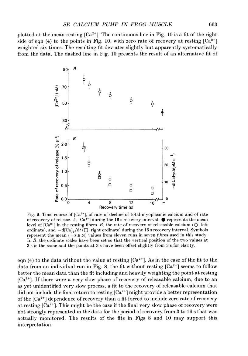
- Schneider M. F., Simon B. J., Szucs G. Depletion of calcium from the sarcoplasmic reticulum during calcium release in frog skeletal muscle. J Physiol. 1987 Nov;392:167–192. doi: 10.1113/jphysiol.1987.sp016775. [DOI] [PMC free article] [PubMed] [Google Scholar]
- Simon B. J., Klein M. G., Schneider M. F. Calcium dependence of inactivation of calcium release from the sarcoplasmic reticulum in skeletal muscle fibers. J Gen Physiol. 1991 Mar;97(3):437–471. doi: 10.1085/jgp.97.3.437. [DOI] [PMC free article] [PubMed] [Google Scholar]
- Squier T. C., Hughes S. E., Thomas D. D. Rotational dynamics and protein-protein interactions in the Ca-ATPase mechanism. J Biol Chem. 1988 Jul 5;263(19):9162–9170. [PubMed] [Google Scholar]
- Taylor K. A., Ho M. H., Martonosi A. Image analysis of the Ca2+-ATPase from sarcoplasmic reticulum. Ann N Y Acad Sci. 1986;483:31–43. doi: 10.1111/j.1749-6632.1986.tb34493.x. [DOI] [PubMed] [Google Scholar]
- Watanabe T., Lewis D., Nakamoto R., Kurzmack M., Fronticelli C., Inesi G. Modulation of calcium binding in sarcoplasmic reticulum adenosinetriphosphatase. Biochemistry. 1981 Nov 10;20(23):6617–6625. doi: 10.1021/bi00526a015. [DOI] [PubMed] [Google Scholar]