

Abstract
This study investigates the antioxidant, antimicrobial, and anticancer properties of Pancratium maritimum L. in Sp. Pl.: 291 (1753) seeds and flowers. Antioxidant activity was assessed using DPPH free radical scavenging and iron chelation assays. Antimicrobial evaluations assessed the efficacy of the extracts against diverse microorganisms. Cell viability assays were conducted on the dukes c colon cancer (SW480), while gas chromatography‐mass spectrometry (GC‐MS) analysis facilitated the identification of bioactive compounds. The ethanol extract of P. maritimum seeds exhibited a total phenolic content of 296.89±14.53 mg GAE/g extract DW and a total flavonoid content of 361.03±20.18 mg QE/g extract DW. Conversely, the flower extract showed a total phenolic content of 95.03±7.22 mg GAE/g extract DW and a total flavonoid content of 272.12±16.42 mg QE/g extract DW. As a result, the ethanol extract of P. maritimum seeds contains higher phenolic and flavonoid contents than the flower extract. Antimicrobial evaluations demonstrated significant inhibitory effects of both seed and flower extracts, with minimum inhibitory concentration (MIC) values ranging from 25 to >50 mg/mL. Notably, the seed extract showed greater activity against E. coli and C. krusei. GC‐MS analysis identified 18 bioactive compounds in the seed extract and 16 in the flower extract, with crucial components including ethyl oleate and 5‐hydroxymethylfurfural. Additionally, cell viability assays revealed that ethanol extracts from seeds and flowers significantly reduced SW480 cell viability, particularly at doses of 750 μg/mL and 250 μg/mL, respectively. These findings underscore the therapeutic potential of P. maritimum in terms of its antioxidant, antimicrobial, and anticancer properties, highlighting its value as a natural source of antioxidants and antimicrobial agents. Furthermore, the molecular docking study emphasises strong binding interactions of key compounds, particularly ethyl oleate and hexadecanoic acid ethyl ester, with the human STARD10 protein. The biological interactions and health implications of P. maritimum provide a significant foundation for future research in drug development and therapeutic applications.
Keywords: Antioxidant activity, GC-MS analysis, Molecular docking, Pancratium maritimum, STARD10 protein, SW480 cell line
This study explores the antioxidant, antimicrobial, and anticancer properties of Pancratium maritimum L. seeds and flowers. Ethanol extracts show high phenolic and flavonoid content, strong antimicrobial activity, and significant cytotoxic effects on SW480 colon cancer cells. GC‐MS analysis identifies bioactive compounds, and molecular docking highlights key interactions with STARD10 protein, underscoring the plant‘s therapeutic potential.

Introduction
Colorectal cancer is described as the second most common cancer worldwide, with 1.7 million cases diagnosed annually. [1] Despite substantial advancements in treatment approaches and early diagnostic technologies, drug resistance remains a major obstacle in cancer therapy. This challenge underscores the urgent need to discover effective and reliable compounds with minimal side effects, particularly in cases where current therapeutic options are limited, and adverse effects are a concern. Herbal products, extensively utilised in traditional medicine, hold significant potential in preventing and treating various diseases, including cancer. Their richness in bioactive compounds and generally lower side effect profiles have made them a focal point of interest in modern medical research. [2]
Pancratium maritimum L. in Sp. Pl.: 291 (1753) belonging to the Amaryllidaceae (J.St.‐Hil.), is one of the 24 species classified within the genus Pancratium Dill. ex L., commonly referred to as the sea daffodil. While the genus Pancratium comprises 24 species globally, only one species, P. maritimum, is naturally distributed in Türkiye (Figure 1).[ 3 , 4 , 5 ] This bulbous and perennial plant has a wide native range, primarily spanning the Canary Islands, the Mediterranean region, and the Black Sea coasts. Its distribution extends from Portugal, Morocco, and the Balearic Islands to Türkiye, Syria, Israel, and the Caucasus. Additionally, it is found along the southern coastline of Bulgaria and the northern shores of Turkey and Georgia. Furthermore, the species has successfully naturalised in certain regions beyond its native range, including southern California, Bermuda, and the Azores, showcasing its ecological adaptability to diverse environmental conditions. [4]
Figure 1.

P. maritimum plant morphological structure.
P. maritimum grows in coastal dunes and beaches, with much of its leaves and flower stems often buried in sand. The plant has a long‐necked bulb and glaucous, broadly linear evergreen leaves, though they often die back in hot summers. Its flower stem can grow up to 40 centimetres tall. The plant produces 3 to 15 white flowers in an umbel, each up to 15 centimetres long. The flower's corona is two‐thirds the length of its tepals. These flowers have a subtle, exotic lily scent most noticeable on calm, windless summer nights. The blooming period is from June to October.[ 6 , 7 ]
P. maritimum found in Sicily, Italy, exhibits antioxidant properties, [8] In Tunisia, P. maritimum demonstrates antioxidant, anticoagulant, biofilm inhibitory, and antibacterial activities. [9] P. maritimum, found in India, has anti‐inflammatory properties. [10] Alkaloids isolated from P. maritimum in Squillace, Italy, display antiviral activity. [11] The alkaloid Pancratistatin, isolated from P. maritimum in Marūḥ, Egypt, exhibits anticancer properties. [12] P. maritimum collected in Matrouh, Egypt, shows both anticancer and antimicrobial activities [13] P. maritimum grown in Djerba, Tunisia, demonstrates antioxidant and antimicrobial activities. [9]
Due to their rich phytochemical content, P. maritimum root, leaf, and fruit extracts are expected to control oxidative stress effectively. Antioxidants protect against reactive oxygen species generated by oxidative stress in the body. [14] Antioxidants are chemical substances in various foods, including fruits and vegetables, that help prevent cell damage under various stress conditions. [15] This study aims to evaluate the antioxidant potential of P. maritimum under in vitro conditions and conduct content analysis to understand which components contribute to this effectiveness. Demonstrating the antioxidant potential of sea daffodil extracts can provide a framework for future studies involving the plant.
Bacterial conditions are one of the determining factors for antimicrobial activity. When exposed to antibiotics, bacterial phenotypes can exhibit various changes; these changes include characteristics such as sensitivity, resistance, tolerance, and persistence. [16] Antimicrobial agets are typically categorised into natural agents and synthetic compounds. Natural antimicrobial agents are derived from living organisms, including curcumin from the Zingiberbuaceae and other substances extracted from filamentous saprophytic microbes or medicinal plants. [17]
In this context, the rich compositions of P. maritimum indicate significant potential for antimicrobial research. Investigating its antimicrobial properties may support the modern applications of this plant, which has long been used in folk medicine. Thus, the importance of researching to enhance the pharmaceutical value of P. maritimum and develop new therapeutic methods to combat microbial resistance becomes even more apparent.
STARD10 is a phospholipid transfer protein that regulates the transport of phosphatidylcholine and phosphatidylethanolamine between intracellular membranes, containing a lipid transfer domain related to the steroidogenic acute regulatory protein (StAR). [18] Erb‐B2 receptor tyrosine kinase 2 (ERBB2), an epidermal growth factor receptor family member, is particularly active in ERBB2‐positive metastatic colorectal cancer patients. The overexpression of ERBB2 leads to an increase in STARD10 expression, which modulates the activity of STARD10 and plays a significant role in cancer progression. [19] Molecular docking studies have revealed that bioactive compounds found in P. maritimum seeds, including Ethyl Oleate, Hexadecanoic acid, ethyl ester, and Hordenine, as well as compounds identified in its flowers, such as 5‐Hydroxymethylfurfural, Hexadecanoic acid, 2‐hydroxy‐1‐(hydroxymethyl), and 1,3,5‐Triazine‐2,4,6‐triamine, exhibit strong interactions with the STARD10 protein. These compounds’ binding energies and interaction profiles highlight their potential to modulate STARD10 activity, emphasising its critical role in cancer progression. These findings provide a significant foundation for the development of novel strategies in cancer therapy.
The different parts of P. maritimum may exhibit distinct biological activity profiles, which is an important consideration. This study aims to investigate the antimicrobial potential of P. maritimum seed and flower extracts and elucidate the plant's role as a natural antimicrobial agent. Limited research has shown that P. maritimum possesses anticancer, antiviral, and antioxidant properties, inhibiting cell proliferation in various cancer cell lines, including breast, lung, and prostate. However, further research is needed to explore its medical applications and efficacy, particularly concerning colon cancer, which remains under‐examined. Such investigations could provide valuable insights into the plant's potential anticancer activity.
A molecular docking study has been conducted on the phytochemical compounds identified in the seeds and flowers of P. maritimum with the STARD10 protein. There are gaps in the literature regarding this topic. Determining the in‐silico interactions of STARD10 proteins with these phytochemical compounds could provide important data on their potential use as therapeutic agents in colorectal cancer treatment.
Results and Discussions
Our study used various methods to analyse the antioxidant activity and phenolic content of P. maritimum seeds and flowers. Our findings are summarised in Table 1. The 2,2‐diphenyl‐1‐picrylhydrazyl (DPPH) IC50 value for P. maritimum flowers in ethanol was determined to be 0.62±0.01 mg/mL. The DPPH IC50 value for the positive control, Butylated hydroxytoluene (BHT), was measured at 0.23±0.01 mg/mL. The iron chelation IC50 value for P. maritimum flowers was 8.56±0.33 mg/mL, while the Ethylenediaminetetraacetic acid (EDTA) iron chelation IC50 value was 5.30±4.44 mg/mL. The total phenolic content (TPC) was 95.03±7.22 mg GAE/g extract DW, and the total flavonoid content (TFC) was 272.12±16.42 mg QE/g extract DW.
Table 1.
DPPH assay activities of P. maritimum seeds and flowers extracts (IC50 (mg/mL)±SD) and TPC and TFC±SD* Values (n=3).
|
Plant Name |
DPPH (IC50 mg/mL) |
Iron Chelating (IC50 mg/mL) |
TPC (mg GAE/g extract DW) |
TFC (mg QE/g extract DW) |
|---|---|---|---|---|
|
P. maritimum seeds ethanol |
0.45±0.01 |
8.72±0.20 |
296.89±14.53 |
361.03±20.18 |
|
P. maritimum flowers ethanol |
0.62±0.01 |
8.56±0.33 |
95.03±7.22 |
272.12±16.42 |
|
BHT (Positive control) |
0.23±0,01 |
|||
|
EDTA (Positive control) |
5.30±4.44 |
In contrast, the DPPH IC50 value for P. maritimum seeds in ethanol was measured at 0.45±0.01 mg/mL. The DPPH IC50 value for the positive control, BHT, was also recorded as 0.23±0.01 mg/mL. The iron chelation IC50 value for P. maritimum seeds was 8.72±0.20 mg/mL, while the EDTA iron chelation IC50 value was 5.30±4.44 mg/mL. The TPC was 296.89±14.53 mg GAE/g extract DW, and the TFC was 361.03±20.18 mg QE/g extract DW. These findings indicate that P. maritimum seeds possess superior antioxidant capacity compared to the flowers, associated with their higher total phenolic and flavonoid content.
In this study, the antimicrobial activities of P. maritimum seed and flower extracts against various microorganisms were compared with standard drugs such as Ampicillin (Amp), Chloramphenicol (C), and Ketoconazole (Keto). According to the antimicrobial screening results, the minimum inhibitory concentration (MIC) values of P. maritimum flowers ranged from 25 to >50 mg/mL against all bacterial and fungal strains. In comparison, Amp exhibited MIC values ranging from 31.25 to >125 mg/mL, C from 15.63 to >125 mg/mL, and Keto from <0.98 to 62.5 mg/mL. Similarly, the MIC values of P. maritimum seeds ranged from 25 to >50 mg/mL against all bacterial and fungal strains, with Amp showing MIC values between 31.25 and >125 mg/mL, C between 15.63 and >125 mg/mL, and Keto between <0.98 and 62.5 mg/mL (Table 2). The seed extract exhibits a lower MIC value against the Escherichia coli bacterial strain, indicating higher antimicrobial activity. Additionally, the seed extract shows a lower MIC value against the Candida krusei fungal strain, demonstrating higher antimicrobial activity.
Table 2.
MIC, MBC, and MFC of the P. maritimum seeds and flowers extracts and controls (mg/ml).
|
Plant Species |
Positive Control |
|||||
|---|---|---|---|---|---|---|
|
Microorganisms |
P. maritimum flowers |
P. maritimum seeds |
Amp |
C |
Keto |
|
|
E. coli ATCC 25922 |
MIC |
50 |
25 |
>125 |
125 |
NS |
|
MBC |
>50 |
25 |
>125 |
>125 |
NS |
|
|
S. aureus ATCC 25923 |
MIC |
>50 |
>50 |
62.5 |
125 |
NS |
|
MBC |
>50 |
>50 |
62.5 |
>125 |
NS |
|
|
K. pneumoniae ATCC 13883 |
MIC |
25 |
25 |
125 |
15.63 |
NS |
|
MBC |
50 |
25 |
125 |
15.63 |
NS |
|
|
P. vulgaris RSKK 96029 |
MIC |
50 |
50 |
>125 |
125 |
NS |
|
MBC |
>50 |
>50 |
>125 |
>125 |
NS |
|
|
B. cereus NRRL B‐3711 |
MIC |
>50 |
>50 |
31.25 |
125 |
NS |
|
MBC |
>50 |
>50 |
>125 |
125 |
NS |
|
|
C. albicans ATCC 10231 |
MIC |
25 |
25 |
NS |
NS |
31.25 |
|
MFC |
50 |
>50 |
NS |
NS |
62.5 |
|
|
C. krusei ATCC 6258 |
MIC |
50 |
25 |
NS |
NS |
<0.98 |
|
MFC |
>50 |
>50 |
NS |
NS |
15.63 |
|
Amp: Ampicillin; C: Chloramphenicol; Keto: Ketoconazole; NS: Not Studies.
Various bioactive compounds have been identified in the ethanol extracts of P. maritimum seeds and flowers. The extracts’ retention times (RT), concentrations (% area), and chemical structures are presented in Tables 3 and 4. The seed extract contains 18, while the flower extract contains 16 bioactive phytochemical compounds. The main components of P. maritimum seeds are Ethyl Oleate (28.51 %), Hexadecanoic acid ethyl ester (palmitic acid) (11.97 %), Hordenine (4.02 %) and in flowers, 5‐Hydroxymethylfurfural (13.44 %), Hexadecanoic acid 2‐hydroxy‐1‐(hydroxymethyl) ethyl ester (Palmitic acid β‐monoglyceride) (5.70 %), 1,3,5‐Triazine‐2,4,6‐triamine (3.15 %), and 4H‐Pyran‐4‐one, 2,3‐dihydro‐3,5‐dihydroxy (2.40 %).
Table 3.
GC‐MS analysis results of P. maritimum seeds.
|
No |
Retention Time (minutes) |
Compound Name |
Molecular Weight (g/mol) |
Base Peak |
Area (%) |
Structure |
|---|---|---|---|---|---|---|
|
1 |
11.864 |
Maltol |
126.11 |
126.05 |
1.02 |
|
|
2 |
19.109 |
5‐Hydroxymethylfurfural |
126.11 |
97.05 |
3.89 |
|
|
3 |
31.356 |
Hordenine |
165.23 |
58.05 |
4.02 |
|
|
4 |
31.717 |
Eicosane |
282.5 |
57.05 |
1.02 |
|
|
5 |
31.872 |
Sucrose |
342.30 |
57.05 |
1.53 |
|
|
6 |
48.463 |
Hexadecanoic acid, ethyl ester |
284.5 |
88.05 |
11.97 |
|
|
7 |
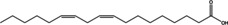
50.306 |
9,12‐Octadecadienoic acid (Z, Z)‐ |
280.4 |
67.10 |
2.07 |
|
|
8 |
50.381 |
Oleic Acid |
282.5 |
55.10 |
2.39 |
|
|
9 |
50.637 |
Ethyl Oleate* |
310.5 |
55.05 |
26.22 |
|
|
10 |
50.890 |
Octadecanoic acid, ethyl ester |
312.5 |
88.10 |
2.49 |
|
|
11 |
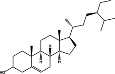
51.609 |
Gamma‐Sitosterol |
414.7 |
225.05 |
1.69 |
|
|
12 |
51.712 |
9,10‐Anthracenedione, 1‐amino‐ |
223.23 |
223.05 |
1.55 |
|
|
13 |
53.480 |
Tetracosamethyl‐ cyclododecasiloxane |
889.8 |
73.05 |
1.66 |
|
|
14 |
53.759 |
1‐(2,4‐Dihydroxyphenyl)‐3‐ phenyl‐2‐propen‐1‐one |
240.25 |
256.10 |
1.23 |
|
|
15 |
54.403 |
Galathan, 1,2,3,12,15,16‐hexadehydro‐9,10‐ [methylenebis(oxy)]‐ |
251.28 |
250.00 |
1.97 |
|
|
16 |
54.602 |
Hexadecanoic acid, 2‐hydroxy‐1‐ (hydroxymethyl)ethyl ester |
330.5 |
98.10 |
3.54 |
|
|
17 |
54.707 |
5,6‐Dihydro‐5,6‐dihydroxy‐ 7‐hydroxymethyl‐12‐ methylbenz(a)anthracene, trans‐ |
306.4 |
257.10 |
1.19 |
|
|
18 |
56.067 |
7,12‐Dimethyl‐8,9,10,11‐ tetrahydrobenz(a)anthracene |
260.4 |
248.05 |
1.28 |
|


Table 4.
GC‐MS analysis results of P. maritimum flowers.
|
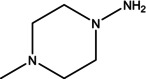
No |
Retention Time (minutes) |
Compound Name |
Molecular Weight (g/mol) |
Base Peak |
Area (%) |
Structure |
|---|---|---|---|---|---|---|
|
1 |
3.285 |
Dihydroxyacetone |
90.08 |
31.00 |
1.54 |
|
|
2 |
12.139 |
1,3,5‐Triazine‐ 2,4,6‐triamine |
126.12 |
126.05 |
3.15 |
|
|
3 |
13.925 |
4H‐Pyran‐4‐one, 2,3‐dihydro‐3,5‐ dihydroxy‐6‐methyl‐ |
144.12 |
43.00 |
2.40 |
|
|
4 |
19.359 |
5‐Hydroxymethylfurfural |
126.11 |
97.00 |
13.44 |
|
|
5 |
19.954 |
1,2,3‐Propanetriol, 1‐acetate |
134.13 |
43.00 |
1.49 |
|
|
6 |
23.395 |
1‐Amino‐4‐ methylpiperazine |
115.18 |
42.00 |
1.63 |
|
|
7 |
24.470 |
Cyclohexasiloxane, dodecamethyl‐ |
444.92 |
340.90 |
1.00 |
|
|
8 |
27.033 |
1‐Pentadecene |
210.40 |
43.05 |
1.06 |
|
|
9 |
48.046 |
n‐Hexadecanoic acid |
256.42 |
88.00 |
2.88 |
|
|
10 |
48.430 |
Hexadecanoic acid, ethyl ester |
284.5 |
43.00 |
1.72 |
|
|
11 |
49.343 |
Gamma‐Sitosterol |
414.7 |
42.00 |
1.88 |
|
|
12 |
50.252 |
9,12‐Octadecadienoic acid (Z,Z)‐ |
280.4 |
255.10 |
1.55 |
|
|
14 |
50.602 |
9,12,15‐Octadecatrienoic acid, ethyl ester |
306.5 |
126.05 |
6.26 |
|
|
15 |
54.613 |
Hexadecanoic acid, 2‐hydroxy‐1‐ (hydroxymethyl)ethyl ester* |
330.5 |
98.10 |
6.90 |
|
|
16 |
55.202 |
Diisooctyl phthalate |
390.6 |
149.05 |
1.57 |
|


When examining the effects of ethanol extracts of P. maritimum flowers on human colon cancer (SW480) cell line viability levels, it was determined that all doses significantly reduced cell viability compared to the control and ethanol groups (Figure 2). A similar trend was observed with ethanol extracts of the seeds of the same plant (Figure 3). According to the data, the dose showing the most significant decrease in cell viability was 750 μg/mL for flower extract and 250 μg/mL for seed extract.
Figure 2.

Cytotoxic effects of P. maritimum flower extract on SW480 cell line.
Figure 3.

Cytotoxic effects of P. maritimum seed extract on SW480 cell line.
The results of our molecular docking study evaluated the interactions and binding energies of various organic compounds with specific amino acids. The findings help us understand potential biological interactions and binding properties. In our study, the simulation of 2D and 3D interactions between molecules and the human STARD10 enzyme in a computer environment is shown in Figures 4, 5 and 6. The interactions between the compounds and the enzymes in the best position are shown in Tables 5 and 6.
Figure 4.

Molecular docking process of A) Ethyl Oleate B) Hexadecanoic acid, ethyl ester C) Hordenin with Human STARD10 (6SER).
Figure 5.

Molecular docking process of D) 5‐Hydroxymethylfurfural E) Hexadecanoic acid, 2‐hydroxy‐1‐(hydroxymethyl) with Human STARD10 (6SER).
Figure 6.

Molecular docking process of F) 1,3,5‐Triazine‐2,4,6‐triamine G) 4H‐Pyran‐4‐one, 2,3‐dihydro‐3,5‐dihydroxy‐with Human STARD10 (6SER).
Table 5.
Molecular docking energy and report of predicted interactions of compounds from P. maritimum seeds against human STARD10 protein (6SER).
|
Component |
Binding Energy (kcal/mol) |
Amino acid |
Interacting |
Distance |
|---|---|---|---|---|
|
Ethyl Oleate |
−4.0 |
A: GLU90 : HN: O1 |
Conventional Hydrogen Bond |
2.17 |
|
H8 ‐ A: ASP88 : OD2 |
Carbon Hydrogen Bond |
2.92 |
||
|
A: ARG107 |
Alkyl |
4.02 |
||
|
A:PRO235 |
Alkyl |
5.33 |
||
|
A:PRO239 |
Alkyl |
4.24 |
||
|
Hexadecanoic acid, ethyl ester |
−5.4 |
A: VAL189 : HN: O2 |
Conventional Hydrogen Bond |
1.91 |
|
A: GLN188: HA: O2 |
Carbon Hydrogen Bond |
2.60 |
||
|
H5 ‐ A: HIS66 : O |
Carbon Hydrogen Bond |
1.96 |
||
|
A: ALA187 |
Alkyl |
4.56 |
||
|
A:PRO191 |
Alkyl |
5.35 |
||
|
A: VAL200 |
Alkyl |
4.67 |
||
|
A: ALA208 |
Alkyl |
4.34 |
||
|
C1‐ A:PRO191 |
Alkyl |
4.02 |
||
|
C1‐ A: LEU195 |
Alkyl |
4.98 |
||
|
A: LEU207 |
Alkyl |
4.59 |
||
|
A: ILE68 |
Alkyl |
4.30 |
||
|
C17‐ A: ILE68 |
Alkyl |
4.37 |
||
|
A: TYR185 |
Alkyl |
5.08 |
||
|
Hordenine |
−5.5 |
A: HIS87 : HD1: O1 |
Conventional Hydrogen Bond |
1.68 |
|
H15‐ A: HIS87 : O |
Conventional Hydrogen Bond |
1.72 |
||
|
H4‐ A: VAL110 : O |
Carbon Hydrogen Bond |
2.07 |
||
|
H6‐ A:PRO235 : O |
Carbon Hydrogen Bond |
2.37 |
||
|
H8‐ A:PRO235 : O |
Carbon Hydrogen Bond |
2.30 |
||
|
A: ASP104 : OD2 |
Pi‐Anion |
3.80 |
Table 6.
Docking energy and report of predicted interactions of compounds from P. maritimum flowers against human STARD10 protein (6SER).
|
Component |
Binding Energy (kcal/mol) |
Amino acid |
Interacting |
Distance |
|---|---|---|---|---|
|
5‐Hydroxymethylfurfural |
−5.0 |
A: HIS87 : HD1: O1 |
Conventional Hydrogen Bond |
2.07 |
|
A: ARG134 : HH11 |
Conventional Hydrogen Bond |
2.11 |
||
|
H4 ‐ A: VAL110 : O |
Conventional Hydrogen Bond |
2.14 |
||
|
A: TRP236: HA: O1 |
Carbon Hydrogen Bond |
2.48 |
||
|
H5 ‐ A:PRO235 : O |
Carbon Hydrogen Bond |
3.09 |
||
|
H6 ‐ A: VAL110 : O |
Carbon Hydrogen Bond |
3.02 |
||
|
A: ASP113 : OD2 |
Pi‐Anion |
4.24 |
||
|
A:PRO235 |
Pi‐Alkyl |
4.93 |
||
|
Hexadecanoic acid, 2‐hydroxy‐1‐(hydroxymethyl) |
−5.2 |
A: ARG92 : HH12: O2 |
Conventional Hydrogen Bond |
1.85 |
|
A: ARG92 : HH22: O1 |
Conventional Hydrogen Bond |
2.27 |
||
|
H1 ‐ A: ASP96 : OD1 |
Conventional Hydrogen Bond |
2.24 |
||
|
H4‐ A: ASP96 : OD2 |
Conventional Hydrogen Bond |
1.92 |
||
|
A: ALA208 |
Alkyl |
4.82 |
||
|
A: ALA211 |
Alkyl |
4.83 |
||
|
A: LEU207 |
Alkyl |
5.11 |
||
|
A: MET212 |
Alkyl |
5.37 |
||
|
A: MET215 |
Alkyl |
4.84 |
||
|
A: TRP119:C1 |
Pi‐Alkyl |
4.29 |
||
|
A: TYR185 |
Pi‐Alkyl |
4.36 |
||
|
1,3,5‐Triazine‐2,4,6‐triamine |
−5.0 |
A: ARG134 : HH21: N3 |
Conventional Hydrogen Bond |
2.73 |
|
H2‐ A: THR132 : O |
Conventional Hydrogen Bond |
2.95 |
||
|
A: GLY115 : HA1: N3 |
Carbon Hydrogen Bond |
2.20 |
||
|
4H‐Pyran‐4‐one, 2,3‐dihydro‐3,5‐dihydroxy‐ |
−5.4 |
A: ARG107: HE: O1 |
Conventional Hydrogen Bond |
2.53 |
|
A: SER242: HG: O1 |
Conventional Hydrogen Bond |
2.37 |
||
|
H1‐ A: ASP113 : OD2 |
Conventional Hydrogen Bond |
1.80 |
||
|
H2‐ A:PRO235 : O |
Conventional Hydrogen Bond |
1.76 |
||
|
A: ARG107 : HD2: O1 |
Carbon Hydrogen Bond |
2.47 |
||
|
A: TRP236: HA: O3 |
Carbon Hydrogen Bond |
2.30 |
||
|
H8‐ A: ASP113 : OD2 |
Carbon Hydrogen Bond |
2.96 |
||
|
C6‐ A:PRO239 |
Alkyl |
4.45 |
This study presents the results of molecular docking analyses of three different compounds (Ethyl Oleate, Hexadecanoic acid ethyl ester, and Hordenine) identified through gas chromatography‐mass spectrometry (GC‐MS) analysis of P. maritumum seed extract against human STARD10 protein. The binding energies obtained indicate strong interactions between these compounds and STARD10 protein, as evidenced by their negative values (Ethyl Oleate: −4.0 kcal/mol, Hexadecanoic acid ethyl ester: −5.4 kcal/mol, Hordenine: −5.5 kcal/mol). Ethyl Oleate forms a traditional hydrogen bond between the NH group of GLU90 amino acid and the O1 atom (2.17 Å), as well as a carbon‐hydrogen bond between the OD2 atom of ASP88 and H8 (2.92 Å). Additionally, alkyl interactions were observed with ARG107, PRO235, and PRO239 (4.02 Å, 5.33 Å, and 4.24 Å, respectively). Hexadecanoic acid ethyl ester uses a traditional hydrogen bond with VAL189 : HN: O2 (1.91 Å) and a carbon‐hydrogen bond with GLN188: HA: O2 (2.60 Å). Furthermore, alkyl interactions were noted with HIS66, ALA187, PRO191, VAL200, ALA208, PRO191, LEU195, LEU207, ILE68, and TYR185 (ranging from 1.96 Å to 5.35 Å). Hordenine forms traditional hydrogen bonds with HIS87 : HD1: O1 and HIS87 (1.68 Å and 1.72 Å, respectively), as well as carbon‐hydrogen bonds with VAL110, PRO235, and PRO235 (ranging from 2.07 Å to 2.37 Å). Additionally, a pi‐anion interaction was observed with ASP104 (3.80 Å). These findings underscore the robust binding capabilities of these compounds to the STARD10 protein, as elucidated through molecular docking methodologies. This approach provides valuable insights into the potential biological impacts of these interactions, which are relevant for further exploration in drug design and therapeutic strategies.
This study demonstrates that the active compounds present in the P. maritumum flower extract, specifically 5‐Hydroxymethylfurfural, Hexadecanoic acid 2‐hydroxy‐1‐(hydroxymethyl), 1,3,5‐Triazine‐2,4,6‐triamine, and 4H‐Pyran‐4‐one 2,3‐dihydro‐3,5‐dihydroxy, exhibit high binding affinities with the STARD10 protein. Molecular docking analyses reveal that each compound forms stable interactions with STARD10, with binding energies of −5.0 kcal/mol, −5.2 kcal/mol, −5.0 kcal/mol, and −5.4 kcal/mol, respectively. 5‐Hydroxymethylfurfural establishes conventional hydrogen bonds with amino acids such as HIS87, ARG134, and VAL110, and forms carbon‐hydrogen bonds with TRP236 and PRO235. Additionally, interactions involving pi‐anion with ASP113 and pi‐alkyl with PRO235 are observed. Hexadecanoic acid, 2‐hydroxy‐1‐(hydroxymethyl), forms conventional hydrogen bonds with ARG92, ASP96, and other amino acids, and exhibits alkyl interactions with ALA208, ALA211, LEU207, MET212, MET215, and pi‐alkyl interactions with TRP119 and TYR185. 1,3,5‐Triazine‐2,4,6‐triamine forms conventional hydrogen bonds with ARG134, THR132, and GLY115, and establishes a carbon‐hydrogen bond with TRP236. Lastly, 4H‐Pyran‐4‐one, 2,3‐dihydro‐3,5‐dihydroxy‐, forms conventional hydrogen bonds with ARG107, SER242, and ASP113, and engages in carbon‐hydrogen bonds with PRO235, ARG107, TRP236, and other amino acids. Also, alkyl interactions with PRO239 and at the C6 position with PRO239 are observed. These findings underscore the potential interactions of compounds isolated from P. maritimum seeds and flower extracts with the STARD10 protein, elucidating their molecular mechanisms. This study highlights the successful application of molecular docking methods in understanding these interactions, providing a foundational basis for drug design, and elucidating biological effects.
When we conducted a literature review on P. maritimum, we found limited studies, particularly noting the absence of research on iron chelation activity related to P. maritimum and its species. Microbial studies are notably sparse based on the literature review. Additionally, limited research concerning antioxidant phenolic contents is associated with P. maritimum. Our study reveals that P. maritimum exhibits excellent antioxidant and antimicrobial effects. Plants with high antioxidant and phenolic contents are considered to have potential benefits in cancer prevention and treatment by neutralising free radicals and reducing cellular damage, thus potentially inhibiting cancer development. Therefore, plants rich in antioxidants and phenolic compounds are believed to hold significant promise in cancer research.
In the study by Melliti et al. [9] the TPC of the above ground methanolic extract of P. maritimum has been reported as 161.31±3.9 mg GAE/g DW, while the TFC is 126.7±3 mg QE/g DW. According to the study by Melliti et al. the methanol extract obtained from the aerial parts of P. maritimum has a higher phenolic content than the ethanol extract of P. maritimum flowers and a lower phenolic content than the seeds. Additionally, compared to the methanol extract from the aerial parts of P. maritimum, the total phenolic and flavonoid contents of the ethanol extracts from the seeds and flowers of P. maritimum are higher.
In the study by Leporini et al. [20] demonstrated that the ethanol extract of P. maritimum flowers had a TPC of 228.6±2.4 mg GAE/g extract DW and a TFC of 45.7±0.2 mg QE/g extract DW. The ethanol extract of P. maritimum fruits had a TPC of 277.8±2.9 mg GAE/g extract DW and a TFC of 52.7±0.3 mg QE/g extract DW. The ethanol extract of P. maritimum bulbs had a TPC of 48.4±1.5 mg GAE/g extract DW and a TFC of 24.4±0.3 mg QE/g extract DW. Additionally, it was reported that the ethanol extracts of P. maritimum flowers, fruits, and stems exhibited IC50 values of 81.3±8.1 μg/mL, 846.1±44.2 μg/mL, and 84.1±8.4 μg/mL, respectively, in the DPPH test. According to the study by Leporini et al., the total phenolic content of the ethanol extract from P. maritimum flowers is higher than ours, while the TFC is also higher.
Similarly, when comparing the TPC of the ethanol extract from P. maritimum fruits in Leporini et al. study with our findings, the TPC of the ethanol extract from P. maritimum flowers in our study is lower, but the TFC is higher. Additionally, compared to the TPC of the ethanol extract from P. maritimum bulbs in Leporini et al. study, the TPC and TFC of the ethanol extract from P. maritimum flowers in our study are higher. In the study by Leporini et al. the TPC and TFC of the ethanol extracts from P. maritimum flowers, bulbs, and fruits are lower than the methanol extracts from P. maritimum flowers and seeds in our study. In the study by Leporini et al. the IC50 values of P. maritimum flowers and stems in the DPPH test indicate that the antioxidant activity of the ethanol extracts from P. maritimum flowers and seeds is high compared to our findings. Additionally, Leporini et al. reported that the antioxidant activity of P. maritimum fruit extract is lower than that of seed and flower extracts.
In the study by Nikolova et al. [21] determined that the IC50 value for P. maritimum methanol bulb extract is more significant than 200 μg/mL. In the study by Nikolova et al. it was observed that the methanol extracts of P. maritimum flowers and seeds exhibited good antioxidant activity compared to the methanol extract of P. maritimum bulbs.
In the study by Taie et al. [22] the TPC and TFC of P. maritimum leaf extract were measured at 5.36 mg GAE/g extract DW and 1.17 mg QE/g extract DW, respectively. Furthermore, the methanol extracts of P. maritimum flowers and leaves demonstrated inhibition rates of 85.2 % and 81.3 %, respectively. Taie et al. found that, compared to our study, the TPC and TFC of the ethanol extracts from P. maritimum flowers and seeds were higher than those of the P. maritimum leaf extract. Additionally, it was observed that both the methanol extracts of P. maritimum flowers and seeds exhibited good antioxidant activity compared to the methanol extracts of P. maritimum flowers and leaves.
In the study by Melliti et al. [9] the MIC of the essential oil of P. maritimum was found to range from 3.25 to 26 mg/mL, whereas the MIC for the microemulsion ranged from 1.625 to 26 mg/mL. Notably, the microemulsion exhibited superior antibacterial activity against the tested bacterial strains compared to the essential oil. The antibacterial effect was particularly pronounced against Gram‐positive bacteria, with significant activity observed against E. faecalis, demonstrating MIC and minimum bactericidal concentration (MBC) values of 1.625 mg/mL and 3.25 mg/mL, respectively. The highest recorded MIC value was 3.25 mg/mL for S. aureus and E. faecalis. Additionally, while the essential oil of P. maritimum showed considerable antibacterial activity against all strains tested, it exhibited a relatively weak potential against Candida glabrata, with an MIC value of 13 mg/mL.
In contrast, the microemulsion of P. maritimum demonstrated enhanced efficacy against Candida albicans and Candida parapsilosis, with recorded MIC values of 0.4 mg/mL. In our study, compared to the work conducted by Melliti et al. the P. maritimum flower and seed extracts demonstrate lower antimicrobial and antifungal activities against the essential oil and microemulsion of P. maritimum. These findings underscore the varying efficacy of P. maritimum extracts and formulations against different microorganisms, highlighting the importance of formulation strategies, such as microemulsion, in enhancing antimicrobial activity.
In the study by Melliti et al. [9] the methanolic extract of P. maritimum demonstrated an MIC of 10 mg/mL against C. albicans ATCC 942. The extract also showed MIC values of 20 mg/mL against Staphylococcus aureus ATCC 6583, 40 mg/mL against Pseudomonas aeruginosa ATCC 27583, and 10 mg/mL against E. coli ATCC 25922. Our study found that the ethanol extract of P. maritimum flowers and seeds exhibited good antifungal activity against the C. albicans ATCC 10231 strain. In contrast, the study by Melliti et al. indicated that the methanol extract of P. maritimum demonstrated stronger antifungal activity. Additionally, the ethanol extract of P. maritimum flowers and seeds showed effective antimicrobial activity against the S. aureus ATCC 25923 strain. In contrast, the methanol extract in the study by Melliti et al. also displayed strong antimicrobial activity against this strain.
The study by Hetta et al. [13] reported that the fruit ethanol extract of P. maritimum exhibits significant inhibition effects on Trichophyton mentagrophytes and Aspergillus fumigatus. It is particularly effective against C. glabrata. The fruit ethanol extract also shows effective inhibition against Streptococcus pneumoniae. Moreover, it demonstrates pronounced inhibition effects on Klebsiella pneumoniae. These findings underscore the strong antimicrobial properties of P. maritimum fruit ethanol extract against various microorganisms. The P. maritimum seed extract exhibits higher antimicrobial activity against the E. coli bacterial strain in our study. The P. maritimum seed extract also demonstrates higher antimicrobial activity against the K. pneumoniae bacterial strain. Furthermore, the seed extract also shows higher antifungal activity against the C. krusei fungal strain.
In the study Bonvicini et al. [23] according to the antimicrobial screening results, the MIC values of Pancratium illyricum bulbs ranged from 19.5 to 156 mg/mL against all bacterial and fungal strains. At the same time, Amp exhibited MIC values ranging from 0.078 to 1.25 mg/mL. The seed extract showed a lower MIC value against the E. coli bacterial strain, indicating higher antimicrobial activity. In our study, compared to the work of Bonvicini et al., the P. maritimum flower and seed extracts exhibit lower antimicrobial and antifungal activities against P. illyricum bulbs.
Determining the TPC in plant extracts is directly linked to the antioxidant capacity of these bioactive compounds. They can act as reducing agents, free radical scavengers, metal chelators, or deactivators of singlet oxygen. [20] Oxidative stress can lead to damage in cell structure, potentially contributing to neoplastic transformation and playing a significant role in cancer initiation and progression. [24] In this context, combating oxidative stress with potent antioxidant agents is a prominent area of research. Studies have investigated the antioxidant activity of P. maritimum extracts using various in vitro methods, demonstrating significant antioxidant effects that are concentration dependent.
In a study by Leporini et al. on P. maritimum, extracts from the plant's stem, flowers, bulbs, and fruits were utilised. It was found that direct ethanol extraction from flowers had higher total phenol and total flavonoid content compared to extractions using ether, ethanol, and water, respectively. Antiproliferative activity studies on flower ethanol extracts were conducted using human breast cancer (MCF‐7), human cervical cancer (HeLa), human breast cancer (MDA‐MB‐231), human melanoma (C32), human lung carcinoma (A549), human prostate cancer (LNCaP, and PC3) cell lines, with IC50 values ranging from 85.5±3.5 to 176.7±3.9 μg/mL. Flower extracts exhibited antioxidant activity according to DPPH, 2,2′‐Azino‐bis (3‐ethylbenzothiazoline‐6‐sulfonic acid (ABTS), β‐carotene bleaching, and FRAP tests. [20] Furthermore, Youssef et al. purified pancratistatin from P. maritimum flower ethanol extracts and applied it to MDA‐MB‐231, HeLa, human colorectal cancer (HCT116), and human dermal fibroblast (NHDF) cell lines, demonstrating selective action against cancer cell lines. [12] McLachlan et al. investigated the cellular mechanisms of pancratistatin, revealing its disruption of mitochondrial membrane potential, increase in caspase 3 and proteasome activities, and augmentation of mitochondrial ROS production. [25] Additionally, GC‐MS analysis detected Hydroxymethylfurfural in plant flowers. In a study by Chow et al. Hydroxymethylfurfural was reported to affect aquaporin‐1 on the HT29 cell line, altering ion conductivity and inhibiting cell migration. [26] Using ethanol extracts of P. maritimum flowers at 100–1000 μg/mL doses, similar effects were observed on SW480 cell line viability in this study. Moreover, findings indicated that cell viability approached approximately 50 % compared to the control group across all application doses.
No information was found in the literature regarding the anticancer effects of P. maritimum seed extracts. According to the data, seed ethanol extracts contain 28.51 % Ethyl Oleate, 11.97 % Hexadecanoic acid ethyl ester (palmitic acid), and 1.01 % Hordenine. Studies suggest that palmitic acid induces endoplasmic reticulum (ER) stress, calcium release from the ER, and transferrin‐dependent ferroptosis. However, palmitic acid alone does not affect Cluster of Differentiation (CD36) expression, indicating its lack of sole efficacy. [27] An investigation with oleic acid reported increased apoptotic cell numbers in the human colon adenocarcinoma (HT‐29) cell line at 48 and 72 hours, [28] yet another study found oleic acid to have mitogenic effects on cancer cells. [29] Additionally, Anwar et al. identified Hordenine's high binding to pyruvate dehydrogenase kinase‐3 (PDK3) in A549 and human lung carcinoma (H1299) cell lines, inhibiting glycolysis metabolism and affecting cellular energy cycles. [30] Considering these findings, seed extracts appear to be as effective as flower extracts, likely due to the synergistic effects of their constituent compounds.
Maltol is a compound known for its anticancer [31] and antioxidant [32] properties. 5‐Hydroxymethylfurfural exhibits antioxidant and anticancer activities, [33] as well as antimicrobial activity. [34] Hordenine exhibits antimicrobial, [35] antioxidant, [36] and anticancer [30] activities. Eicosane exhibits antifungal [37] activity. Sucrose demonstrates antimicrobial [38] and antioxidant [39] activities. Hexadecanoic acid, ethyl ester, has been reported to possess antioxidant and antimicrobial activities. [40] 9,12‐Octadecadienoic acid (Z, Z) has been reported to possess antioxidant and anticancer activities. [41] Additionally, 9,12‐Octadecadienoic acid (Z, Z) possesses antimicrobial activity.[ 42 , 43 ] Oleic acid has been reported to exhibit antibacterial, antifungal, antioxidant, and anticancer activities. [44] Ethyl oleate exhibits antioxidant and antibacterial activity. [45] 9‐octadecenoic acid, methyl ester, possesses antioxidant and anticancer activity. [46] Gamma‐sitosterol exhibits antibacterial, anticancer, antifungal and antioxidant activity. [47] 9,10‐Anthracenedione, 1‐amino, has been reported to possess antimicrobial, anticancer, antioxidant, and cytotoxic activities. [48] Hexadecanoic acid, 2‐hydroxy‐1‐(hydroxymethyl) ethyl ester exhibits antioxidant activity. [49]
Dihydroxyacetone is an antifungal agent. [50] 1,3,5‐Triazine‐2,4,6‐triamine exhibits antimicrobial, antioxidant, and anticancer properties.[ 51 , 52 ] 4H‐Pyran‐4‐one, 2,3‐dihydro‐3,5‐dihydroxy‐6‐methyl exhibits antioxidant activity. [53] 1‐Amino‐4‐methylpiperazine has been found to exhibit antioxidant, anticancer, and antimicrobial activities.[ 54 , 55 , 56 ] n‐Hexadecanoic Acid exhibits antioxidant, antimicrobial, and anticancer properties.[ 57 , 58 ] 9,12,15‐Octadecatrienoic acid, ethyl ester antioxidant and anticancer properties. [59] Diisooctyl phthalate has been reported to exhibit antioxidant, antibacterial, and antifungal effects. [60]
P. maritimum seed and flower extracts have been traditionally used in medicine and are known for their diverse biomedical effects. These effects include antiviral, antimicrobial, immunostimulant, analgesic, antimalarial, antitumor, antifungal, and antioxidant activities. Additionally, the plant has been reported to play significant roles in the treatment of neurological disorders such as Alzheimer's and Parkinson's diseases. [61] In conclusion, the antioxidant, anticancer, and antimicrobial activities observed in P. maritimum seed and flower extracts are thought to be attributed to their rich phytochemical content. These phytochemicals include compounds such as Maltol, 5‐Hydroxymethylfurfural, Hordenine, Eicosane, Sucrose, Hexadecanoic acid ethyl ester, 9,12‐Octadecadienoic acid (Z, Z), Oleic acid, Ethyl oleate, Gamma‐sitosterol, 9,10‐Anthracenedione, 1‐Amino, 1,3,5‐triazine‐2,4,6‐triamine, 4H‐Pyran‐4‐one, 2,3‐dihydro‐3,5‐dihydroxy‐6‐methyl, 1‐Amino‐4‐methylpiperazine, and Diisooctyl phthalate. These compounds have been reported in various studies to exhibit significant antioxidant, antimicrobial, and anticancer activities, which likely contribute to the observed biological effects of the extracts. The bioactive compounds identified through GC‐MS profiling hold potential as therapeutic agents for the treatment of colon cancer and could also serve as valuable candidates for rational drug design.
Conclusions
This study highlights the therapeutic potential of P. maritimum, a member of the Amaryllidaceae, by focusing on its antioxidant, antimicrobial, and anticancer activities. Ethanol extracts of P. maritimum seeds and flowers demonstrated strong antioxidant activity, as determined by DPPH assays and total phenolic and flavonoid content measurements. The high levels of phenolic and flavonoid compounds suggest that these extracts can effectively combat oxidative stress by neutralising reactive oxygen species. Antimicrobial tests revealed that P. maritimum extracts exhibit significant antimicrobial properties against various bacterial and fungal strains. Specifically, the seed extracts showed higher antimicrobial activity against E. coli and C. krusei, as indicated by lower MIC values. This indicates the potential use of P. maritimum extracts as natural antimicrobial agents. Ethanol extracts of P. maritimum significantly reduced the viability of SW480 colon cancer cells. The extracts induced a dose‐dependent decrease in cell viability, with the most pronounced effects observed at concentrations of 750 μg/mL for the flower extract and 250 μg/mL for the seed extract. This suggests that P. maritimum extracts have potential anticancer properties, particularly against colorectal cancer. Molecular docking studies revealed that bioactive compounds in P. maritimum extracts exhibit strong interactions with the human STARD10 protein. The binding energies and interaction profiles suggest that these compounds can modulate the activity of STARD10, which plays a significant role in cancer progression. This provides a molecular basis for the anticancer effects observed in cell culture studies. The findings of this study underscore the potential of P. maritimum as a source of natural bioactive compounds for therapeutic use.
Materials and Methods
Collection of plant material. The plant materials of P. maritimum used in the study were collected from homogeneous populations on the coastal dunes of Samsun province, particularly in the Costal and Hürriyet areas of the Çarşamba district, the beach dunes in front of the Kızılay camp, the beach dunes of the 19 Mayıs district and the Kızılırmak delta, as well as the beach dunes of Alaçam and Bafra (Figure 7). The sampling was conducted in September‐October. Dr. Alper DURMAZ identified the plant samples that were collected. The P. maritimum samples are registered in the Herbarium of the Department of Biology at Ondokuz Mayıs University (OMUB) under the accession number OMUB‐8363.
Figure 7.

Map image of the collection site of the plant in P. maritimum.
Plant extraction. The flowers and seeds of P. maritimum were dried at 40 °C in an oven and ground into a powder using a blender. Extraction was performed using the maceration method recommended by Aytar and Aydın . [62] The aboveground parts were extracted with ethanol at room temperature in a dark environment for two days. The obtained ethanol extracts were filtered through filter paper. Following filtration, the solvent was evaporated under reduced pressure at 40 °C using a rotary evaporator, and the solid extracts were stored at 4 °C.
An appropriate extraction method is crucial for obtaining high yields of bioactive compounds and enhancing antioxidant activity. Extraction methods and processing parameters directly influence the extract's yield, composition, and activity. Non‐toxic solvents are preferred for plant extractions; while water is effective for extracting polar compounds, organic solvents may provide better results for less polar compounds. Although methanol is effective for extracting phenolic compounds among organic solvents, it raises safety concerns. Ethanol, with its lower toxicity and safety for pharmaceutical use, is preferred. Ethanol extracts, particularly in antioxidant assays like DPPH, have demonstrated superior performance, indicating a higher antioxidant potential than other solvents. [63]
Gas chromatography‐mass spectrometry analysis. GC‐MS analysis was conducted by the methodology outlined by Aytar and Aydın. [62] The analysis utilised a SHIMADZU GCMS‐QP2010 Mass Spectrometer and an AOC‐5000 Auto‐Injector. An Rxi‐5MS column (30 m×0.25 mm×0.25 μm) was employed with a 30–450 Da scanning range. The electron ionisation system was set to 70 eV ionisation energy, and helium gas was used at a constant flow rate of 1 ml/min. The injection volume was 1.5 μl with a split ratio 10 : 1. The injector temperature was maintained at 250 °C, while the ion source temperature was set at 200 °C. The oven temperature was initially held at 70 °C for 10 minutes, then gradually increased at a rate of 3 °C/min to 150 °C, which was maintained for 5 minutes. Subsequently, the oven temperature was raised by 10 °C/min to 250 °C, where it was held for an additional 5 minutes. The solvent delay ranged from 0 to 2 minutes, and the total GC/MS run time was 56.67 minutes. For the liquid sampling method, orchid samples extracted with methanol were diluted 100 times and placed in 1.5 ml vials. The available NIST 2017 databases were used for this chemical analysis.
Spectroscopic Analysis of Secondary Metabolites
Total phenolic content. The TPC was quantified using a modified version of the method described by Singleton and Rossi. [64] In this process, the extract with a concentration of 1 mg/mL was diluted with distilled water in a 1 : 1 ratio, and 200 μL of Folin‐Ciocalteu reagent was added to achieve a homogeneous mixture. This mixture was incubated at room temperature for 3 minutes, after which 1 mL of 2 % sodium carbonate solution was added to stop the reaction, and it was subsequently incubated in the dark for an additional hour. The absorbance of the resulting solution was measured at 765 nm using an ultraviolet (UV) spectrophotometer (Thermo Scientific Varioskan). The results were expressed as milligrams of gallic acid equivalents per gram of dry weight extract (mg GAE/g extract DW), and all measurements were repeated in triplicate.
Total flavonoid content. Some modifications were made based on the method of by Osuna‐Ruiz et al. [65] The extract's TFC was determined using the AlCl3 method. Firstly, 1 mL of extract (1 mg/mL) was diluted with 6.4 mL of distilled water. Subsequently, 0.3 mL of NaNO2 (5 %) was added, and the mixture was allowed to stand for 5 minutes. Next, 0.3 mL of AlCl3 (10 %) was added, and the solution was incubated for 6 minutes. In the second step, 2 mL of NaOH (1 M) was added, and the solution was allowed to stand at room temperature for 30 minutes. The absorbance of the resulting solution was measured at 510 nm using a UV spectrophotometer. The TFC was expressed as milligrams of quercetin equivalents per gram of dry weight extract (mg QE/g extract DW). All measurements were repeated three times.
Determination of Antioxidant Capacity
DPPH assay. Modifications were made based on the methodology of Braca et al. [66] focusing on reducing DPPH radical (0.6 mmol stock) in an alcoholic solution facilitated by an antioxidant acting as a hydrogen donor. The absorbance for the blank was recorded at 0 minutes using 1 ml of DPPH and 2 ml of ethanol without sample addition.
The DPPH free radical scavenging activity was determined through a series of steps: DPPH (8 mg) was dissolved in ethanol (100 ml) to achieve a concentration of 80 μg/ml. Stock solutions of plant extracts were prepared at 1 mg/ml, followed by serial dilutions. Each solution (2 ml) was mixed with 2 ml of DPPH solution and incubated in the dark at room temperature for 30 minutes.
The reaction of antioxidants with DPPH, in the presence of a hydrogen donor, resulted in the conversion to the DPPH‐H form, causing a decrease in absorbance and a colour change indicative of electron capture. The absorbance was measured at 517 nm using a UV spectrophotometer. DPPH scavenging activity (%) was calculated using the formula:
A concentration curve was plotted to determine the extract concentration that would cause a 50 % reduction in initial DPPH concentration, and the IC50 value was obtained via linear regression analysis. A lower IC50 value signifies a stronger antioxidant capacity. BHT was used as a reference standard, with all measurements conducted in triplicate. The results are expressed in the format of (IC50 mg/mL).
Determination of ferrous ion chelating capacity. The extract's ferr ous ion chelating capacity was measured based on the method of Dinis et al. [67] Varying concentrations of the extract were mixed with 135 μL of the solvent. 2 mM FeCl2 was added to the solution and incubated for 5 min. After that, five mM ferrozine solution was added and lasted 10 min. After incubation, absorbance was measured at 562 nm by using a spectrophotometer (Thermo Scientific Varioskan Flash) against a blank.
The calculation was performed using the following formula:
 |
(A0: OD of FeCl2 and Ferrozine solution without extract or standard A1: OD of FeCl2 and Ferrozine solution with extract or standard)
EDTA was used as a reference standard. All measurements were performed in triplicate. The results are expressed in the format of (IC50 mg/mL).
Determination of Antimicrobial Activity. Gram‐positive strains S. aureus ATCC 25923, B. cereus NRRL B‐3711, Gram‐negative strains K. pneumoniae ATCC 13883, Proteus vulgaris RSKK 96029, and E. coli ATCC 25922 yeast strains C. albicans ATCC 10231 and C. krusei ATCC 6258 strain was cultured in sterile Sabouraud Dextrose Broth.
The antimicrobial activity of P. maritimum extract against bacteria and yeast strains was screened by microdilution method according to recommendations of clinical and laboratory standards institute (CLSI) reference methods for bacteria with M07‐A7, [68] and for fungi with M27‐A3. [69] MIC, MBC, and minimum fungicidal concentrations (MFC) were determined by using 96‐well plates, and the analysis was carried out in triplicate. [70] Amp and C were positive controls for bacteria and yeast, respectively.
Cell culture studies. SW480 cell line was obtained from ATCC. Experiments were conducted at the Laboratory of Cannabis Research Institute, Ondokuz Mayıs University. Cells stored at −80 °C were thawed at room temperature, centrifuged, and the supernatant was discarded. Cells were then incubated in flasks at 37 °C with 5 % CO2 in Dulbecco's Modified Eagle Medium (DMEM) growth medium supplemented with 10 % Fetal Bovine Serum (FBS) and 1 % antibiotics (penicillin, streptomycin, and amphotericin B). Upon reaching sufficient cell density, cells were passaged and seeded into 96‐well plates for further incubation. [71] When wells reached 80 % confluence, extracts were applied at specified doses (100, 250, 500, 750, and 1000 μg/mL). After 24 hours of treatment, MTT (2,3‐bis‐(2‐methoxy‐4‐nitro‐5‐sulfophenyl)‐2H‐tetrazolium‐5‐carboxanilide) reagent was added to the wells and incubated for 4 hours at 37 °C in the dark. Formazan crystals were dissolved in 100 μL Dimetilsülfoksit (DMSO), and spectrophotometric measurement was performed at 570 nm (reference wavelength 630 nm) to determine cell viability. [72]
Molecular docking studies. Molecular docking is an essential computational tool in drug design that employs algorithms to predict the binding affinities and interactions of ligands such as luteolin, cynaroside, and isoorientin with target proteins. This method contributes to identifying potential drug candidates, optimising molecular structures, and understanding complex molecular recognition mechanisms critical for effective pharmaceutical design.
The protein (PDB ID: 6SER) was downloaded from the Protein Data Bank (PDB) and prepared by removing water molecules and heteroatoms and adding polar hydrogens using BIOVIA Discovery Studio Visualizer 2021. After selecting the embedded ligand, a binding site grid was generated using the “Define and Edit Binding Site – From Current Selection” tool in BIOVIA Discovery Studio Visualizer 2021. The protein was saved in.pdb format and converted to. pdbqt format using AutoDock Tools (v‐1.5.7). The internal ligand was extracted from the grid box, pasted into a new window, and saved in.pdb format. This ligand was also converted to. pdbqt format using AutoDock Tools (v‐1.5.7). The drawn ligand underwent energy minimisation, was saved in.pdb format, and converted to. pdbqt format using AutoDock Tools (v‐1.5.7). Docking scores were obtained for specific poses and utilised for scoring analysis from the initial pose. Log and output files were generated. [73] BIOVIA Discovery Studio Visualizer 2021 analysed amino acid interactions. [74]
Statistical analyses. Correlation coefficients (R) were calculated using the CORREL function in MS Excel to assess the relationship between two variables. Data are presented as mean±SD from three independent observations, with analysis conducted using SPSS 21.
Author Contributions
EİT Writing – manuscript, visualisation, research. ECA Analysis, writing – manuscript, data analysis, research, experiments, interpretation. BA Experiments, data analysis, interpretation, research. AD Material collection and extraction. ÖB Experiments, data analysis, interpretation. MSE Experiments, data analysis. ARV Article review
Conflict of Interests
The authors declare that no conflicts of interest exist.
1.
Acknowledgments
This research received no specific grant from funding agencies in the public, commercial, or not‐for‐profit sectors.
Sait Ertuğrul M., Balpınar Ö., Can Aytar E., Aydın B., Incilay Torunoglu E., Durmaz A., Rossato Viana A., ChemistryOpen 2025, 14, e202400407. 10.1002/open.202400407
Data Availability Statement
The datasets used and/or analysed during the current study are available from the corresponding author upon reasonable request.
References
- 1. Morton D., Seymour M., Magill L., Handley K., Glasbey J., Glimelius B., Palmer A., Seligmann J., Laurberg S., Murakami K., West N., Quirke P., Gray R., J. Clin. Oncol. 2023, 41, 1541–1552. [DOI] [PMC free article] [PubMed] [Google Scholar]
- 2. Shanehbandi D., Zarredar H., Asadi M., Zafari V., Esmaeili S., Seyedrezazadeh E., Soleimani Z., Sabagh Jadid H., Eyvazi S., Feyziniya S., Moghadam S. B., Khalili M., J. Gastrointest. Cancer 2021, 52, 99–105. [DOI] [PubMed] [Google Scholar]
- 3.POWO, “Pancratium Dill. ex L.|Plants of the World Online | Kew Science,” can be found under https://powo.science.kew.org/taxon/urn:lsid:ipni.org:names:327251-2, 2024.
- 4.POWO, “Pancratium maritimum L.|Plants of the World Online|Kew Science,” can be found under https://powo.science.kew.org/taxon/urn:lsid:ipni.org:names:66466-1, 2024.
- 5.P. H. Davis (ed), Flora of Turkey and the East Aegean Islands, Edinburgh University Press, Edinburgh, 1967, 2, pp. 1–581.
- 6. Redhwan A., Acemi A., Özen F., Plant Cell Tissue Organ Cult. 2023, 154, 97–110. [Google Scholar]
- 7. Alexopoulos A. A., Mavrommati E., Kartsonas E., Petropoulos S. A., Agronomy 2022, Vol. 12, Page 2786 2022, 12, 2786. [Google Scholar]
- 8. Cicio A., Sut S., Dall'Acqua S., Bruno M., Luparello C., Serio R., Zizzo M. G., Molecules 2023, 28, 3986. [DOI] [PMC free article] [PubMed] [Google Scholar]
- 9. Melliti M., Horchani M., Alsaiari N. A., Hamdi A., Ben Jannet H., Mastouri M., Hamoudi M., Edziri H., Chem. Afr. 2024, 1–17. [Google Scholar]
- 10. Patil D. N., Yadav S. R., Patil S., Bapat V. A., Jadhav J. P., J. Am. Coll. Nutr. 2020, 39, 601–618. [DOI] [PubMed] [Google Scholar]
- 11. Masi M., Di Lecce R., Mérindol N., Girard M. P., Berthoux L., Desgagné-Penix I., Calabrò V., Evidente A., Toxins 2022, Vol. 14, Page 262 2022, 14, 262. [DOI] [PMC free article] [PubMed] [Google Scholar]
- 12. Youssef D. T. A., Shaala L. A., Altyar A. E., Plants 2022, 11, 476. [DOI] [PMC free article] [PubMed] [Google Scholar]
- 13. Hetta M. H., Shafei A. A., J. Am. Sci. 2013, 9(7), 104–109. [Google Scholar]
- 14. Chaudhary P., Janmeda P., Docea A. O., Yeskaliyeva B., Abdull Razis A. F., Modu B., Calina D., Sharifi-Rad J., Front. Chem. 2023, 11, 1158198. [DOI] [PMC free article] [PubMed] [Google Scholar]
- 15. Kumar N., Singh H., Sharma S. K., Sustainable Agriculture in the Era of Climate Change 2020, 1, 251–264. [Google Scholar]
- 16. Li J., Xie S., Ahmed S., Wang F., Gu Y., Zhang C., Chai X., Wu Y., Cai J., Cheng G., Front. Pharmacol. 2017, 8, 364. [DOI] [PMC free article] [PubMed] [Google Scholar]
- 17. Skwarczynski M., Bashiri S., Yuan Y., Ziora Z. M., Nabil O., Masuda K., Khongkow M., Rimsueb N., Cabral H., Ruktanonchai U., Blaskovich M. A. T., Toth I., Antibiotics 2022, 11. DOI: 10.3390/ANTIBIOTICS11030412. [DOI] [PMC free article] [PubMed] [Google Scholar]
- 18. Carrat G. R., Haythorne E., Tomas A., Haataja L., Müller A., Arvan P., Piunti A., Cheng K., Huang M., Pullen T. J., Georgiadou E., Stylianides T., Amirruddin N. S., Salem V., Distaso W., Cakebread A., Heesom K. J., Lewis P. A., Hodson D. J., Briant L. J., Fung A. C. H., Sessions R. B., Alpy F., Kong A. P. S., Benke P. I., Torta F., Teo A. K. K., Leclerc I., Solimena M., Wigley D. B., Rutter G. A., Mol. Metab. 2020, 40, 101015. [DOI] [PMC free article] [PubMed] [Google Scholar]
- 19. Strickler J. H., Yoshino T., Graham R. P., Siena S., Bekaii-Saab T., JAMA Oncol. 2022, 8, 760–769. [DOI] [PubMed] [Google Scholar]
- 20. Leporini M., Catinella G., Bruno M., Falco T., Tundis R., Loizzo M. R., Evid.-Based Complement. Altern. Med. 2018, 2018, 9301247. [DOI] [PMC free article] [PubMed] [Google Scholar]
- 21. Nikolova M., Evstatieva L., Uan T., Nguyen D., Nikolova M., Evstatieva L., Nguyen T. D., Botanica Serbica 2011, 35, 43–48. [Google Scholar]
- 22. Taie H. A. A., Esawy M. A. E. T., Mohamed M. A. E. M., Planta Med. 2015, 81, 166–167. [Google Scholar]
- 23. Bonvicini F., Antognoni F., Iannello C., Maxia A., Poli F., Gentilomi G. A., BMC Complement. Altern. Med. 2014, 14, 409. [DOI] [PMC free article] [PubMed] [Google Scholar]
- 24. Reuter S., Gupta S. C., Chaturvedi M. M., Aggarwal B. B., Free Radic. Biol. Med. 2010, 49, 1603–1616. [DOI] [PMC free article] [PubMed] [Google Scholar]
- 25. McLachlan A., Kekre N., McNulty J., Pandey S., Apoptosis 2005, 10, 619–630. [DOI] [PubMed] [Google Scholar]
- 26. Chow P. H., Kourghi M., Pei J. V., Nourmohammadi S., Yool A. J., Mol. Pharmacol. 2020, 98, 38–48. [DOI] [PubMed] [Google Scholar]
- 27. Kuang H., Sun X., Liu Y., Tang M., Wei Y., Shi Y., Li R., Xiao G., Kang J., Wang F., Peng J., Xu H., Zhou F., FEBS J. 2023, 290, 3664–3687. [DOI] [PubMed] [Google Scholar]
- 28. Llor X., Pons E., Roca A., Àlvarez M., Mañé J., Fernández-Bañares F., Gassull M. A., Clin. Nutr. 2003, 22, 71–79. [DOI] [PubMed] [Google Scholar]
- 29. Storniolo C. E., Martínez-Hovelman N., Martínez-Huélamo M., Lamuela-Raventos R. M., Moreno J. J., J. Agric. Food Chem. 2019, 67, 11420–11427. [DOI] [PubMed] [Google Scholar]
- 30. Anwar S., Mohammad T., Shamsi A., Queen A., Parveen S., Luqman S., Hasan G. M., Alamry K. A., Azum N., Asiri A. M., Hassan M. I., Biomedicines 2020, 8, 119. [DOI] [PMC free article] [PubMed] [Google Scholar]
- 31. Han N. R., Park H. J., Ko S. G., Moon P. D., Front. Pharmacol. 2023, 14, 1255586. [DOI] [PMC free article] [PubMed] [Google Scholar]
- 32. Ahn H., Lee G., Han B. C., Lee S. H., Lee G. S., Antioxidants 2022, 11, 1923. [DOI] [PMC free article] [PubMed] [Google Scholar]
- 33. El Bohi K. M., Ghoniem M. H., Azab H. H., Ali H., Farag M. R., Environ. Sci. Pollut. Res. 2020, 27, 11882–11891. [DOI] [PubMed] [Google Scholar]
- 34. Steglińska A., Bekhter A., Wawrzyniak P., Kunicka-Styczyńska A., Jastrząbek K., Fidler M., Śmigielski K., Gutarowska B., Molecule 2022, 27, 1579. [DOI] [PMC free article] [PubMed] [Google Scholar]
- 35. Vidyavarsha S., Kariyil B., Unni V., Vergis J., Juliet S., Journal of Veterinary and Animal Sciences 2022, 53, 407–412. [Google Scholar]
- 36. Xie Y., Li X., Xu D., He D., Wang J., Bi J., Liu J., Fu S., J. Agric. Food Chem. 2024. DOI: 10.1021/ACS.JAFC.4C02867/SUPPL_FILE/JF4C02867_SI_001.PDF. [DOI] [PubMed] [Google Scholar]
- 37. Bhat M. P., Kumar R. S., Chakraborty B., Nagaraja S. K., Gireesh Babu K., Nayaka S., Environ. Res. 2024, 251, 118666. [DOI] [PubMed] [Google Scholar]
- 38. Yu Z., Guo J., J. Hazard. Mater. 2022, 433, 128840. [DOI] [PubMed] [Google Scholar]
- 39. da Silva B. S., Paulino A. M. B., Taffarel M., Borba I. G., Telles L. O., Lima V. V., Aguiar D. H., Dias M. C., Nascimento A. F., Sinhorin V. D. G., de A. M. Luvizotto R., Bomfim G. F., Life Sci. 2021, 267, 118944. [DOI] [PubMed] [Google Scholar]
- 40. Siswadi S., Saragih G. S., AIP Conf. Proc. 2021, 2353. DOI: 10.1063/5.0053057/636554. [DOI] [Google Scholar]
- 41. Elwekeel A., Hassan M. H. A., Almutairi E., AlHammad M., Alwhbi F., Abdel-Bakky M. S., Amin E., Mohamed E. I. A., Separations 2023, 10, 72. [Google Scholar]
- 42. Sarkar S., Khan M. F., Kaphalia B. S., Ansari G. A. S., J. Biochem. Mol. Toxicol. 2006, 20, 302–308. [DOI] [PubMed] [Google Scholar]
- 43. Cai P., Kaphalia B. S., Ansari G. A. S., Toxicology 2005, 210(2–3), 197–204. [DOI] [PubMed] [Google Scholar]
- 44. Janik-Hazuka M., Szafraniec-Szczęsny J., Kamiński K., Odrobińska J., Zapotoczny S., Int. J. Biol. Macromol. 2020, 164, 2000–2009. [DOI] [PubMed] [Google Scholar]
- 45. Saravanakumar K., Chelliah R., Ramakrishnan S. R., Kathiresan K., Oh D. H., Wang M. H., Microb. Pathog. 2018, 115, 338–342. [DOI] [PubMed] [Google Scholar]
- 46. Belakhdar G., Benjouad A., Abdennebi E. H., J. Mater. Environ. Sci. 2015, 6(10), 2778–2783. [Google Scholar]
- 47. Verma V. P., Kumar S. H., Rani K. V., Sehgal N., Prakash O., Glob. J. Adv. Biol. Sci. 2015, 1, 38–49. [Google Scholar]
- 48. Fouillaud M., Venkatachalam M., Girard-Valenciennes E., Caro Y., Dufossé L., Mar. Drugs 2016, 14, 64. [DOI] [PMC free article] [PubMed] [Google Scholar]
- 49. Mgbeje B., Abu C., Ugoanyanwu F., Ebong P., Merit Research Journal of Medicine and Medical Sciences 2020, 8, 537–547. [Google Scholar]
- 50. Stopiglia C. D. O., Vieira F. J., Mondadori A. G., Oppe T. P., Scroferneker M. L., Mycopathologia 2011, 171, 267–271. [DOI] [PubMed] [Google Scholar]
- 51. Patil V., Noonikara-Poyil A., Joshi S. D., Patil S. A., Patil S. A., Lewis A. M., Bugarin A., J. Mol. Struct. 2020, 1220, 128687. [Google Scholar]
- 52. Singh S., Mandal M. K., Masih A., Saha A., Ghosh S. K., Bhat H. R., Singh U. P., Arch. Pharm. (Weinheim) 2021, 354. DOI: 10.1002/ARDP.202000363. [DOI] [PubMed] [Google Scholar]
- 53. Chen Z., Liu Q., Zhao Z., Bai B., Sun Z., Cai L., Fu Y., Ma Y., Wang Q., Xi G., RSC Adv. 2021, 11, 34456–34461. [DOI] [PMC free article] [PubMed] [Google Scholar]
- 54. Kumar D., Aggarwal N., Kumar V., Kumar H., Deep A., Bibi S., Chopra H., Kumar Marwaha R., Pharmaceuticals 2023, 16(4), 517. [DOI] [PMC free article] [PubMed] [Google Scholar]
- 55. Tumosienė I., Kantminienė K., Klevinskas A., Petrikaitė V., Jonuškienė I., Mickevičius V., Int. J. Mol. Sci. 2020, 22(15), 7799. [DOI] [PMC free article] [PubMed] [Google Scholar]
- 56. Konovalenko A. S., Zhirnov V. V., Shablykin O. V., Shablykina O. V., Moskvina V. S., S V., Biopolym. Cell 2022, 38(1), 37–47. [Google Scholar]
- 57. Moglad E. H., Alnoor A. A., Eltayeb N. M., Abdalkareem E. A., Ali A., Oraiby M. E., Sultana S., Khalid A., Abdalla A. N., J. Spectrosc. 2024, 2024(1), 8733990. [Google Scholar]
- 58. Purushothaman R., Vishnuram M. G., Ramanathan D. T., SSRN Electron. J. 2024. DOI: 10.2139/SSRN.4886224. [DOI] [PubMed] [Google Scholar]
- 59. Madkour H. M. F., Ghareeb M. A., Abdel-Aziz M. S., Khalaf O. M., Saad A. M., El-Ziaty A. K., J. Appl. Pharm. Sci. 2017, 7, 023–032. [Google Scholar]
- 60. Khalid A., Abdalgadir E. A., Gadir I. K. A., Abdalla A. N., Homeida H. E., Sultana S., ur Rehman Z., Hassani R., Alqahtani A. S., Alhazmi H. A., J. Spectrosc. 2024, 1121745. [Google Scholar]
- 61. Tayoub G., Al-Odat M., Amer A., Aljapawe A., Ekhtiar A., Iran J. Med. Sci. 2018, 43, 52. [PMC free article] [PubMed] [Google Scholar]
- 62. Aytar E. C., Aydın B., Food Bioproc. Tech. 2024, 1–19. [Google Scholar]
- 63. Sarı Z. B., Sarı M. E., Torunoğlu E. I., Demirel G., Aydın B., Aytar E. C., Chem. Afr. 2024, 7, 4741–4755. [Google Scholar]
- 64. Singleton V. L., Rossi J. A., Am. J. Enol. Vitic. 1965, 16, 144–158. [Google Scholar]
- 65. Osuna-Ruiz I., López-Saiz C. M., Burgos-Hernández A., Velázquez C., Nieves-Soto M., Hurtado-Oliva M. A., Pharm. Biol. 2016, 54, 2196–2210. [DOI] [PubMed] [Google Scholar]
- 66. Braca A., De Tommasi N., Di Bari L., Pizza C., Politi M., Morelli I., J. Nat. Prod. 2001, 64, 892–895. [DOI] [PubMed] [Google Scholar]
- 67. Dinis T. C. P., Madeira V. M. C., Almeida L. M., Arch. Biochem. Biophys. 1994, 315, 161–169. [DOI] [PubMed] [Google Scholar]
- 68.CLSI. Methods for Dilution Antimicrobial Susceptibility Tests for Bacteria That Grow Aerobically 11th edition. CLSI standard M07 2018.
- 69.Nccls, 2002, 22, 1–30.
- 70. Aydın B., Gönder L. Y., Çerçi N. A., Ateş Y. C., Yalçınkaya S., Canbolat N., Açık L., Karacan N., Turumtay E. A., Turumtay H., Int. J. Plant Based Pharm. 2023, 3, 73–85. [Google Scholar]
- 71. Balpınar Ö., Nadaroğlu H., Hacımüftüoğlu A., Mol. Biol. Rep. 2023, 50, 9143–9151. [DOI] [PubMed] [Google Scholar]
- 72. Ertugrul M. S., Nadaroglu H., Nalci O. B., Hacimuftuoglu A., Alayli A., Cytotechnology 2020, 72, 885–896. [DOI] [PMC free article] [PubMed] [Google Scholar]
- 73. Trott O., Olson A. J., J. Comput. Chem. 2010, 31, 455–461. [DOI] [PMC free article] [PubMed] [Google Scholar]
- 74.BIOVIA, D. S. Discovery Studio, version 21.1. 0. San Diego: Dassault Systemes‘ 2021.
Associated Data
This section collects any data citations, data availability statements, or supplementary materials included in this article.
Data Availability Statement
The datasets used and/or analysed during the current study are available from the corresponding author upon reasonable request.