

Abstract
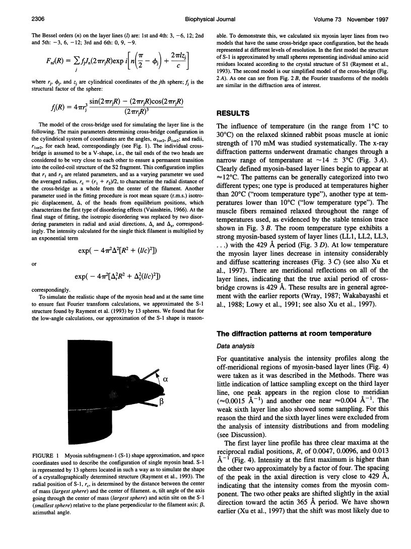
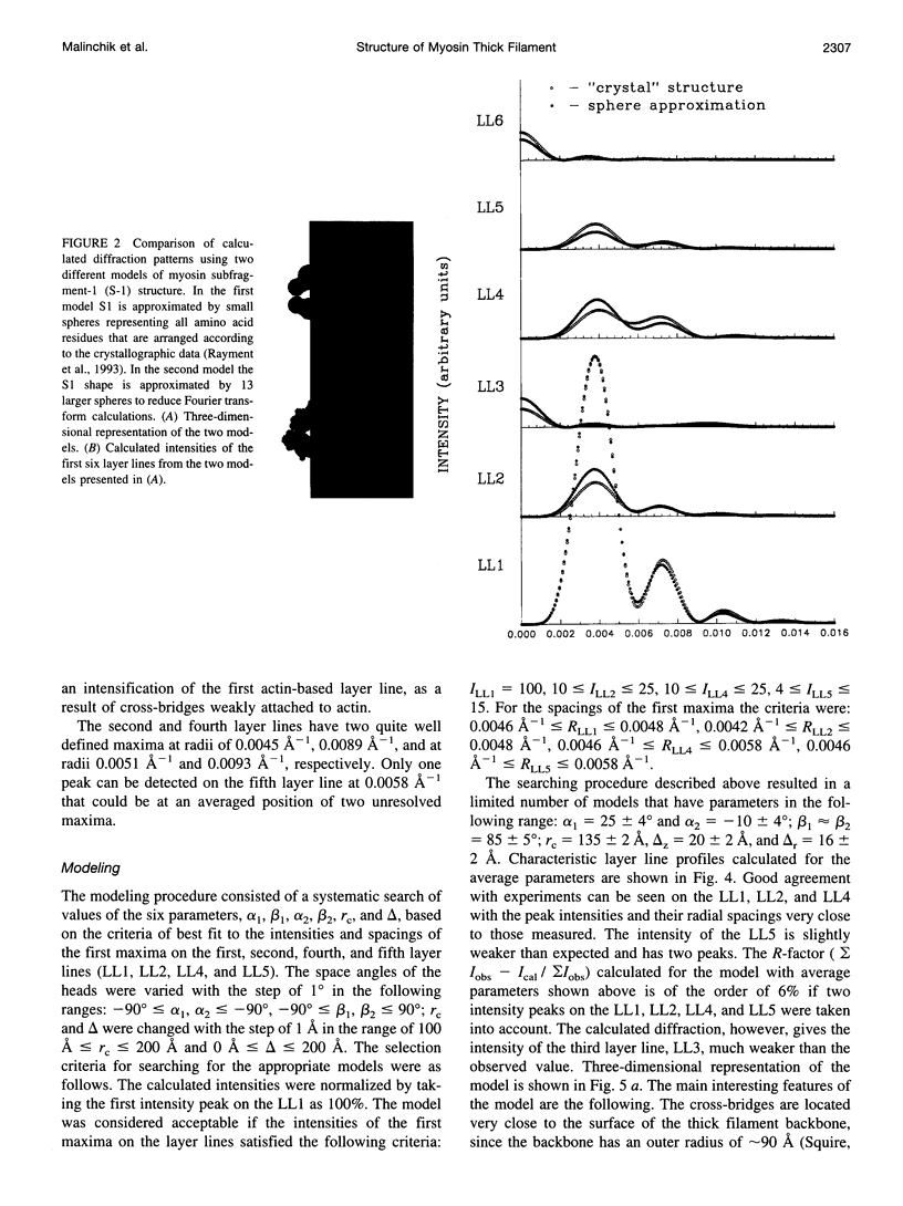
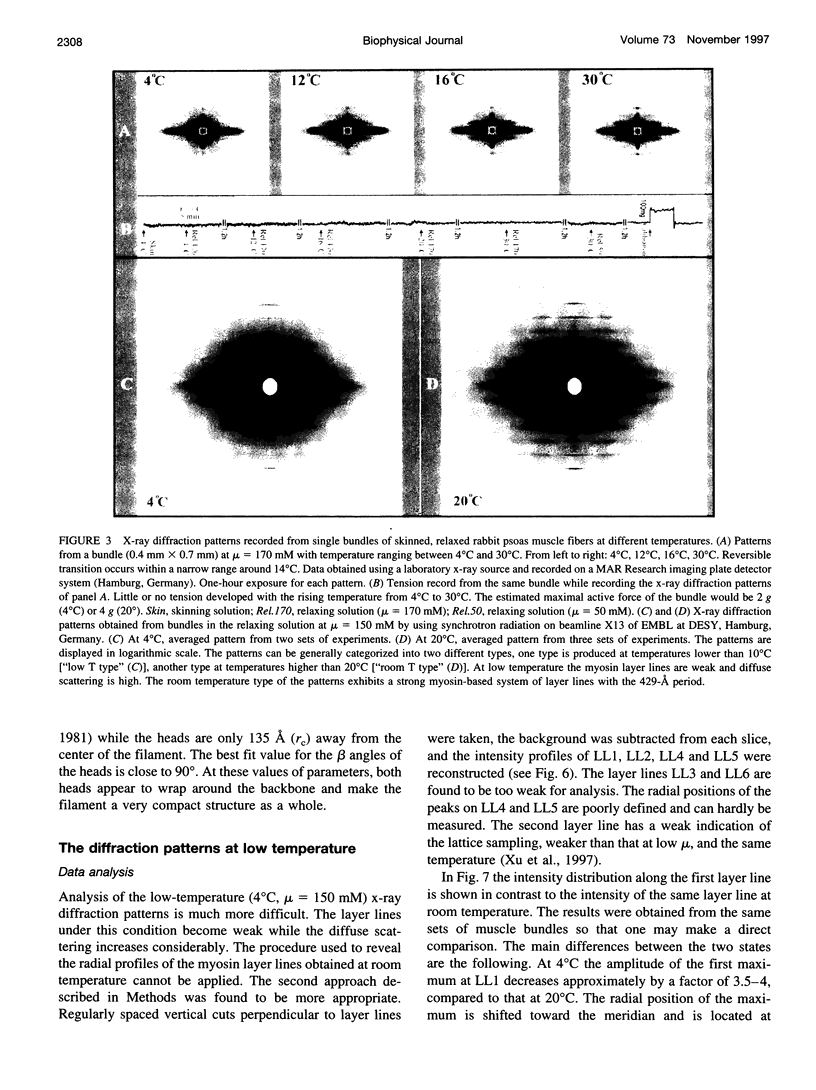
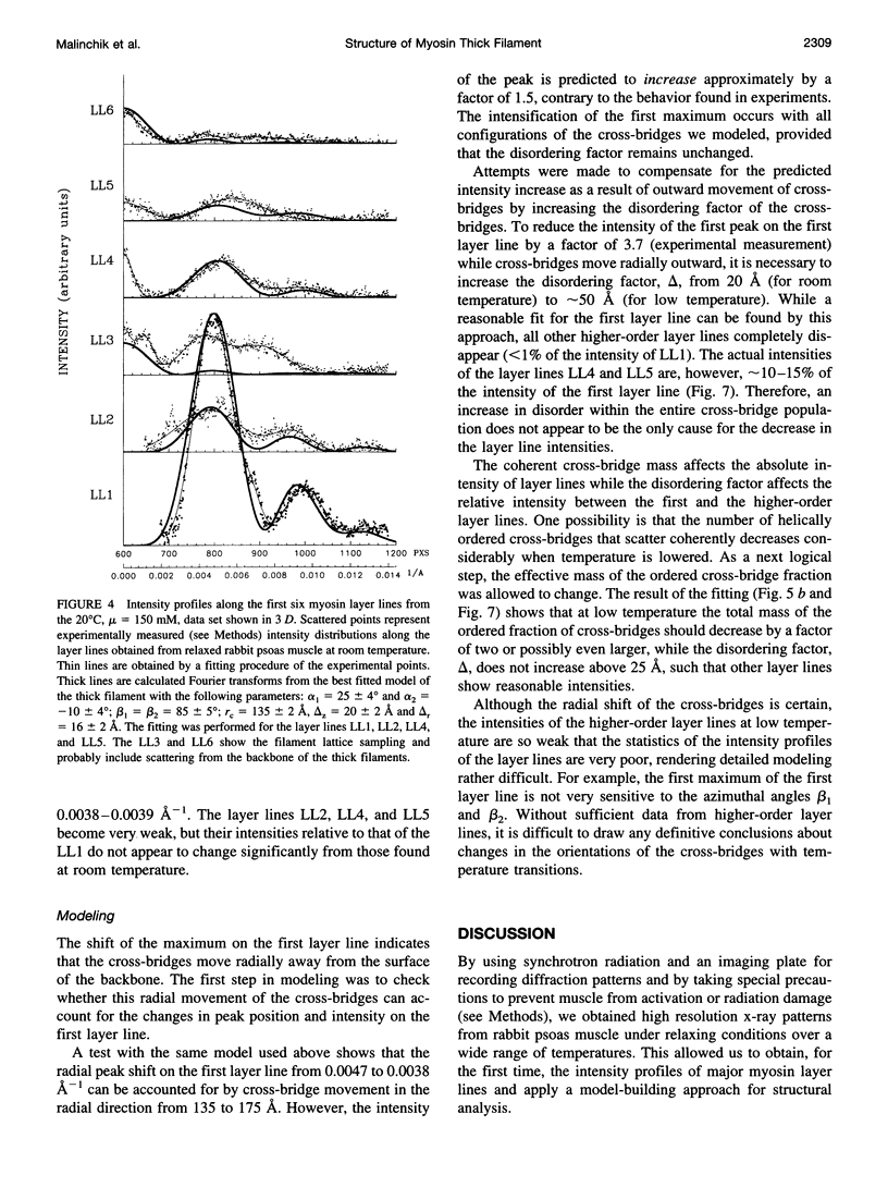
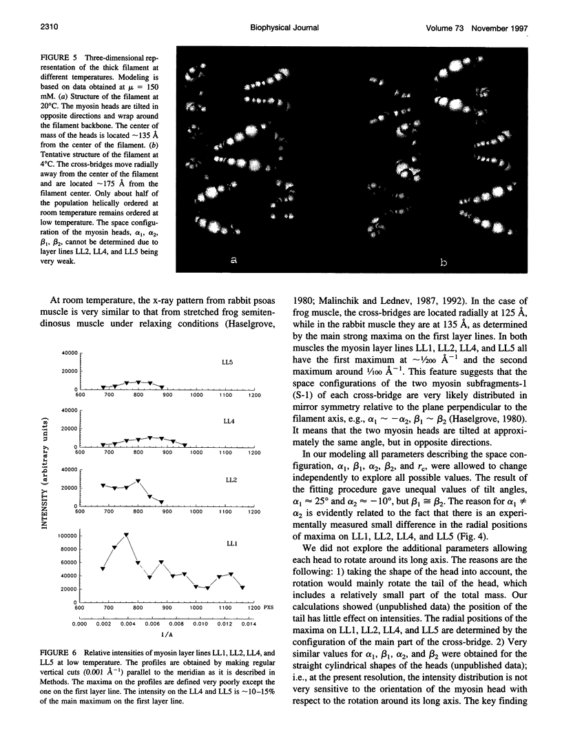
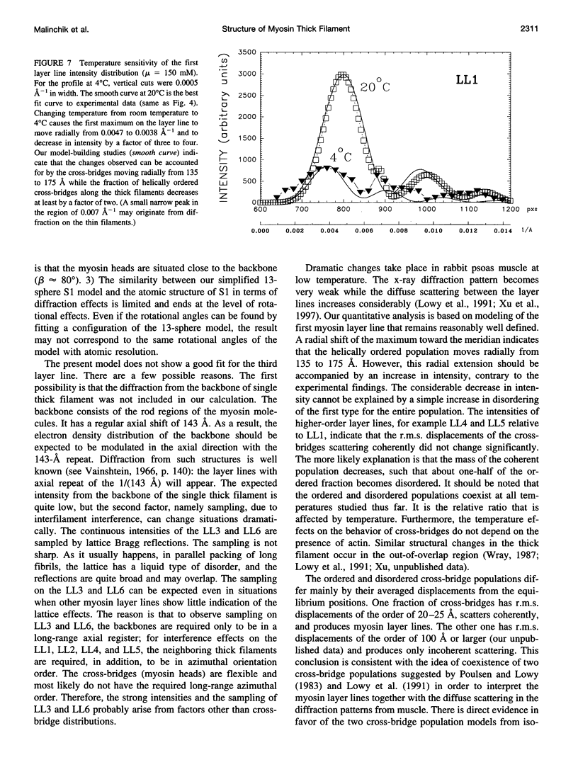
By using synchrotron radiation and an imaging plate for recording diffraction patterns, we have obtained high-resolution x-ray patterns from relaxed rabbit psoas muscle at temperatures ranging from 1 degree C to 30 degrees C. This allowed us to obtain intensity profiles of the first six myosin layer lines and apply a model-building approach for structural analysis. At temperatures 20 degrees C and higher, the layer lines are sharp with clearly defined maxima. Modeling based on the data obtained at 20 degrees C reveals that the average center of the cross-bridges is at 135 A from the center of the thick filament and both of the myosin heads appear to wrap around the backbone. At 10 degrees C and lower, the layer lines become very weak and diffuse scattering increases considerably. At 4 degrees C, the peak of the first layer line shifts toward the meridian from 0.0047 to 0.0038 A(-1) and decreases in intensity approximately by a factor of four compared to that at 20 degrees C, although the intensities of higher-order layer lines remain approximately 10-15% of the first layer line. Our modeling suggests that as the temperature is lowered from 20 degrees C to 4 degrees C the center of cross-bridges extends radially away from the center of the filament (135 A to 175 A). Furthermore, the fraction of helically ordered cross-bridges decreases at least by a factor of two, while the isotropic disorder (the temperature factor) remains approximately unchanged. Our results on the order/disordering effects of temperature are in general agreement with earlier results of Wray [Wray, J. 1987. Structure of relaxed myosin filaments in relation to nucleotide state in vertebrate skeletal muscle. J. Muscle Res. Cell Motil. 8:62a (Abstr.)] and Lowy et al. (Lowy, J., D. Popp, and A. A. Stewart. 1991. X-ray studies of order-disorder transitions in the myosin heads of skinned rabbit psoas muscles. Biophys. J. 60:812-824). and support Poulsen and Lowy's hypothesis of coexistence of ordered and disordered cross-bridge populations in muscle (Poulsen, F. R., and J. Lowy. 1983. Small angle scattering from myosin heads in relaxed and rigor frog skeletal muscle. Nature (Lond.). 303:146-152.). However, our results added new insights into the disordered population. Present modeling together with data analysis (Xu, S., S. Malinchik, Th. Kraft, B. Brenner, and L. C. Yu. 1997. X-ray diffraction studies of cross-bridges weakly bound to actin in relaxed skinned fibers of rabbit psoas muscle. Biophys. J. 73:000-000) indicate that in a relaxed muscle, cross-bridges are distributed in three populations: those that are ordered on the thick filament helix and those that are disordered; and within the disordered population, some cross-bridges are detached and some are weakly attached to actin. One critical conclusion of the present study is that the apparent order <--> disorder transition as a function of temperature is not due to an increase/decrease in thermal motion (temperature factor) for the entire population, but a redistribution of cross-bridges among the three populations. Changing the temperature leads to a change in the fraction of cross-bridges located on the helix, while changing the ionic strength at a given temperature affects the disordered population leading to a change in the relative fraction of cross-bridges detached from and weakly attached to actin. Since the redistribution is reversible, we suggest that there is an equilibrium among the three populations of cross-bridges.
Full text
PDF
Images in this article
Selected References
These references are in PubMed. This may not be the complete list of references from this article.
- Harford J., Squire J. "Crystalline" myosin cross-bridge array in relaxed bony fish muscle. Low-angle x-ray diffraction from plaice fin muscle and its interpretation. Biophys J. 1986 Jul;50(1):145–155. doi: 10.1016/S0006-3495(86)83447-6. [DOI] [PMC free article] [PubMed] [Google Scholar]
- Haselgrove J. C. A model of myosin crossbridge structure consistent with the low-angle x-ray diffraction pattern of vertebrate muscle. J Muscle Res Cell Motil. 1980 Jun;1(2):177–191. doi: 10.1007/BF00711798. [DOI] [PubMed] [Google Scholar]
- Holmes K. C., Popp D., Gebhard W., Kabsch W. Atomic model of the actin filament. Nature. 1990 Sep 6;347(6288):44–49. doi: 10.1038/347044a0. [DOI] [PubMed] [Google Scholar]
- Huxley H. E., Brown W. The low-angle x-ray diagram of vertebrate striated muscle and its behaviour during contraction and rigor. J Mol Biol. 1967 Dec 14;30(2):383–434. doi: 10.1016/s0022-2836(67)80046-9. [DOI] [PubMed] [Google Scholar]
- Ip W., Heuser J. Direct visualization of the myosin crossbridge helices on relaxed rabbit psoas thick filaments. J Mol Biol. 1983 Nov 25;171(1):105–109. doi: 10.1016/s0022-2836(83)80317-9. [DOI] [PubMed] [Google Scholar]
- Kabsch W., Mannherz H. G., Suck D., Pai E. F., Holmes K. C. Atomic structure of the actin:DNase I complex. Nature. 1990 Sep 6;347(6288):37–44. doi: 10.1038/347037a0. [DOI] [PubMed] [Google Scholar]
- Knight P., Trinick J. Structure of the myosin projections on native thick filaments from vertebrate skeletal muscle. J Mol Biol. 1984 Aug 15;177(3):461–482. doi: 10.1016/0022-2836(84)90295-x. [DOI] [PubMed] [Google Scholar]
- Lowy J., Popp D., Stewart A. A. X-ray studies of order-disorder transitions in the myosin heads of skinned rabbit psoas muscles. Biophys J. 1991 Oct;60(4):812–824. doi: 10.1016/S0006-3495(91)82116-6. [DOI] [PMC free article] [PubMed] [Google Scholar]
- Malinchik S. B., Lednev V. V. Interpretation of the X-ray diffraction pattern from relaxed skeletal muscle and modelling of the thick filament structure. J Muscle Res Cell Motil. 1992 Aug;13(4):406–419. doi: 10.1007/BF01738036. [DOI] [PubMed] [Google Scholar]
- Poulsen F. R., Lowy J. Small-angle X-ray scattering from myosin heads in relaxed and rigor frog skeletal muscles. Nature. 1983 May 12;303(5913):146–152. doi: 10.1038/303146a0. [DOI] [PubMed] [Google Scholar]
- Rayment I., Rypniewski W. R., Schmidt-Bäse K., Smith R., Tomchick D. R., Benning M. M., Winkelmann D. A., Wesenberg G., Holden H. M. Three-dimensional structure of myosin subfragment-1: a molecular motor. Science. 1993 Jul 2;261(5117):50–58. doi: 10.1126/science.8316857. [DOI] [PubMed] [Google Scholar]
- Stewart M., Kensler R. W. Arrangement of myosin heads in relaxed thick filaments from frog skeletal muscle. J Mol Biol. 1986 Dec 20;192(4):831–851. doi: 10.1016/0022-2836(86)90032-x. [DOI] [PubMed] [Google Scholar]
- Wakabayashi T., Akiba T., Hirose K., Tomioka A., Tokunaga M., Suzuki M., Toyoshima C., Sutoh K., Yamamoto K., Matsumoto T. Temperature-induced change of thick filament and location of the functional sites of myosin. Adv Exp Med Biol. 1988;226:39–48. [PubMed] [Google Scholar]