Abstract
An inactivated SVDV antigen is used in current enzyme-linked immunosorbent assays (ELISAs) for the detection of antibodies to swine vesicular disease virus (SVDV). To develop a noninfectious recombinant alternative, we produced SVDV-like particles (VLPs) morphologically and antigenically resembling authentic SVDV particles by using a dual baculovirus recombinant, which expresses simultaneously the P1 and 3CD protein genes of SVDV under different promoters. Antigenic differences between recombinant VLPs and SVDV particles were not statistically significant in results obtained with a 5B7-ELISA kit, indicating that the VLPs could be used in the place of SVDV antigen in ELISA kits. We developed a blocking ELISA using the VLPs and SVDV-specific neutralizing monoclonal antibody 3H10 (VLP-ELISA) for detection of SVDV serum antibodies in pigs. The VLP-ELISA showed a high specificity of 99.9% when tested with pig sera that are negative for SVDV neutralization (n = 1,041). When tested using sera (n = 186) collected periodically from pigs (n = 19) with experimental infection with each of three different strains of SVDV, the VLP-ELISA detected SVDV serum antibodies as early as 3 days postinfection and continued to detect the antibodies from all infected pigs until termination of the experiments (up to 121 days postinfection). This test performance was similar to that of the gold standard virus neutralization test and indicates that the VLP-ELISA is a highly specific and sensitive method for the detection of SVDV serum antibodies in pigs. This is the first report of the production and diagnostic application of recombinant VLPs of SVDV. Further potential uses of the VLPs are discussed.
Swine vesicular disease (SVD) is a highly contagious disease of pigs, characterized by vesicles on the coronary bands, heels of the feet, and occasionally on the lips, tongue, snout, and teats. These clinical signs make the disease indistinguishable from that caused by foot-and-mouth disease in pigs. Epidemics of SVD have occurred in Europe and eastern Asia (26) since the disease was recognized for the first time in Italy in 1966 (28). However, in the last five years, infection with SVDV has been reported only in Portugal and Italy.
Swine vesicular disease virus (SVDV) is a member of the genus Enterovirus (diameter of 24 to 30 nm) and family Picornaviridae (36, 37). The virus is antigenically related to the human coxsackievirus B5 (8, 16, 41). The virus has a positive-sense single-stranded RNA approximately 7,400 nucleotides long encoding four capsid proteins (VP2, VP4, VP3, and VP1) and seven nonstructural proteins (2A, 2B, 2C, 3A, 3B, 3C, and 3D) (22, 26, 37). The assembly of SVDV in host cells is believed to follow a scheme similar to that of other enteroviruses such as poliovirus. The first cleavage of the polyprotein occurs cotranslationally by viral protein 2A, resulting in a structural protein precursor (P1) and the remaining polyprotein P2-P3 (38). Subsequent proteolytic processing of the precursor P1 into VP0 (VP2 plus VP4), VP3, and VP1 occurs by means of a viral protease (3C) or its precursor (3CD) (6, 19, 32, 40). These structural proteins can then self-assemble to form empty capsid particles, which are composed of 60 copies of each structural protein (4, 36). The final step in virion assembly involves cleavage of structural protein VP0 into VP4 and VP2 during encapsidation of viral RNA (4, 34, 36).
Effective control of SVD depends on rapid detection of infected animals and those in contact with them. Severe cases in which animals show marked clinical signs can be detected easily through clinical surveillance and the direct detection of virus in clinical samples, while diagnosis of SVDV in subclinically infected animals can be mainly achieved by serological surveillance. In recent years, most cases of SVDV infection have been mild or subclinical and therefore serological surveillance has become of greater importance.
A virus neutralization test (VNT), the Office International des Epizooties “prescribed test” for international trade (31), is laborious, and expensive, since the test should be carried out within a biosafety level 3 (BSL3) laboratory. For these reasons, VNT is not ideal for large-scale routine testing. Due to their simplicity, high sensitivity, and economy, several enzyme-linked immunosorbent assays (ELISAs) have been developed as serological screening tools (1, 7, 9, 12, 13). All current ELISAs utilize inactivated SVDV antigen that has been prepared by killing infectious SVDV by use of a biohazard chemical such as binary ethyleneimine after viral propagation in cell cultures in a BSL3 laboratory. Despite efforts to replace the viral antigen by use of a recombinant P1 polypeptide in an indirect ELISA (24), for example, a satisfactory recombinant antigen alternative has not yet been developed.
Virus-like particles (VLPs) resemble authentic virus particles antigenically and morphologically but lack infectious genetic material (30). For this reason, they have been used as a highly effective type of alternative antigen in several viral diseases (15, 17, 18, 33) as well as to provide subunit vaccines (3, 14, 20, 27, 30). In the present work, we generated a dual baculovirus recombinant expressing simultaneously the P1 and 3CD protein genes of SVDV to induce formation of noninfectious VLP aggregates that mimic the antigenic structure of authentic SVDV particles. By using the recombinant VLPs, we developed a blocking ELISA (VLP-ELISA) for detection of SVDV serum antibodies and evaluated the test using pig sera.
MATERIALS AND METHODS
Cells and virus.
IBRS-2 cells were grown in Eagle's minimal essential medium alpha (GibcoBRL) supplemented with 10% heat-inactivated fetal bovine serum (GibcoBRL) and antibiotic-antimycotic solution (Invitrogen) at 37°C in an incubator. SVDV (strain UKG/27/72), from the Institute for Animal Health, Pirbright, United Kingdom, propagated in IBRS-2 cell culture was used for SVDV genomic RNA extraction. Spodoptera frugiperda (Sf9) insect cells (GibcoBRL) for propagation of recombinant baculovirus were grown in Grace's medium supplemented with 10% heat-inactivated fetal bovine serum, lactalbumin hydrolysates, and antibiotic-antimycotic solution (GibcoBRL) at 27°C.
MAbs.
The monoclonal antibody (MAb) 5B7, which is the indicator antibody in an internationally accepted SVD competitive ELISA (c-ELISA) kit (5B7-ELISA) (7, 31), was used as the reference antibody in this study. The 5B7 antibody is known to recognize a conformational neutralizing epitope (antigenic site 2A) on the VP2 protein of SVDV (29). 3H10, another neutralizing MAb with immunoglobulin M (IgM) heavy chains, was produced at our laboratory from BALB/c mice immunized with inactivated SVDV antigen (derived from a 5B7-ELISA kit) by use of a previously described protocol (10). Preliminary work using a comprehensive binding assay with labeled MAbs demonstrated a two-way competition between the 3H10 and 5B7 antibodies. The MAb 3H10 was biotin labeled for use as an indicator in the VLP-ELISA using a biotin-labeling kit (Roche Diagnostics Co., Indianapolis, Ind.) after being purified through an ImmunoPure IgM purification kit (Pierce) according to the manufacturer's instructions.
Sera.
A panel of standardized SVD reference pig sera (produced at the Institute for Animal Health, Pirbright, United Kingdom) was used that consisted of the following sera: RS1 (normal pig serum), RS2 (a strongly positive serum collected 21 days postinfection [dpi] from a SVDV strain UKG/27/72-infected pig), RS3 (a weakly positive pig serum collected from a SVDV strain ITL8/94-infected pig at 5 dpi and diluted 1:10 in normal pig serum RS1), RS4 (a weakly positive pig serum obtained by diluting RS2 1:40 in RS1), RS5 (a weakly positive pig serum collected 4 dpi from a SVDV strain UKG/27/72-infected pig), and RS6 (a weakly positive pig serum collected 5 dpi from a SVDV strain UKG/27/72-infected pig). All pigs used for reference positive sera were negative for SVDV in VNT prior to challenge. Three control sera (strongly positive, weakly positive, and negative) enclosed in the 5B7-ELISA kit were used as control sera for the VLP-ELISA. Hyperimmune anti-SVDV guinea pig serum supplied by the Institute for Animal Health, Pirbright, United Kingdom, was also used.
Sera specific for VP1 of SVDV were produced at our laboratory from guinea pigs that received two consecutive subcutaneous inoculations of keyhole limpet hemocyanin coupled with synthetic peptides mixed with Freund's adjuvant and given 2 weeks apart. Two synthetic peptides (C76YTTYKNHDSDGDNFA90 and C201RFDKQGTYGIST211) coupled to keyhole limpet hemocyanin (100 μg/dose) were synthesized for immunization based on amino acid sequences of the VP1 protein of SVDV UKG/27/72 (37). Two guinea pigs immunized with each peptide were bled at 2 weeks after their last inoculation, and then the sera were pooled.
SVDV antibody-negative pig sera (n = 1,041) from South Korea (n = 865) and Malta (n = 176) were used to test for the specificity of VLP-ELISA developed in this study. All sera used were negative in virus neutralization (VN) testing for SVDV.
Experimental pig serum samples (n = 186) that had been collected from pigs (n = 19) kept at the Institute for Animal Health, Pirbright, United Kingdom, following experimental infection with each of three different strains of SVDV were used to evaluate the sensitivity of the VLP-ELISA. Group A sera (n = 42) were collected from pigs (TM65, TM77, and TM78) infected with SVDV strain ITL/9/93, group B sera (n = 77) from pigs (TV1 to TV4 and TV14 to 16) infected with SVDV strain ITL/11/98, group C sera (n = 31) from pigs (UQ68, UQ69, and UQ70) infected with SVDV strain UKG/27/72, and group D sera (n = 36) from pigs (TN80 to TN85) immunized with a recombinant SVDV P1 polypeptide and then challenged with SVDV (strain ITL/9/93) 42 days later.
Construction of recombinant baculovirus transfer vectors and generation of recombinant baculovirus.
The viral RNA was extracted from SVDV (UKG/27/72 strain)-infected IBRS-2 cells by use of an RNeasy extraction Minikit (QIAGEN). The P1 and 3CD genes were amplified separately using the viral RNA as a template and using a one-step RT-PCR kit (QIAGEN) according to the manufacturer's instructions. Synthetic primer set 5′-GGATCC733ATGGGAGCTCAAGTGTCAAC772-3′ and 5′-AAGCTT3295AAGTGGTTTTCATGGTTGTTA3276-3′ and synthetic primer set 5′-GCCCTCGAGA5361TGGGTCCAGCGTTTGAGTTC5380-3′ and 5′-GCTCGGTACC7299TAAAAGGAGTCCAACCACTT7279-3′ used for amplification of full-length P1 and 3CD genes, respectively, were designed on the basis of the genomic sequence of SVDV strain UKG/27/72 (37). Restriction enzyme sites (underlined) were incorporated at the 5′ ends of each primer to facilitate cloning as indicated by the underlined nucleotides.
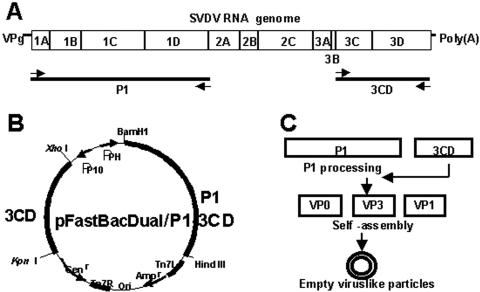
After amplification the genes were separately cloned into a pGEMTeasy vector (Promega) and then subcloned into a pFastBacDual vector (Invitrogen), after which the P1 and the 3CD were inserted downstream of the polyhedrin promoter (PPH) and the P10 promoter, respectively (Fig. 1). Recombinant baculovirus DNA was generated by site-specific transposition of pFastBac/SVDV-P1-3CD into a baculovirus shuttle vector (bacmid) propagated in Escherichia coli strain DH10Bac cells (Invitrogen) by using the Bac-to-Bac baculovirus expression system (GibcoBRL) according to the manufacturer's protocol. Dual recombinant baculovirus containing SVDV P1 and 3CD DNAs was generated by transfecting recombinant baculovirus DNA into Sf9 insect cells. The recombinant baculovirus was plaque purified, and then the presence of the specific SVDV DNA sequences was confirmed by PCR using the primer sets described above.
FIG. 1.
(A) Amplification of P1 and 3CD protein genes of SVDV by using reverse transcription-PCR. (B) Construction of a recombinant dual expression vector containing P1 and 3CD protein genes of SVDV. (C) A schematic strategy for production of virus-like particles induced by recombinant baculovirus in insect cells.
Production and purification of the SVD VLPs.
The Sf9 cells (at a density of 3 × 10⁶ cells/ml) grown in 200-ml spinner cultures were infected at a high multiplicity of infection of 10 with plaque-purified baculovirus and harvested at 5 days postinfection. The cells were frozen and thawed three times, clarified by centrifugation at 10,000 × g for 40 min, and concentrated by using polyethylene glycol precipitation as previously described (11). Following dialysis against 0.01 M phosphate-buffered saline (PBS), pH 7.4, a 0.5-ml volume of recombinant proteins was further purified by discontinuous CsCl gradient centrifugation (using 5.5-ml volume layers of 4% and 40% CsCl in PBS) at 150,000 ×g for 6 h at 4°C (Beckman). Each 0.5-ml volume fraction was collected from the CsCl gradient and subjected to an indirect sandwich ELISA. Fractions with a peak of antigenic materials were examined by electron microscopy for the presence of the recombinant proteins. The antigenic materials were pooled and concentrated using a Centricon apparatus (Millipore) for ELISA antigen. The concentration of recombinant proteins in the fractions was measured by using a BCA protein assay kit (Pierce). The VLP antigen was stored in 50% (vol/vol) glycerol at −70°C at a final concentration of 100 μg/ml. As a control, inactivated SVDV harvested from IBRS cells was purified by the same procedure described above.
Indirect sandwich ELISA.
MaxiSorp ELISA plates (Nalge Nunc International, Roskilde, Denmark) were coated with 50 μl of MAb 5B7 at the optimal concentration in 0.01 M phosphate-buffered saline (PBS), pH 7.4, at 37°C for 1 h with constant shaking. Plates were washed three times with PBST (0.002 M PBS containing 0.05% Tween 20) and then incubated with 50 μl of each fraction from a CsCl gradient in a blocking buffer (0.01 M PBS containing 1% yeast extract and 0.05% Tween 20) for 1 h at 37°C. Inactivated SVDV antigen (enclosed in a reference c-ELISA kit) and mock-infected antigen were used as positive and negative controls, respectively. Following a washing step, the plates were incubated at 37°C for 1 h with optimally diluted peroxidase-conjugated MAb 5B7 (enclosed in a reference c-ELISA kit) in the blocking buffer. Following another washing step, the plates were incubated for 10 min with the substrate O-phenylenediamine (Sigma) in 0.05 M citrate phosphate buffer (pH 5.0) containing 0.015% hydrogen peroxidase. The colorimetric reaction was stopped by adding a 50-μl volume of 1.25 M sulfuric acid to all wells. Optical density (OD) readings were measured at the 492-nm wavelength.
Electron microscopy.
Antigenic materials from CsCl gradients were used for morphological examination by electron microscopy. A 20-μl volume of purified proteins was placed on a carbon-coated cupper grid. It was then blot dried, negatively stained with 1% uranyl acetate, and immediately blot dried again. The stained grids were air dried for 40 min before examination by transmission electron microscopy (Hitachi).
Western immunoblotting.
Protein preparations (i.e., VLPs and viral proteins) were electrophoretically separated through NuPAGE Novex Bis-Tris gels by use of an Xcell SureLock Mini-cell (Invitrogen) according to the manufacturer's instructions. Separated polypeptides were transferred from the gels onto nitrocellulose membranes by use of an Xcell II blot module (Invitrogen) according to the manufacturer's instructions. Immunoblotting was then performed by standard techniques using guinea pig sera against SVDV or VP1 peptides (1:100 dilution). Specific antigen-antibody reactions on each membrane were visualized by applying anti-species IgG (heavy plus light chains) conjugated with alkaline phosphatase (1:1,000 dilution) followed by BCIP (5-bromo-4-chloro-3-indolylphosphate)/Nitro Blue Tetrazolium substrate solution (Kirkegaard-Perry Laboratories Inc., Gaithersburg, MD).
VLP-ELISA.
Optimal concentrations of all reagents, including antigen, MAb, and serum, were predetermined by checkerboard titration. All buffers used were the same as in the indirect ELISA described above. MaxiSorp ELISA plates were coated with 50-μl volumes of purified MAb 3H10 (1.0 μg/ml) in 0.05 M carbonate/bicarbonate buffer, pH 9.6, overnight 4°C. Plates were washed three times with PBST and then incubated with 50 μl of the VLP antigen (2.5 μg/ml in the blocking buffer) for 1 h at 37°C. Following a washing step, plates were incubated with 50-μl volumes of test sera (final dilution of 1:20 in the blocking buffer) for 1 h at 37°C. Control sera (strongly positive, weakly positive, and negative) were added to each plate. Plates were washed three times with PBST and then incubated with 50-μl volumes of biotin-labeled 3H10 antibody (1:500 dilution) for 1 h at 37°C. Following a washing step, the amount of biotin bound was detected by 1-h incubation with 50-μl volumes of a peroxidase-streptavidin conjugate (Kirkegaard-Perry Laboratories Inc.) at 1:200 dilution. The optical density (OD) values for each well after colorimetric reaction were converted to percent inhibition (PI) values for each MAb pair with the following formula: PI = 100 − [(OD of MAb-serum mixture)/(OD of MAb alone)] × 100. The threshold cutoff was determined by calculation (mean PI value minus 3 standard deviations) using European Union (EU) SVD reference pig serum 4 (RS4) (Institute for Animal Health, Pirbright, United Kingdom) with the lowest level of SVDV antibodies.
Other tests.
The 5B7-ELISA (7) was carried out according to the protocols supplied by the manufacturer. Briefly, the SVD viral antigen is trapped to the solid phase by use of the MAb 5B7. The ability of test sera to inhibit the binding of peroxidase-conjugated MAb 5B7 to the trapped antigen is then evaluated. Finally, the amount of conjugated MAb bound is detected by the addition of substrate and OPD chromogen. Sera are considered positive when PI values are higher than 70% at the 1/7.5 serum dilution.
The virus neutralization (VN) test was performed in flat-bottomed microtiter plates with IBRS-2 cells, according to a published Office International des Epizooties protocol (31). VN titers of 1/45 or more were regarded as positive.
RESULTS
Identification of recombinant VLPs induced by simultaneous expression of SVDV P1 and 3CD proteins.
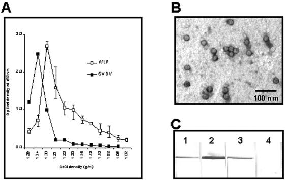
The P1 and 3CD genes of SVDV were separately amplified and cloned into a pFastBacDual vector under each of two different promoters. A dual baculovirus recombinant containing the P1 and 3CD genes of SVDV was generated from the recombinant pFastBacDual vector by using the Bac-to-Bac expression system. Insect cells infected with the recombinant baculovirus were harvested after 5 days when most of the cells showed cytopathic effects. Recombinant proteins reactive to the 5B7 antibody were detected from both the cell lysates (1:50 dilution) and the supernatant (1:5 dilution) in the indirect ELISA (data not shown). Recombinant proteins from cell lysates were concentrated, purified, and then fractionated by use of a discontinuous CsCl gradient centrifugation. The peak of antigenic materials was observed in a fraction with a buoyant density of approximately 1.30 g/ml in an indirect ELISA using 5B7 antibody (Fig. 2A). This was slightly different from the results obtained from inactivated SVDV, which showed the antigenic peak in a fraction with a buoyant density of 1.34 g/ml. The yield of the VLP protein purified was usually 2.0 mg or more per liter of culture volume. Electron microscopy revealed that the purified proteins from the fraction displayed the structure of SVD virus-like particles (VLPs) with diameters of 25 nm to 26 nm (Fig. 2B).
FIG. 2.
Characterization of recombinant SVDV-like particles. (A) Antigenic reactivity of each fraction from CsCl gradients. The reactivity of each fraction was measured by an indirect ELISA using 5B7 antibody. (B) Electron microscopy of SVDV-like particles from antigenic materials made by a dual baculovirus recombinant. (C) Western immunoblot analyses of SVDV-like particles. Proteins from SVDV-like particles (lane 1 and 2), SVDV (lane 3), and mock-infected cells (lane 4) were electrophoretically separated, transferred onto nitrocellulose membranes, and detected using guinea pig sera specific for SVDV (lanes 1, 3, and 4) and VP1 peptide (lane 2).
A protein band with a molecular mass of approximately 31 kDa was detected from the VLP fraction in Western immunoblot analyses using anti-SVDV guinea pig serum (Fig. 2C). A protein band with the same size was also visualized by using guinea pig sera specific for VP1 peptides, indicating the presence of VP1 protein. However, the P1 protein (precursor of viral capsid proteins) and other viral proteins VP0, VP2, VP4, and VP3 were not detected in either the VLP fraction or the proteins from SVDV-infected cells in Western immunoblotting (Fig. 2C).
Replacement of recombinant VLPs with SVDV antigen in the 5B7-ELISA kit.
Competitive ELISAs were performed using the 5B7-ELISA kit according to the kit protocol to investigate whether VLPs can be used as alternative antigen. Purified VLPs (2.5 μg/ml) were used as the ELISA antigen. For comparison, SVDV antigen (of the 5B7-ELISA kit) was used as positive control. Six EU SVD reference sera (RS1 to RS6) were tested at least three times by the 5B7-ELISA using each of three antigens. The 5B7-ELISA using VLP antigen had average PI values of 4.0, 98.9, 91.9, 70.0, 91, and 95.3 for RS1, RS2, RS3, RS4, RS5 and RS6, respectively. The 5B7-ELISA using SVDV antigen had average PI values of 1.5, 93.7, 93.7, 71.7, 85.0, and 94.5 for RS1, RS2, RS3, RS4, RS5, and RS6, respectively. The VLP antigen correlated well (correlation coefficient r = 0.995) with the SVDV antigen, and the difference of the reactivity between the two antigens was not statistically significant by Student's t test (P > 0.05).
Specificity and sensitivity of VLP-ELISA.
A monoclonal antibody-based blocking ELISA using recombinant VLPs (designated VLP-ELISA) was developed for detection of SVDV antibodies in pigs. Purified VLPs from the antigenic peak fraction of the CsCl gradients described above were used as an ELISA antigen. Purified MAb 3H10 (IgM class) was used as coating antibody (unlabeled) and indicator (biotin labeled). Serum controls of the 5B7-ELISA kit were used for optimization of the VLP-ELISA.
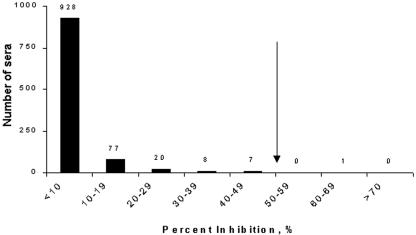
When tested using VLP-ELISA, EU SVD reference sera RS1, RS2, RS3, RS4, RS5, and RS6 had PI values ± standard deviations of −0.2 ± 7.7, 95.0 ± 2.6, 84.4 ± 2.3, 62.4 ± 4.2, 87.1 ± 1.7, and 88.2 ± 2.8, respectively. Here, the cutoff value (mean PI value − 3× the standard deviation) of the VLP-ELISA was set at a PI value of 50 which allowed the weakest of the EU SVD reference sera (RS4) to be scored as positive. Based on the cutoff PI value of 50, known SVD-negative serum samples (n = 1,041) were tested by the VLP-ELISA. All sera except only one were negative in VLP-ELISA (Fig. 3), indicating a specificity of 99.9% (Fig. 3). Most of the pig sera (928/1,041; 89.1%) had PI values of <10. The PI value of the pig serum with nonspecific positive reaction in the VLP-ELISA was 62, similar to that (62.4 ± 4.2) of the RS4 control, which has been available as the lowest positive-level serum in the EU.
FIG. 3.
Frequency distribution of PI values obtained from VLP-ELISA using known SVDV-negative pig sera (n = 1,041). The cutoff value of the ELISA was set at a PI value of 50 (↓), based on the mean PI value of (62.4) of the most weakly positive serum (EU SVD RS4) minus 3× the standard deviation (3 × 4.2).
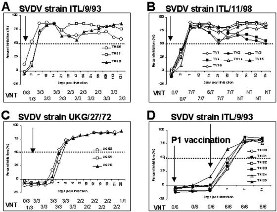
In VLP-ELISA, we tested a total of 186 sera collected sequentially from four groups of pigs (groups A to D) that had been inoculated with each of three different strains of SVDV (Fig. 4). VLP-ELISA detected SVDV antibodies from 3 dpi (TM65) to 7 dpi (TM77) from pigs infected with SVDV strain ITL/9/93 (group A), 7 dpi from all pigs (TV1 to TV4 and TV14 to TV16) with SVDV strain ITL/11/98 (group B), and 4 dpi (UQ68, UQ70) to 5 dpi (UQ69) from pigs infected with SVDV strain UKG/27/72 (group C). All pigs in group D, which had been vaccinated with recombinant SVDV P1 polyprotein, were negative in the VLP-ELISA, as in the VN test. However, SVDV antibodies were detected as early as 4 (TN85) or 7 (TN80 to TN84) dpi when pigs were challenged with the ITL/9/93 strain of SVDV 42 days after the vaccination. VLP-ELISA continued to detect SVDV antibodies from all infected pigs until termination of each experiment (up to 121 dpi).
FIG. 4.
Kinetics of early antibody development as determined by VLP-ELISA and virus neutralization (VN) test in pigs (n = 19) with induced SVDV infections. Pigs of the four groups A, B, C, and D received inoculations with different SVDV strains of ITL/9/93, ITL/11/98, UKG/27/72, and ITL/9/93 (42 days postvaccination with SVDV P1), respectively. The dotted lines represent the cutoff PI value. Arrows represent the time of SVDV challenge (A to D) or P1 vaccination (D). Results obtained from VN test were presented as number positive/number tested.
Most animals (15/19) seroconverted on the same day in both VLP-ELISA and VN test, although some individuals (TM65, TM78, and TN80 to TN84) showed differences between the tests. However, overall, there was no indication that the VN test was detecting animals sooner after infection than the VLP-ELISA and the kinetics of the antibody responses were similar in the both tests.
DISCUSSION
Recombinant SVD virus-like particles (VLPs) were induced by simultaneous expression in insect cells of P1 and 3CD genes by a recombinant baculovirus. To our knowledge, this is the first time that this has been reported for SVDV, although it has been done for other enteroviruses, by use of baculovirus recombinants in insect cells (6, 21, 39), vaccinia recombinants in mammalian cells (2), and the yeast Saccharomyces cerevisiae (35). Recombinant VLPs have been produced on the basis of the expression of full-length polyprotein (39), the P1 and 3CD proteins (21, 35), the P1 and P3 proteins (2), or the individual VP0, VP3, and VP1 proteins (6) in several expression systems. The yield (20 to 180 μg/liter) of VLP obtained by single recombinant (based on the expression of full-length polyprotein) (39) was enhanced compared to that obtained by multiple baculovirus recombinants (6, 21) but was still not satisfactory for development of a subunit vaccine or diagnostic antigen. For efficient VLP production in insect cells, we generated a dual baculovirus recombinant carrying the P1 (approximately 3.3 kb) and 3CD (approximately 1.9 kb) genes of SVDV, of which the total size of the inserts was reduced to 2.1 kb compared to the size of the full-length polyprotein gene (6.6 kb) in a recombinant baculovirus previously reported. The high yield (2 mg or more per liter of culture) of VLPs produced in this study indicates that use of a single recombinant baculovirus containing the P1 and 3CD genes might be a more efficient and economic method of producing VLPs than the use of other, more complex baculovirus recombinants described previously.
Processing of the P1 protein into the VP1 protein was identified in Western immunoblotting and indicates capsid protein cleavage in insect cells after the simultaneous appearance of P1 and 3CD proteins, due to strong enzymatic activity of coexpressed 3CD protease rather than Sf9 cellular proteases, as demonstrated in insect cells with coexpression of P1 and 3CD genes of enterovirus 71 (21). Our failure to detect the other capsid proteins VP0 (precursor of VP4 and VP2) and VP3 in the Western immunoblotting may be due to a lack of sera able to recognize linear epitopes, as previously reported for SVDV-specific sera (26). This seems to result from a lack of detectable serum antibodies to linear epitopes present on these proteins. The peak of antigenic materials from CsCl gradients contained the VLPs (diameter of 25 to 26 nm), which are morphologically indistinguishable from authentic enterovirus particles (36). The buoyant density of the antigenic materials was 1.30 g/ml, which was the same as reported for the empty shell (75S) of enterovirus (6, 36). Taken together, these results support the conclusion that the simultaneous expression of P1 and 3CD proteins in insect cells has resulted in the production of empty SVD virus-like particles through a series of capsid-assembly steps (i.e., a 5S protomer, a 14S pentameric subunit, and then an 75S icosahedral procapsid).
To our knowledge, our report provides the first demonstration of recombinant SVD virus-like particles as alternatives for SVDV antigen in competitive ELISAs for detection of antibodies to SVDV in pigs. The 5B7-ELISA kit is an internationally accepted c-ELISA kit and a standard screening test for detection of SVDV antibodies in the EU. In the present study, we attempted to replace the SVDV antigen with VLPs by use of the same protocol specified in the kit. The results obtained for six EU SVD reference sera (RS1 to RS6) were not significantly different using VLP antigen from those obtained with the unmodified 5B7-ELISA kit (using inactivated SVDV antigen). This indicates that the VLPs retain the antigenicity of the epitope of the 5B7 antibody, making them applicable as alternative antigens in the 5B7-ELISA kit, although further evaluation with a more extensive panel of sera would be needed to reach a definite conclusion on this.
A new blocking ELISA was developed based on the recombinant VLPs and SVDV-specific monoclonal antibody 3H10. Our recombinant VLP protein provides many advantages over the inactivated SVDV antigen used in currently available SVD ELISA kits. Recombinant VLP antigen can be prepared at any laboratories with cell culture facilities, since the protein is noninfectious and does not represent a hazard to susceptible animals. To prepare inactivated SVDV antigen used in the reference c-ELISA kit, highly contagious SVDV should be grown in cell cultures and killed by a biohazardous chemical, binary ethyleneimine, within a BSL3 laboratory. Significantly, VLP production is much more cost effective and requires less regulatory control than production of inactivated SVDV antigen. Purified VLPs used as ELISA antigens in the VLP-ELISA demonstrated an excellent specificity of 99.9% with SVDV-negative sera (n = 1,041). Meanwhile, we biotin labeled the 3H10 antibody to optimize the sensitivity of the VLP-ELISA since one molecule of the biotin catches up to five molecules of a streptavidin conjugated to a peroxidase. The VLP-ELISA correctly scored the EU SVD reference serum panel (RS1 to RS6). Strongly positive (RS2), weakly positive (RS3, RS5, and RS6), the most weakly positive (RS4), and negative (RS1) sera had differential PI values of 95.0, 84.4 to 88.2, 62.4, and −0.2, respectively. This indicates that our VLP-ELISA can detect SVDV antibodies as early as 4 dpi from infected pigs. Good sensitivity of the VLP-ELISA was also demonstrated in this study by using sera from pigs with experimental SVDV infection. The VLP-ELISA detected SVDV antibodies in all experimental pigs as early as 3 to 4 dpi and continued to detect them until the termination of the experiments. Therefore, the VLP-ELISA can be used for diagnostic purposes (e.g., to supplement virological tests such as virus isolation and antigen detection ELISA or in their place if unavailable or if appropriate samples cannot be obtained) as well as for serological surveillance. The high specificity and sensitivity of the VLP-ELISA combined with ease of performance and rapid provision of results should make the method an appropriate screening test for surveillance programs to rapidly detect infected/exposed animals, although the test remains to be fully evaluated in the field.
As shown in Fig. 4, our VLP-ELISA using neutralizing antibody 3H10 did not detect SVDV-specific antibodies in the sera of pigs immunized with the P1 polyprotein of SVDV. No SVDV neutralizing antibody was detected in these pig sera when tested by VN test. This is therefore not surprising, since the P1 protein failed to induce immune response in pigs involved in neutralization, as reported previously (5, 24). Several epitopes involved in neutralization of SVDV have been identified on the capsid surface (5, 23, 25, 29, 34), and although immune response in pigs is induced by linear epitopes (24), conformational epitopes are known to be the most immunogenic (5). In preliminary work, neutralizing antibodies (VN titers of up to 1:1,000) were developed in BALB/c mice vaccinated with our VLPs in Freund's adjuvant, although high neutralizing antibody titers were elicited by inactivated SVDV antigen (VN titers > 1:3,200) in mice (data not shown). This suggests that a VLP-based vaccine could feasibly be developed for SVDV, as studied with several viral diseases (3, 14, 20, 27). Therefore, we are carrying out further studies to investigate whether VLPs can elicit protective immune responses in pigs.
Acknowledgments
Sang-Mi Kang and Chan-Jin Woo are gratefully acknowledged for their excellent technical assistance. The authors also acknowledge technical assistance from Jung-Won Park for the transmission electron microscopy work.
This study was supported by a grant from the National Veterinary Research and Quarantine Service, Korean Ministry of Agriculture and Fishery.
REFERENCES
- 1.Armstrong, R. M., and I. T. Barnett. 1989. An enzyme-linked immunosorbent assay (ELISA) for the detection and quantification of antibodies against swine vesicular disease virus (SVDV). J. Virol. Methods 25:71-79. [DOI] [PubMed] [Google Scholar]
- 2.Ansardi, D. C., D. C. Porter, and C. D. Morrow. 1991. Coinfection with recombinant vaccinia virus expressing poliovirus P1 and P3 proteins results in polyprotein processing and formation of empty capsid structures. J. Virol. 65:2088-2092. [DOI] [PMC free article] [PubMed] [Google Scholar]
- 3.Ball, J. M., D. Y. Graham, A. R. Opekun, M. A. Gilger, R. A. Guerrero, and M. K. Estes. 1999. Recombinant Norwalk virus-like particles given orally to volunteers: phase I study. Gastroenterology 117:40-48. [DOI] [PubMed] [Google Scholar]
- 4.Basavappa, R., R. Syed, O. Flore, J. P. Icenogle, D. J. Filman, and J. M. Hogle. 1994. Role and mechanism of the maturation cleavage of VP0 in poliovirus assembly: structure of the empty capsid assembly intermediate at 2.9 Å resolution. Protein Sci. 3:1651-1669. [DOI] [PMC free article] [PubMed] [Google Scholar]
- 5.Borrego, B., E. Carra, J. A. Garcia-Ranea, and E. Brocchi. 2002. Characterization of neutralization sites on the circulating variant of swine vesicular disease virus (SVDV): a new site is shared by SVDV and the related coxsackie B5 virus. J. Gen. Virol. 83:35-44. [DOI] [PubMed] [Google Scholar]
- 6.Bräutigam, S., E. Snezhkov, and D. H. L. Bishop. 1993. Formation of poliovirus-like particles by recombinant baculovirus expressing the individual VP0, VP3, and VP1 proteins by comparison to particles derived from the expressed poliovirus polyprotein. Virology 192:512-524. [DOI] [PubMed] [Google Scholar]
- 7.Brocchi, E., A. Berlinzani, D. Gamba, and F. De Simone. 1995. Development of two novel monoclonal antibody-based ELISAs for the detection of antibodies and the identification of swine isotypes against swine vesicular disease virus. J. Virol. Methods 52:155-167. [DOI] [PubMed] [Google Scholar]
- 8.Brown, F., P. Talbot, and R. Burrows. 1973. Antigenic differences between isolates of swine vesicular disease virus and their relationship to coxsackie B5 virus. Nature 245:315-316. [DOI] [PubMed] [Google Scholar]
- 9.Chenard, G., M. Bloemraad, J. A. Kramps, C. Terpstra, and A. Dekker. 1998. Validation of a monoclonal antibody-based ELISA to detect antibodies directed against swine vesicular disease virus. J. Virol. Methods 75:105-112. [DOI] [PubMed] [Google Scholar]
- 10.Choi, K. S., J. J. Nah, Y. J. Ko, S. Y. Kang, and Y. S. Joo. 2003. Localization of antigenic sites at the amino-terminus of rinderpest virus N protein using deleted N mutants and monoclonal antibody. J. Vet. Sci. 4:167-173. [PubMed] [Google Scholar]
- 11.Choi, K. S., J. J. Nah, C. U. Choi, Y. J. Ko, H. J. Sohn, G. Libeau, S. Y. Kang, and Y. S. Joo. 2003. Monoclonal antibody-based competitive ELISA for simultaneous detection of rinderpest virus and peste des petits ruminants virus antibodies. Vet. Microbiol. 96:1-16. [DOI] [PubMed] [Google Scholar]
- 12.Dekker, A., P. L. Moonen, and C. Terpstra. 1995. Validation of a screening liquid phase blocking ELISA for swine vesicular disease. J. Virol. Methods 51:343-348. [DOI] [PubMed] [Google Scholar]
- 13.Dekker, A., F. van Hemert-Kluitenberg, C. Baars, and C. Terpstra. 2002. Isotype specific ELISAs to detect antibodies against swine vesicular disease virus and their use in epidemiology. Epidemiol. Infect. 128:277-284. [DOI] [PMC free article] [PubMed] [Google Scholar]
- 14.Evans, T. G., W. Bonnez, R. C. Rose, S. Koenig, L. Demeter, J. A. Suzich, D. O'Brien, M. Campbell, W. I. White, J. Balsley, and R. C. Reichman. 2001. A Phase 1 study of a recombinant viruslike particle vaccine against human papillomavirus type 11 in healthy adult volunteers. J. Infect. Dis. 183:1485-1493. [DOI] [PubMed] [Google Scholar]
- 15.Graham, D. Y., X. Jiang, T. Tanaka, A. R. Opekun, H. P. Madore, and M. K. Estes. 1994. Norwalk virus infection of volunteers: new insights based on improved assays. J. Infect. Dis. 170:34-43. [DOI] [PubMed] [Google Scholar]
- 16.Graves, J. H. 1973. Serological relationships of swine vesicular disease and coxsackie B5 viruses. Nature 245:314-315. [DOI] [PubMed] [Google Scholar]
- 17.Green, K. Y., J. F. Lew, X. Jiang, A. Z. Kapikian, and M. K. Estes. 1993. Comparison of the reactivities of baculovirus-expressed recombinant Norwalk virus capsid antigen with those of the native Norwalk virus antigen in serologic assays and some epidemiologic observations. J. Clin. Microbiol. 31:2185-2191. [DOI] [PMC free article] [PubMed] [Google Scholar]
- 18.Guo, M., Y. Qian, K. O. Chang, and L. J. Saif. 2001. Expression and self-assembly in baculovirus of porcine enteric calicivirus capsids into virus-like particles and their use in an enzyme-linked immunosorbent assay for antibody detection in swine. J. Clin. Microbiol. 39:1487-1493. [DOI] [PMC free article] [PubMed] [Google Scholar]
- 19.Harris, K. S., S. R. Reddigari, M. J. Nicklin, T. Hammerle, and E. Wimmer. 1992. Purification and characterization of poliovirus polypeptide 3CD, a proteinase and a precursor for RNA polymerase. J. Virol. 66:7481-7489. [DOI] [PMC free article] [PubMed] [Google Scholar]
- 20.Harro, C. D., Y. Y. Pang, R. B. Roden, A. Hildesheim, Z. Wang, M. J. Reynolds, T. C. Mast, R. Robinson, B. R. Murphy, R. A. Karron, J. Dillner, J. T. Schiller, and D. R. Lowy. 2001. Safety and immunogenicity trial in adult volunteers of a human papillomavirus 16 L1 virus-like particle vaccine. J. Natl. Cancer Inst. 93:284-292. [DOI] [PubMed] [Google Scholar]
- 21.Hu, Y. C., J. T. Hsu, J. H. Huang, M. S. Ho, and Y. C. Ho. 2003. Formation of enterovirus-like particle aggregates by recombinant baculoviruses co-expressing P1 and 3CD in insect cells. Biotechnol. Lett. 25:919-925. [DOI] [PubMed] [Google Scholar]
- 22.Inoue, T., T. Suzuki, and K. Sekiguchi. 1989. The complete nucleotide sequence of swine vesicular disease virus. J. Gen. Virol. 70:919-934. [DOI] [PubMed] [Google Scholar]
- 23.Jimenez-Clavero, M. A., A. Douglas, T. Lavery, J. A. Garcia-Ranea, and V. Ley. 2000. Immune recognition of swine vesicular disease virus structural proteins: novel antigenic regions that are not exposed in the capsid. Virology 270:76-83. [DOI] [PubMed] [Google Scholar]
- 24.Jimenez-Clavero, M. A., E. Escribano-Romero, J. M. Sanchez-Vizcaino, and V. Ley. 1998. Molecular cloning, expression and immunological analysis of the capsid precursor polypeptide (P1) from swine vesicular disease virus. Virus Res. 57:163-170. [DOI] [PubMed] [Google Scholar]
- 25.Kanno, T., T. Inoue, Y. Wang, A. Sarai, and S. Yamaguchi. 1995. Identification of the location of antigenic sites of swine vesicular disease virus with neutralization-resistant mutants. J. Gen. Virol. 76:3099-3106. [DOI] [PubMed] [Google Scholar]
- 26.Lin, F., and R. P. Kitching. 2000. Swine vesicular disease: an overview. Vet. J. 160:192-201. [DOI] [PubMed] [Google Scholar]
- 27.Maranga, L., P. E. Cruz, J. G. Aunins, and M. J. Carrondo. 2002. Production of core and virus-like particles with baculovirus infected insect cells. Adv. Biochem. Eng. Biotechnol. 74:183-206. [DOI] [PubMed] [Google Scholar]
- 28.Nardelli, L., E. Lodetti, G. L. Gualandi, R. Burrows, D. Goodridge, F. Brown, and B. Cartwright. 1968. A foot-and-mouth disease syndrome in pigs caused by an enterovirus. Nature 219:1275-1276. [DOI] [PubMed] [Google Scholar]
- 29.Nijhar, S. K., D. K. Mackay, E. Brocchi, N. P. Ferris, R. P. Kitching, and N. J. Knowles. 1999. Identification of neutralizing epitopes on a European strain of swine vesicular disease virus. J. Gen. Virol. 80:277-282. [DOI] [PubMed] [Google Scholar]
- 30.Noad, R., and P. Roy. 2003. Virus-like particles as immunogens. Trends Microbiol. 11:438-444. [DOI] [PubMed] [Google Scholar]
- 31.Office International des Epizooties. 2000. Swine vesicular disease, p100-104, In Manual of standards for diagnostic tests and vaccines, 4th ed. Office International des Epizooties, Paris, France.
- 32.Parsley, T. B., C. T. Cornell, and B. L. Semler. 1999. Modulation of the RNA binding and protein processing activities of poliovirus polypeptide 3CD by the viral RNA polymerase domain. J. Biol. Chem. 274:12867-12876. [DOI] [PubMed] [Google Scholar]
- 33.Purdy, D. E., A. J. Noga, and G. J. Chang. 2004. Noninfectious recombinant antigen for detection of St. Louis encephalitis virus-specific antibodies in serum by enzyme-linked immunosorbent assay. J. Clin. Microbiol. 42:4709-4717. [DOI] [PMC free article] [PubMed] [Google Scholar]
- 34.Rebel, J. M., C. H. Leendertse, A. Dekker, and R. J. Moormann. 2003. Effects of mutations in the VP2/VP4 cleavage site of Swine vesicular disease virus on RNA encapsidation and viral infectivity. Arch. Virol. 148:1747-1756. [DOI] [PubMed] [Google Scholar]
- 35.Rombaut, B., and J. P. Jore. 1997. Immunogenic, non-infectious polio subviral particles synthesized in Saccharomyces cerevisiae. J. Gen. Virol. 78:1829-1832. [DOI] [PubMed] [Google Scholar]
- 36.Rueckert, R. R. 1996. Picornaviridae: the viruses and their replication, p. 609-654. In B. N. Fields, D. M. Knipe, and P. M. Howey (ed.), Virology. Lippincott-Raven Publishers, Philadelphia, Pa.
- 37.Seechurn, P., N. W. Knowles, and J. W. McCauley. 1990. The complete nucleotide sequence of a pathogenic swine vesicular disease virus. Virus Res. 16:255-274. [DOI] [PubMed] [Google Scholar]
- 38.Toyoda, H., M. J. Nicklin, M. G. Murray, C. W. Anderson, J. J. Dunn, F. W. Studier, and E. Wimmer. 1986. A second virus-encoded proteinase involved in proteolytic processing of poliovirus polyprotein. Cell 45:761-770. [DOI] [PubMed] [Google Scholar]
- 39.Urakawa, T., M. Ferguson, P. D. Minor, J. Cooper, M. Sullivan, J. W. Almond, and D. H. Bishop. 1989. Synthesis of immunogenic, but non-infectious, poliovirus particles in insect cells by a baculovirus expression vector. J. Gen. Virol. 70:1453-1463. [DOI] [PubMed] [Google Scholar]
- 40.Ypma-Wong, M. F., P. G. Dewalt, V. H. Johnson, J. G. Lamb, and B. L. Semler. 1988. Protein 3CD is the major poliovirus proteinase responsible for cleavage of the P1 capsid precursor. Virology 166:265-270. [DOI] [PubMed] [Google Scholar]
- 41.Zhang, G., D. T. Haydon, N. J. Knowles, and J. W. McCauley. 1999. Molecular evolution of swine vesicular disease virus. J. Gen. Virol. 80:639-651. [DOI] [PubMed] [Google Scholar]


