Abstract
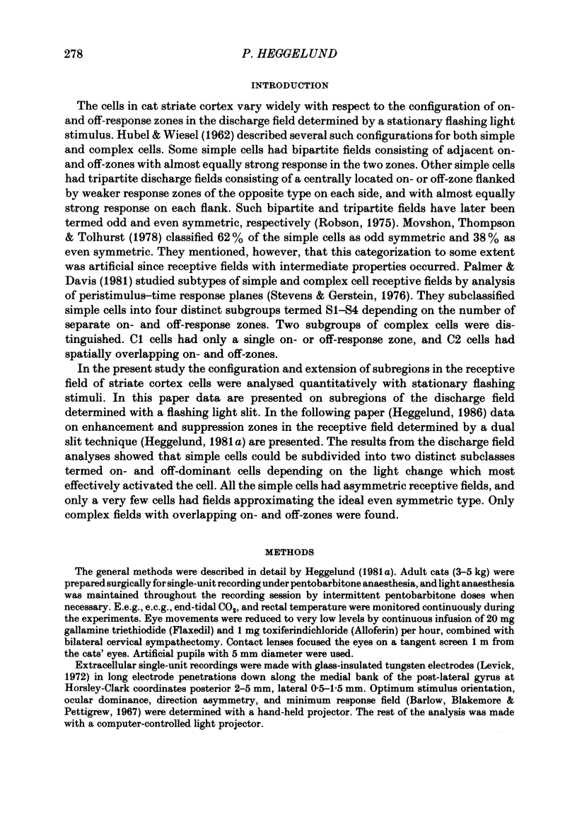
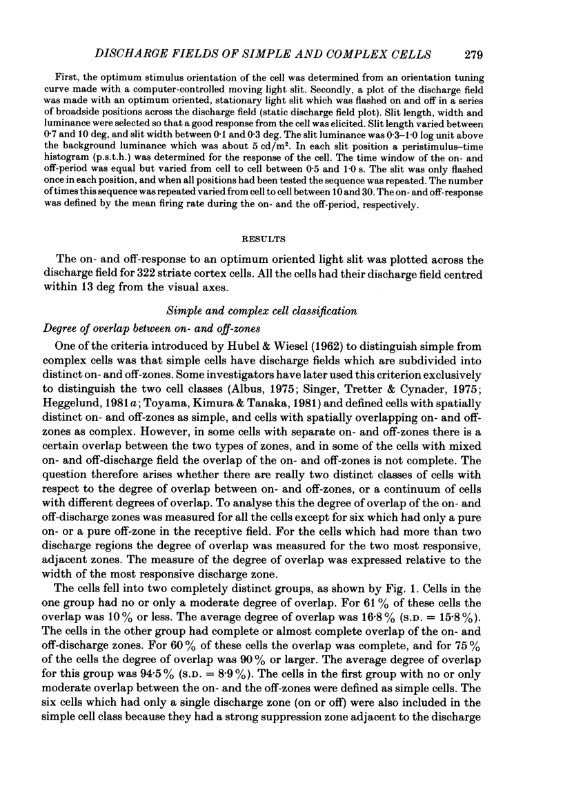
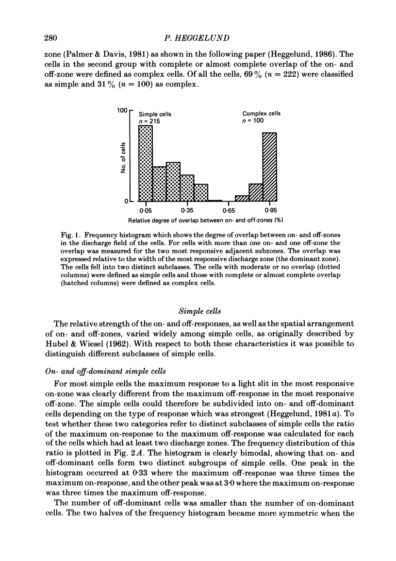
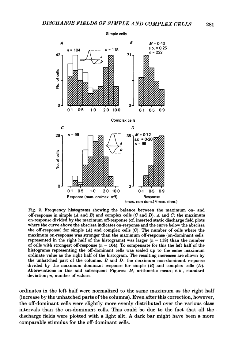
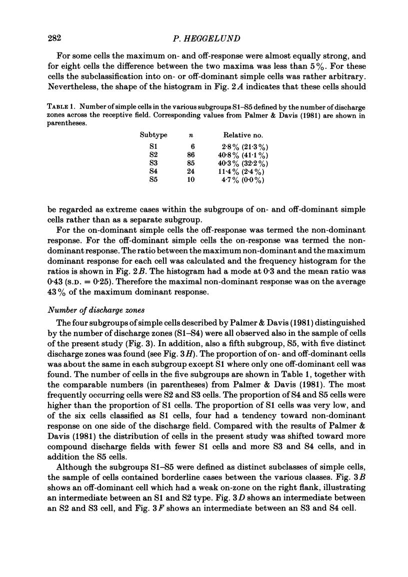
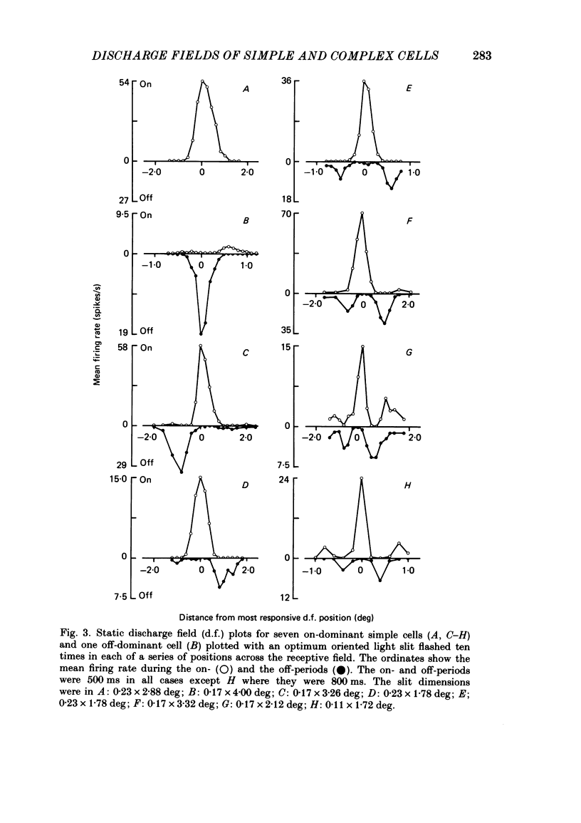
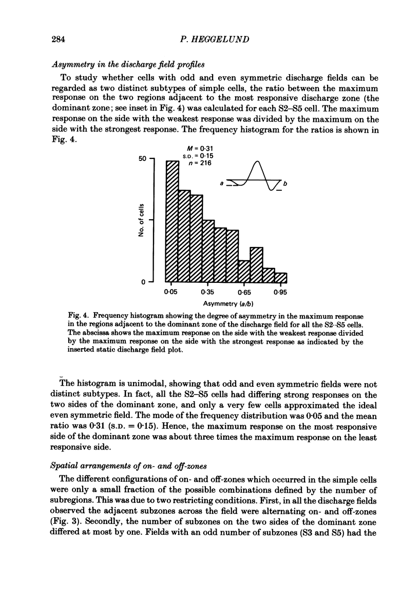
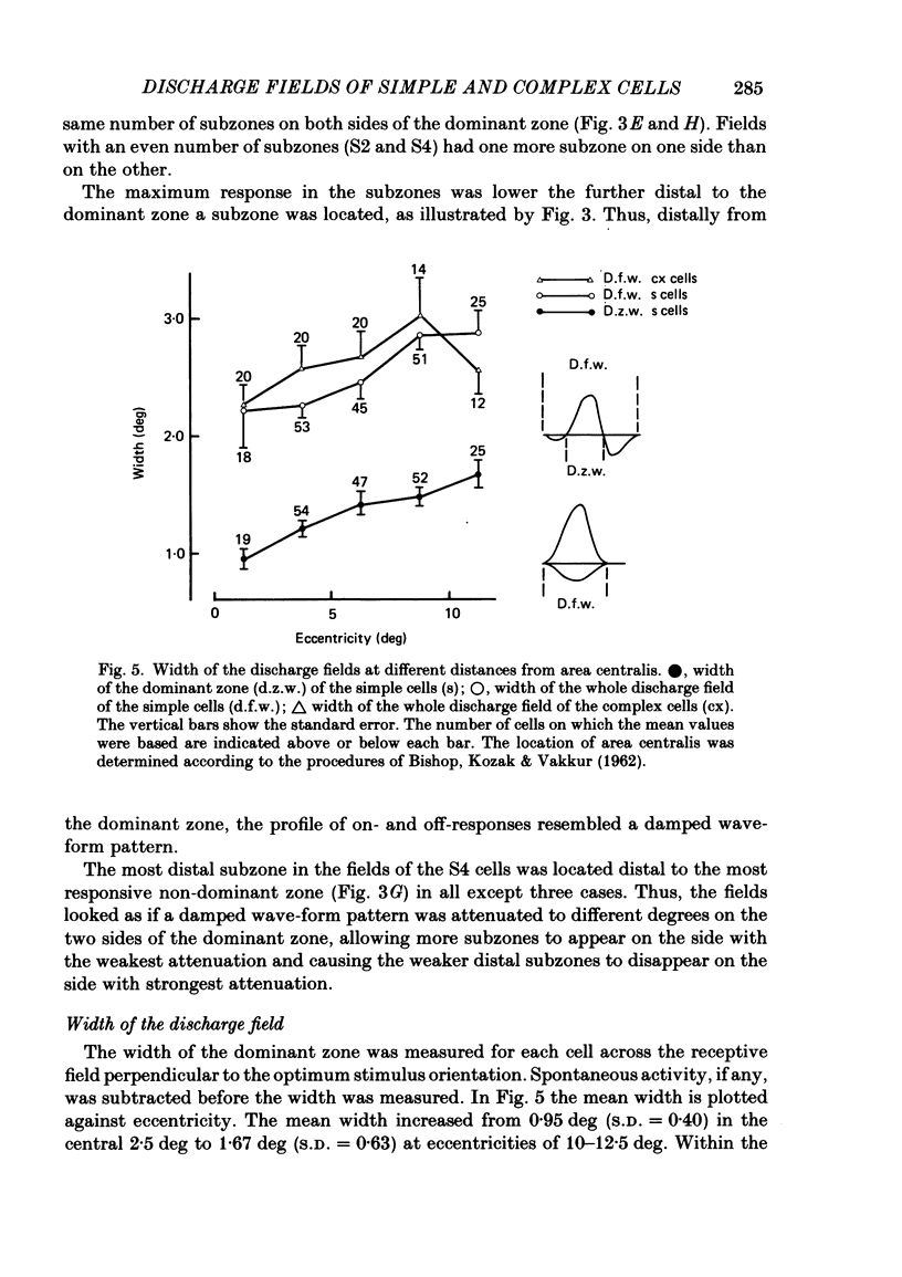
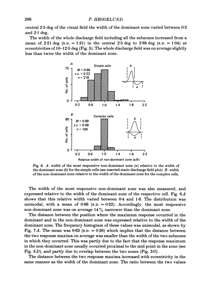
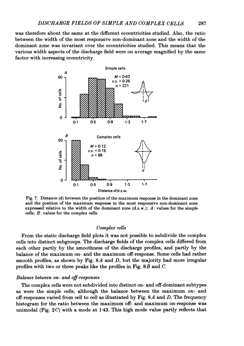
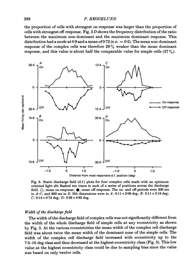
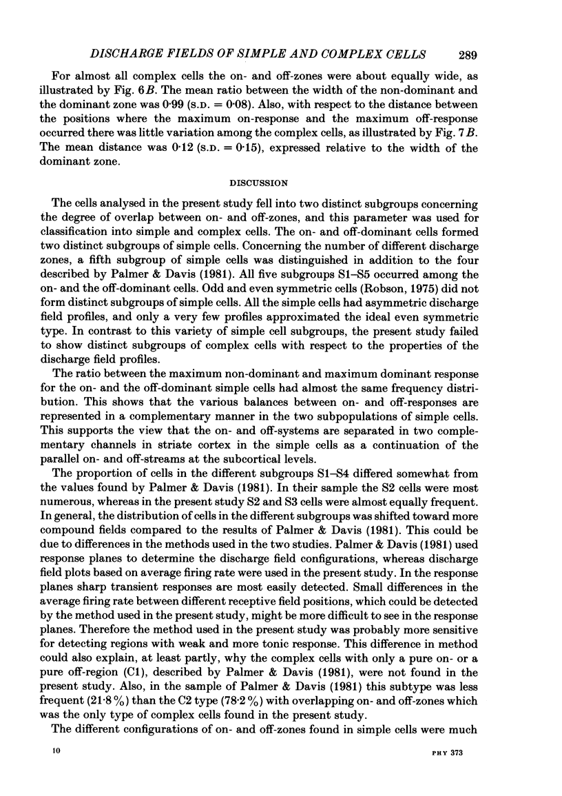
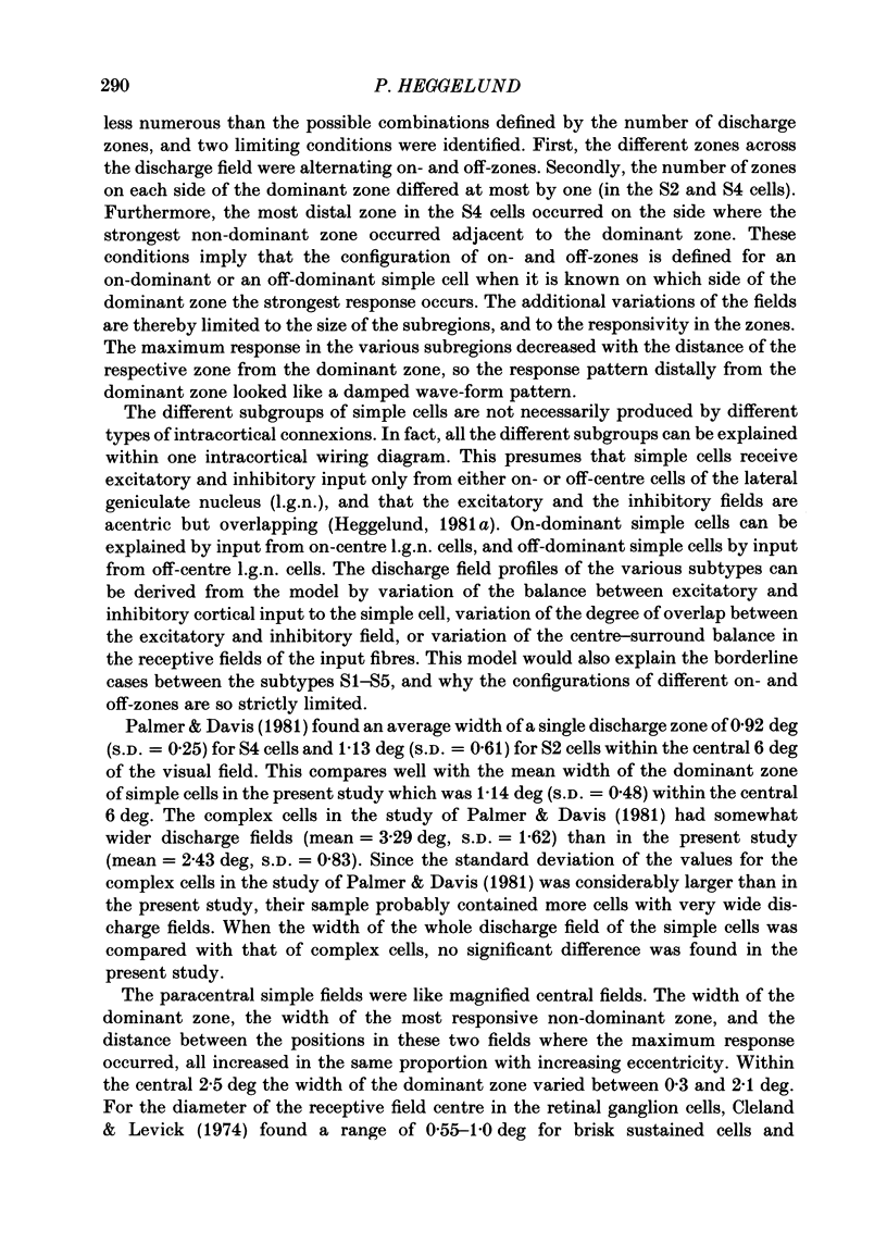
The configuration and width of on- and off-response zones in the discharge field of single cells in cat striate cortex was analysed by quantitative methods. The responses across on- and off-zones were plotted for 321 cells with a stationary optimum oriented light slit. The cells fell into two completely distinct subgroups with respect to the degree of overlap between adjacent on- and off-zones. The simple cells had a mean overlap of 16.8%, the complex cells 94.5%. For simple cells the ratio between the maximum off- and maximum on-response in the discharge field was bimodal, showing that two distinct subgroups termed on- and off-dominant cells could be distinguished. For the complex cells the corresponding frequency distribution was unimodal. The maximum response on the two regions adjacent to the most responsive discharge zone (the dominant zone) differed markedly for most simple cells, and only a very few cells had discharge fields approximating an ideal even symmetric field. The frequency distribution of the ratio between the maximum response in the two regions was unimodal showing that odd and even symmetric fields did not form distinct subgroups of simple cells. The number of different discharge zones in simple cells varied from one to five. The zones were arranged as alternating on- and off-zones across the discharge field. The maximum response in the subzones decreased with increasing sequential distance from the dominant zone, so the response pattern across each side of the discharge field resembled a damped wave-form pattern. All the complex cells had one on- and off-zone which overlapped. The mean width of the subregions in the simple cell discharge field and the mean distance between the response maxima in the subzones increased in the same proportion with increasing eccentricity. The paracentral fields were therefore like magnified central fields. The average width of the whole discharge field was not significantly different for the simple and the complex cells at the various eccentricities.
Full text
PDF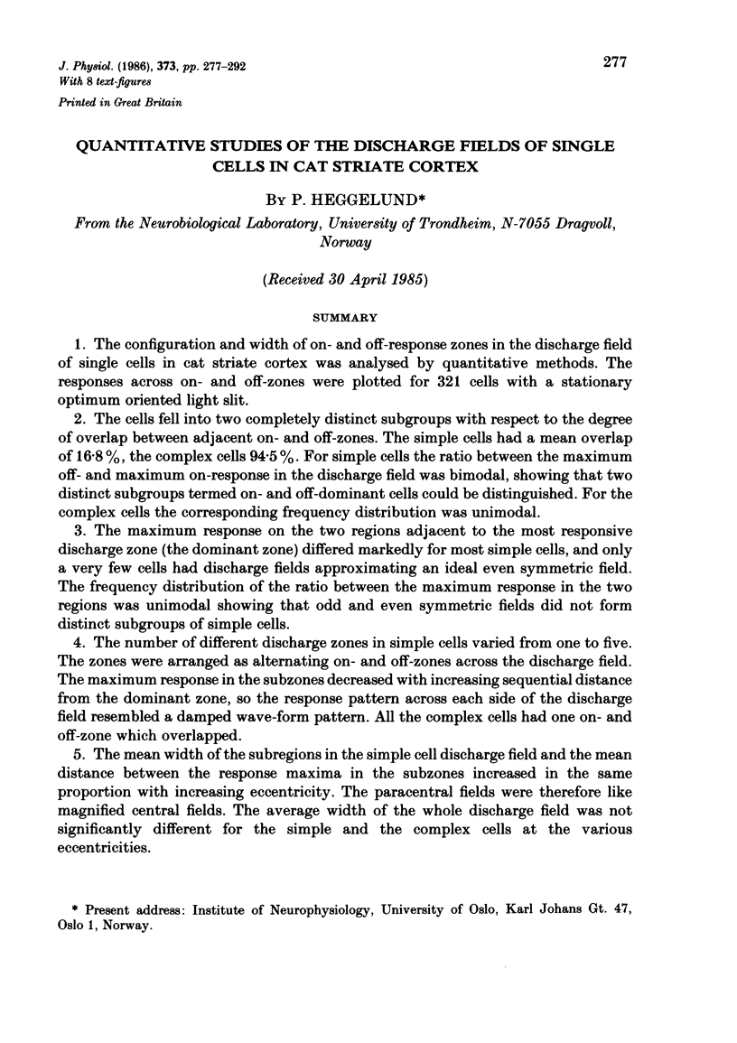
Selected References
These references are in PubMed. This may not be the complete list of references from this article.
- Albus K. A quantitative study of the projection area of the central and the paracentral visual field in area 17 of the cat. I. The precision of the topography. Exp Brain Res. 1975 Dec 22;24(2):159–179. doi: 10.1007/BF00234061. [DOI] [PubMed] [Google Scholar]
- BISHOP P. O., KOZAK W., VAKKUR G. J. Some quantitative aspects of the cat's eye: axis and plane of reference, visual field co-ordinates and optics. J Physiol. 1962 Oct;163:466–502. doi: 10.1113/jphysiol.1962.sp006990. [DOI] [PMC free article] [PubMed] [Google Scholar]
- Barlow H. B., Blakemore C., Pettigrew J. D. The neural mechanism of binocular depth discrimination. J Physiol. 1967 Nov;193(2):327–342. doi: 10.1113/jphysiol.1967.sp008360. [DOI] [PMC free article] [PubMed] [Google Scholar]
- Cleland B. G., Levick W. R. Brisk and sluggish concentrically organized ganglion cells in the cat's retina. J Physiol. 1974 Jul;240(2):421–456. doi: 10.1113/jphysiol.1974.sp010617. [DOI] [PMC free article] [PubMed] [Google Scholar]
- HUBEL D. H., WIESEL T. N. Receptive fields, binocular interaction and functional architecture in the cat's visual cortex. J Physiol. 1962 Jan;160:106–154. doi: 10.1113/jphysiol.1962.sp006837. [DOI] [PMC free article] [PubMed] [Google Scholar]
- Heggelund P. Quantitative studies of enhancement and suppression zones in the receptive field of simple cells in cat striate cortex. J Physiol. 1986 Apr;373:293–310. doi: 10.1113/jphysiol.1986.sp016048. [DOI] [PMC free article] [PubMed] [Google Scholar]
- Heggelund P. Receptive field organization of complex cells in cat striate cortex. Exp Brain Res. 1981;42(1):90–107. [PubMed] [Google Scholar]
- Heggelund P. Receptive field organization of simple cells in cat striate cortex. Exp Brain Res. 1981;42(1):89–98. doi: 10.1007/BF00235733. [DOI] [PubMed] [Google Scholar]
- Levick W. R. Another tungsten microelectrode. Med Biol Eng. 1972 Jul;10(4):510–515. doi: 10.1007/BF02474199. [DOI] [PubMed] [Google Scholar]
- Movshon J. A., Thompson I. D., Tolhurst D. J. Spatial summation in the receptive fields of simple cells in the cat's striate cortex. J Physiol. 1978 Oct;283:53–77. doi: 10.1113/jphysiol.1978.sp012488. [DOI] [PMC free article] [PubMed] [Google Scholar]
- Palmer L. A., Davis T. L. Receptive-field structure in cat striate cortex. J Neurophysiol. 1981 Aug;46(2):260–276. doi: 10.1152/jn.1981.46.2.260. [DOI] [PubMed] [Google Scholar]
- Peichl L., Wässle H. Size, scatter and coverage of ganglion cell receptive field centres in the cat retina. J Physiol. 1979 Jun;291:117–141. doi: 10.1113/jphysiol.1979.sp012803. [DOI] [PMC free article] [PubMed] [Google Scholar]
- Singer W., Tretter F., Cynader M. Organization of cat striate cortex: a correlation of receptive-field properties with afferent and efferent connections. J Neurophysiol. 1975 Sep;38(5):1080–1098. doi: 10.1152/jn.1975.38.5.1080. [DOI] [PubMed] [Google Scholar]
- Stevens J. K., Gerstein G. L. Spatiotemporal organization of cat lateral geniculate receptive fields. J Neurophysiol. 1976 Mar;39(2):213–238. doi: 10.1152/jn.1976.39.2.213. [DOI] [PubMed] [Google Scholar]
- Toyama K., Kimura M., Tanaka K. Cross-Correlation Analysis of Interneuronal Connectivity in cat visual cortex. J Neurophysiol. 1981 Aug;46(2):191–201. doi: 10.1152/jn.1981.46.2.191. [DOI] [PubMed] [Google Scholar]
- Wilson P. D., Rowe M. H., Stone J. Properties of relay cells in cat's lateral geniculate nucleus: a comparison of W-cells with X- and Y-cells. J Neurophysiol. 1976 Nov;39(6):1193–1209. doi: 10.1152/jn.1976.39.6.1193. [DOI] [PubMed] [Google Scholar]


