Abstract
Ebola virus particle formation and budding are mediated by the VP40 protein, which possesses overlapping PTAP and PPXY late domain motifs (7-PTAPPXY-13). These late domain motifs have also been found in the Gag proteins of retroviruses and the matrix proteins of rhabdo- and arenaviruses. While in vitro studies suggest a critical role for late domain motifs in the budding of these viruses, including Ebola virus, it remains unclear as to whether the VP40 late domains play a role in Ebola virus replication. Alteration of both late domain motifs drastically reduced VP40 particle formation in vitro. However, using reverse genetics, we were able to generate recombinant Ebola virus containing mutations in either or both of the late domains. Viruses containing mutations in one or both of their late domain motifs were attenuated by one log unit. Transmission and scanning electron microscopy did not reveal appreciable differences between the mutant and wild-type viruses released from infected cells. These findings indicate that the Ebola VP40 late domain motifs enhance virus replication but are not absolutely required for virus replication in cell culture.
Ebola virus and its close relative the Marburg virus comprise the family Filoviridae. This name derives from the filamentous form of virus particles that are approximately 80 nm in diameter but lack uniform length. The surfaces of filovirus particles are decorated with the glycoprotein GP, which binds to cellular receptors and mediates the fusion of viral and cellular membranes (reviewed in references 6, 19, and 32). The VP40 matrix protein, a major structural component of the virion, is associated with the inner leaflet of the membrane, and a third membrane-associated protein, VP24, was recently shown to be involved in nucleocapsid formation (15). The interior of the virion accommodates a single-stranded, nonsegmented, negative-sense RNA genome, which is encapsidated by the nucleoprotein NP and is associated with the polymerase protein L, and the VP30 and VP35 proteins, both of which participate in the regulation of replication and transcription (29, 51).
The VP40 protein is the driving force in Ebola virus particle formation and budding (reviewed in reference 32). Its expression in eukaryotic cells induces the formation and release of virus-like particles (13, 20, 34, 44). We (34) along with others (44) have found that these particles resemble GP-deficient (spikeless) filoviruses, whereas another group of investigators (1) found only membrane fragments. The reasons for this discrepancy are unclear but may reflect different levels of protein expression. GP expression also induces particle formation; however, these particles are neither filamentous nor uniform in diameter (34, 46). Coexpression of VP40 and GP results in the formation of spiked, virus-like particles of uniform diameter that are indistinguishable from Ebola virus particles on electron microscopic evaluation (34). These particles are also more efficiently released from cells than particles formed as a result of VP40 expression alone (1, 26, 34), indicating that Ebola virus particle formation is primarily regulated by VP40 and that GP augments this process.
Late domains—so called because of their role late in viral infection (52)—are small, conserved motifs that are crucial for the budding of virus-like particles (reviewed in reference 7). Late domains have been identified in the matrix proteins of rhabdo-, arena-, and filoviruses and in the Gag proteins of retroviruses. Three distinct classes have been described to date: PPXY, P(T/S)AP, and YXXL (where X is any amino acid). The YXXL motif appears in the Gag protein of equine infectious anemia virus (37) and in the gp30 protein of bovine leukemia virus (16). While most lentiviruses, including human immunodeficiency virus type 1 (HIV-1) (10) and HIV-2 (30), carry P(T/S)AP motifs in their Gag proteins, the Gag proteins of oncoretroviruses, such as Rous sarcoma virus (53) and Moloney murine leukemia virus (57), are characterized by a PPXY motif. For a growing number of viruses including rhabdo-, arena-, filo- and oncoretroviruses, closely spaced PPXY and P(T/S)AP motifs have been identified (Table 1). The contributions of both motifs to viral budding have been assessed for Mason-Pfizer monkey virus (11, 54), human T-cell leukemia virus type 1 (HTLV-1) (2, 3, 14, 23, 49), Lassa virus (35, 43), and vesicular stomatitis virus (VSV) (17, 18).
TABLE 1.
Comparison of late domain motifsa
| Virus | Sequence | Protein |
|---|---|---|
| Ebola virus | PTAPPEY | VP40 |
| Marburg virus | XXXPPXY | VP40 |
| VSV | XXXPPPYXXXXXXXXXPSAP | M |
| Lassa virus | PTAPXXXXXXXXPPPY | Z |
| HTLV-1 | XXXPPPYXXPTAP | Gag |
| M-PMV | XXXPPPYXXXXPSAP | Gag |
Alignment of the PTAP (italics) and PPXY (bold) late domain motifs and the respective proteins in which they appear. M-PMV, Mason-Pfizer monkey virus.
Recent studies suggest that late domain-containing viruses usurp the cellular ubiquitination and endosomal sorting machinery to promote particle formation and release, although the exact mechanism(s) by which they achieve this remains unknown. Sequential interactions between cellular cargo proteins and ESCRT-I, -II, and -III (endosomal sorting complex required for transport) deliver cargo to vesicles that bud into late endosomes, thereby forming multivesicular bodies (reviewed in reference 39). Viruses are believed to commandeer this cellular protein sorting pathway at different entry points: (i) mono-ubiquitination of cargo protein, which is critical for the recognition of these cargo proteins by the endosomal sorting machinery and is achieved through the interaction of the PPXY motif with Nedd4-like E3 ubiquitin ligases (12, 13, 41, 55); (ii) interaction of the P(T/S)AP motif with Tsg101 (tumor susceptibility gene 101) (9, 28, 45), a component of the ESCRT-I complex; (iii) recognition of the YXXL motif by a subunit of the clathrin adaptor complex type 2 (AP2) (38), which is involved in the endocytosis of proteins into clathrin-coated pits, and by the class E vacuolar protein sorting protein AIP1 (42, 48), which interacts with a component of the ESCRT-III complex.
The VP40 proteins of both the Ebola and Marburg viruses contain late domains at the amino termini. Marburg virus VP40 harbors a PPXY motif, whereas Ebola virus VP40 is unique in possessing overlapping PTAP and PPXY motifs (PTAPPEY at amino acids 7 to 13) (Table 1). Several studies have assessed the role of plasmid-expressed Ebola VP40 protein in particle formation and found that both motifs are critical to the efficiency of this process (13, 20, 25, 27, 28). Yet none of these studies examined the significance of the Ebola VP40 late domain motifs for the viral life cycle, leaving in question their impact on virus replication. We therefore exploited reverse genetics (that is, the generation of Ebola virus entirely from cloned cDNA [31, 47]) to assess the role of the VP40 late domain motifs of Ebola virus for budding in the context of replicating Ebola virus.
MATERIALS AND METHODS
Cells.
Human embryonic kidney (293T) or African green monkey kidney (Vero E6) cells were maintained in Dulbecco's modified Eagle's medium supplemented with 10% fetal calf serum, 2% l-glutamine, and a penicillin-streptomycin solution (Sigma). Cells were maintained at 37°C in 5% CO2.
Construction of plasmids.
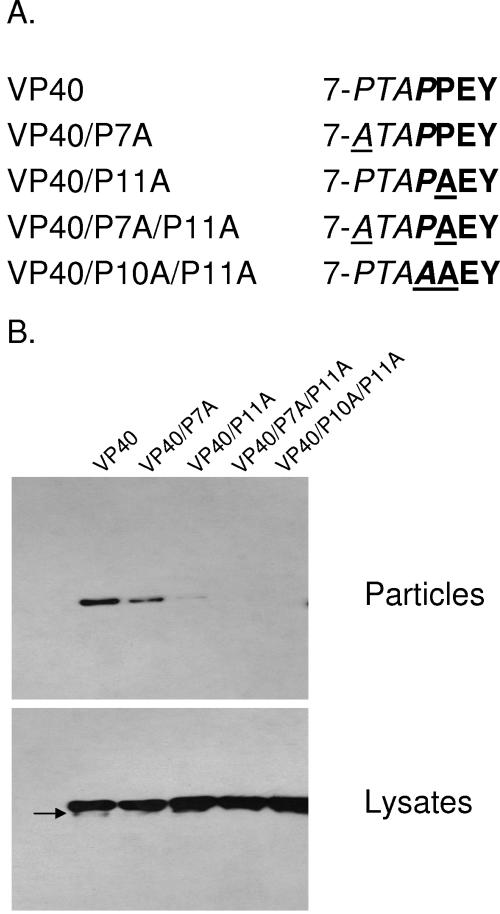
We described plasmid pCEboZVP40 in detail in a previous report (20). Briefly, it contains the coding sequence for the VP40 protein of Ebola virus Zaire (Mayinga strain) under the control of the chicken β-actin promoter in the eukaryotic protein expression vector pCAGGS/MCS (22, 33). Derivatives thereof containing amino acid substitutions in the late domain motifs were generated by site-directed mutagenesis using a QuikChange site-directed mutagenesis kit (Stratagene). The names of the resulting plasmids reflect the amino acid substitution(s) in VP40; for example, pCEboZVP40/P7A encodes a VP40 protein in which the proline at position 7 was replaced with alanine (see Fig. 1A).
FIG. 1.
Ebola VP40 late domain mutants and their effects on particle formation. (A) Ebola virus VP40 PTAP (italics) and PPXY (bold) late domain motifs and mutants thereof. Mutated amino acids are underlined. (B) Western blot analysis. Cells were transfected with plasmids expressing wild-type VP40 or late domain mutants. Cell lysates and supernatants were assayed with an antibody to VP40 for protein expression and particle formation, respectively.
Plasmids pCEZ-NP, pCEZ-VP30, pCEZ-VP35, pCEZ-L, pCEboZGP, and pCEboVP24 are described in our previous publications (31, 50). The plasmids contain the respective Ebola virus protein in the eukaryotic expression vector pCAGGS/MCS; plasmid pC-T7pol contains the gene encoding the T7 RNA polymerase in pCAGGS/MCS.
Plasmid pTM-T7G-Ebo-Rib (31) contains a T7 RNA polymerase promoter, a cDNA encoding the full-length genome of Ebola virus Zaire (Mayinga strain), and a ribozyme sequence. To alter the VP40 late domain motif(s), the respective amino acid(s) was replaced with alanine by site-directed mutagenesis (QuikChange Site-Directed Mutagenesis Kit; Stratagene), resulting in pTM-T7G-Ebo-Rib-P7A, pTM-T7G-Ebo-Rib-P10A, pTM-T7G-Ebo-Rib-P7A-P11A, and pTM-T7G-Ebo-Rib-P10A-P11A. Modification of the wild-type sequences to create the desired mutant counterparts was verified by sequence analysis. For all constructs used in this study, PCR-amplified regions were also subjected to sequence analysis to verify the absence of unwanted mutations.
Transfection of cells with plasmids for the expression of wild-type or mutant VP40 protein.
DNA and transfection reagent (2 μl per μg DNA) (Trans IT-LT1; Panvera) were mixed in Opti-MEM (Gibco-BRL), incubated for 30 min at room temperature, and added to the cells. Transfected cells were incubated at 37°C until the cell monolayer or supernatant was harvested.
Particle formation assay.
Twenty-four hours posttransfection of 293T cells with plasmids pCEboZVP40, pCEboZVP40-P7A, pCEboZVP40-P11A, pCEboZVP40-P7A-P11A, pCEboZVP40-P10A-P11A, or pCEboZVP40-M14A, the culture medium was collected and cleared of cellular debris by centrifugation at 2,000 rpm in a tabletop Allegra 6R centrifuge (Beckman Coulter, Fullerton, Calif.) at 4°C for 10 min. The cleared supernatant was layered onto a 20% sucrose (in phosphate-buffered saline) cushion and centrifuged at ∼92,000 × g for 90 min at 4°C. The particle-containing pellet was then lysed in cold cell lysis buffer (50 mM Tris, 150 mM NaCl, 0.5% Triton X-100, pH 7.4) and mixed with 2× Endurance loading buffer (ISC BioExpress, Kaysville, Utah). The sample was then heated at 100°C for 5 min before 20% of the sample was separated on a 12% polyacrylamide gel. The cell monolayer was also lysed in cold cell lysis buffer and then centrifuged at ∼10,600 × g for 10 min to clear cellular debris. This lysate was then mixed with 2× Endurance loading buffer, and 2% of the total sample was separated by gel electrophoresis. Resolved proteins were detected with a rabbit polyclonal anti-VP40 antibody by Western blotting as described previously (20).
Generation of infectious Ebola virus.
Ebola virus was generated as described previously (31). Briefly, 5 × 10⁵ cells of a Vero E6/293T coculture were transfected with 1 μg of the appropriate plasmid for the synthesis of wild-type or mutant Ebola virus cRNA, together with the following amounts of protein expression plasmids: 1 μg of pCEZ-NP, 0.3 μg of pCEZ-VP30, 0.5 μg of pCEZ-VP35, 2 μg of pCEZ-L, and 1 μg of pC-T7Pol. Four days after transfection, supernatants were collected and used to infect fresh Vero E6 cells. All experiments for the generation of Ebola virus as well as the characterization of recombinant Ebola virus were carried out in the BSL-4 facility at the National Microbiology Laboratory, Winnipeg, Canada.
TEM and SEM.
Vero E6 cells infected with wild-type or mutant Ebola virus were fixed and inactivated with 2.5% glutaraldehyde in 0.1 M cacodylate buffer (pH 7.4) prior to removal from the BSL-4 facility and postfixed with 2% osmium tetroxide. Samples were stained en bloc with 1% aqueous uranyl acetate, and then processed for transmission electron microscopy (TEM) as described elsewhere (34). For scanning electron microscopy (SEM), samples were dehydrated via a series of ethanol gradients, substituted with t-butanol, and dried in a Hitachi ES-2030 freeze dryer. Dried specimens were then coated with osmium tetroxide (HPC-1S osmium coater; Vacuum Device, Inc.) and examined under a Hitachi S-4200 microscope.
RESULTS
VP40 late domains promote efficient particle formation in VP40-expressing cells.
Previously, we reported that substitution of the proline residues at positions 10 and 11 of VP40 significantly decreased VP40-induced particle formation in transfected cells (20). These mutations affected both the PTAP and PPEY motifs (Fig. 1A); therefore, the individual contributions of these motifs to VP40-induced particle formation could not be assessed. We therefore generated the following VP40 late domain mutants (Fig. 1A): VP40-P7A, in which the proline residue at position 7 was replaced with alanine to alter the PTAP motif without affecting the PPEY motif; VP40-P11A, in which the proline at position 11 was replaced with alanine to alter the PPEY motif while leaving the PTAP motif intact; and VP40-P7A-P11A, in which both late domains were altered. For all experiments, we also included plasmid VP40-P10A-P11A (formerly referred to as VP40AAXY [20]) in which both late domain motifs were mutated. A protein expression plasmid (pCEboZVP40) containing authentic late domain motifs served as a control. Cell lysate prepared from plasmid-transfected cells served as a control for VP40 expression levels. Expression of wild-type VP40 induced the formation of virus-like particles (Fig. 1B, lane 1). The second band of lower molecular weight that was observed for the cell lysate (Fig. 1B, arrow) has been described previously by us (20) and by others (13, 25). Mass spectrometry of this molecule showed it to be a carboxyl terminal truncation product (L. Jasenosky and Y. Kawaoka, unpublished data), not a product of internal initiation of translation, as we had previously speculated. Alteration of the PTAP motif (mutant VP40-P7A) had only modest effects on particle formation (lane 2), in accordance with the findings of others (13, 25, 28); however, mutation of the PPXY motif (mutant VP40-P11A) had a pronounced effect on budding efficiencies (lane 3). Modification of both late domain motifs (i.e., VP40-P7A-P11A or VP40-P10A-P11A) further reduced budding efficiencies (lanes 4 and 5). Taken together, these results confirmed that both of the late domain motifs play critical roles in VP40 particle production and that the PPEY motif is more imperative to this process than is the PTAP motif.
Generation of recombinant Ebola viruses containing mutations in the VP40 late domain(s).
To determine the significance of the VP40 late domain motifs of Ebola virus for virus replication, we made use of our system for the generation of Ebola virus entirely from cloned cDNA (31). We modified plasmid pTM-T7G-Ebo-Rib, which contains ribozyme sequences and the full-length Ebola viral cDNA in the positive-sense orientation under the control of a T7 RNA polymerase promoter, by altering the VP40 late domains individually and in combination, as outlined in Fig. 1A. The resulting plasmids were designated pTM-T7G-Ebo-Rib-P7A, pTM-T7G-Ebo-Rib-P11A, pTM-T7G-Ebo-Rib-P7A-P11A, and pTM-T7G-Ebo-Rib-P10A-P11A. Mixtures of Vero E6 and 293T cells were then transfected with the respective plasmid for the transcription of wild-type or mutant viral RNA, with plasmids expressing the Ebola viral proteins that are required for the replication/transcription of viral RNA (i.e., NP, VP30, VP35, and L), and with pC-T7pol for the expression of the T7 RNA polymerase. Four days later, supernatants derived from plasmid-transfected cells were used to infect fresh Vero E6 cells. All four late domain mutant viruses were recovered, indicating that these motifs are not essential for growth of Ebola virus in cell culture. Sequence analyses of these mutant viruses confirmed the presence of the desired mutation(s) in VP40. To ensure that the loss of late domain functions was not compensated for by mutations in other regions of VP40 and/or mutations in GP and/or VP24 (which likely interact with VP40 during virion assembly and budding), we sequenced these genes after three consecutive passages in Vero E6 cells. In addition, we also determined the sequences of the NP, VP30, and VP35 genes. For the VP40 late domain mutants, we did not detect mutations in either of these genes. These findings indicated that the VP40 late domains are indeed dispensable for Ebola virus replication in cell culture.
Are Ebola viruses containing mutant VP40 late domain motifs attenuated in cell culture?
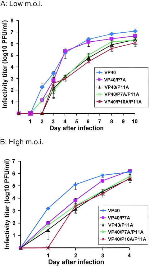
To determine whether Ebola viruses with altered VP40 late domain motifs are impaired in their assembly and/or budding efficiencies, we assessed their growth competency. Vero E6 cells were infected at a multiplicity of infection (MOI) of 0.01 or 1, and supernatants were collected on days 0 (i.e., at 2 h postinfection) to 4 postinfection (p.i.); for cells infected at a low MOI, we collected additional samples on days 6, 8, and 10 p.i. All experiments were carried out in triplicate. Wild-type virus grew to 1.2 × 10⁷ PFU at 10 days p.i. (Fig. 2A). The VP40-P7A mutant, which bears one functional late domain, grew to slightly reduced titers (4.5 × 10⁶ PFU). The remaining mutants, i.e., VP40-P11A, VP40-P7A-P11A, and VP40-P10A-P11A were modestly attenuated, reaching 2.3 × 10⁶, 1.9 × 10⁶, and 1.1 × 10⁶ PFU, respectively. To determine if the slight delay in replication could be relieved by infecting cells at a higher MOI, we examined viral growth kinetics in cells infected with an MOI of 1. Wild-type Ebola virus reached 1.3 × 10⁶ PFU at 4 days p.i. (Fig. 2B). Mutant viruses VP40-P7A, VP40-P11A, and VP40-P7A-P11A were slightly attenuated early in infection but reached titers ranging from 3.5 × 10⁵ to 1.5 × 10⁶ PFU at 4 days p.i. The VP40-P10A-P11A mutant was modestly attenuated early in infection but reached a final titer of 3.9 × 10⁵, which is comparable to titers obtained for the other mutant viruses. These results indicated that while functional VP40 late domain motifs are not essential for Ebola virus replication in cell culture, they do support efficient virus growth.
FIG. 2.
Growth curves of wild-type and mutant Ebola viruses in cell culture. Vero E6 cells were infected at an MOI of 0.01 (A) or 1 (B) and virus titers were determined at the time points indicated. All experiments were carried out in triplicate. Error bars represent the standard deviations.
Are Ebola viruses that lack functional late domains impaired in their budding ability?
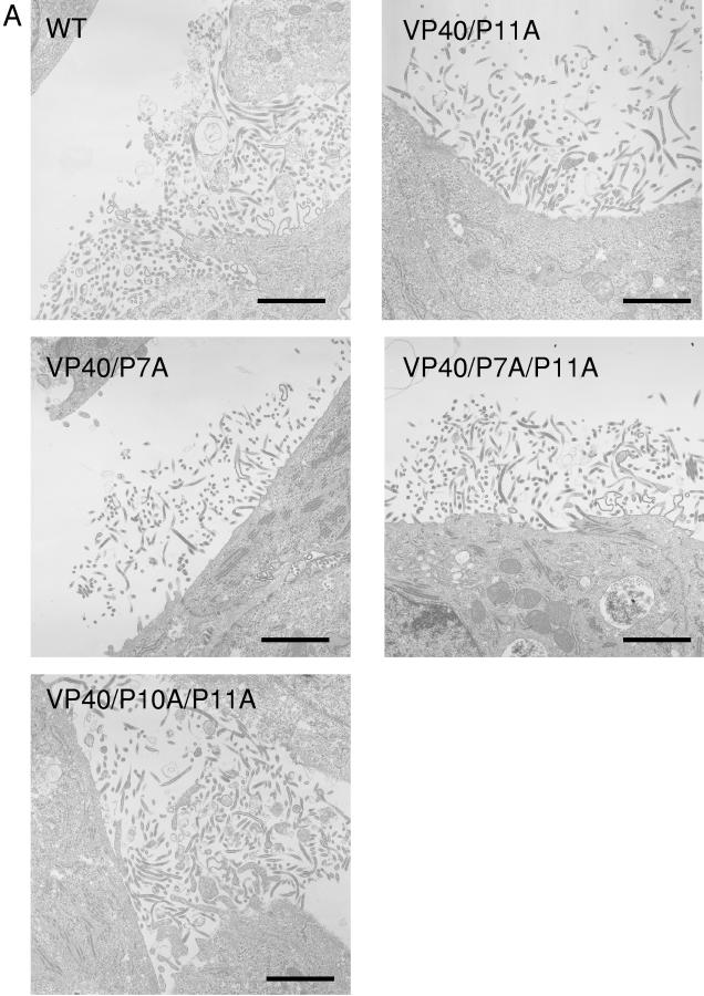
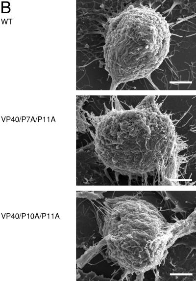
Mutations in viral late domains have been reported to affect particle size (8) and to arrest viruses at various stages of the budding process (2, 11, 14, 21, 23). To assess whether mutation of the VP40 late domains of Ebola virus similarly affect virus particle formation, we performed TEM and SEM. TEM on ultrathin sections of Vero E6 cells infected with wild-type virus revealed virions budding from the plasma membrane (Fig. 3A). Similar budding scenes were observed for all mutants, including the double mutants VP40-P7A-P10A and VP40-P10A-P11A, which suggests that alteration of the late domain motifs does not appreciably alter the shape of budding virions. Furthermore, we did not observe “tethered” viruses, which have been observed for mutants with altered P(T/S)AP motifs (2, 11, 14, 23). Of note, fewer of the cells infected with the double mutants produced virus, even though all of the cells were infected at the same MOI (data not shown). SEM studies also demonstrated no drastic differences in the shape of budding virions between cells infected with wild-type virus or mutants lacking both late domain motifs (VP40-P7A-P10A and VP40-P10A-P11A) (Fig. 3B). Thus, neither TEM nor SEM revealed an appreciable effect of VP40 late domain mutations on the morphology of budding virions.
FIG. 3.
Electron micrographs of cells infected with wild-type or mutant Ebola viruses. Vero E6 cells were infected with wild-type or mutant Ebola viruses and processed for TEM (A) or SEM (B) as described. In panel B, images are of cells infected with wild-type virus (top), VP40-P7A-P11A (middle), or VP40-P10-P11A (bottom). Bar, 200 nm.
DISCUSSION
While many studies have examined and demonstrated a role for late domains in virus-like particle formation and release in vitro, relatively few of them went on to assess the impact of mutations in these domains on the replication competency of the mutant viruses. With respect to Ebola virus, no such studies have been done. For VSV, alteration of either the PPXY or the PSAP motif attenuated mutant viruses by 1 to 2 log units (17), comparable to our findings with Ebola virus. One study has shown that alteration of the PTAP motif of the HIV Gag protein markedly reduces particle formation and results in viruses that fail to replicate efficiently (4). In other studies of the YXXL motif of equine infectious anemia virus (24) or bovine leukemia virus (16), the authors found no strict correlation between virion production and the replication efficiency of the mutant viruses. They did find that some late domain mutations that had no effect on particle formation rendered the mutant viruses replication deficient, suggesting that processes other than particle formation and release (such as virion incorporation of viral genomes and/or other viral proteins) were affected by these mutations. Together, these studies demonstrate that late domains play a critical role in virus replication but that in vitro budding assays may not fully reveal the significance of late domain(s) to the viral life cycle. To overcome this potential limitation, we used a reverse genetics system that allows the artificial generation of Ebola virus from cloned cDNA to generate viruses mutated in their VP40 PTAP and/or PPXY motifs and assessed the growth kinetics of these viruses in cell culture. Compared to wild-type virus, late domain mutants were attenuated by one log unit but still reached appreciable titers (ranging from 1.1 × 10⁶ to 4.5 × 10⁶ PFU), suggesting that the VP40 late domains are not absolutely required for Ebola virus replication in cell culture. Our finding that a virus with mutations in both late domain motifs was slightly more attenuated early in infection than those bearing one functional motif suggests a contribution of both domains to efficient viral replication. Mutant VP40-P10A-P11A appeared to be somewhat more attenuated than VP40-P7A-P11A, which may reflect structural differences between the two mutant proteins or a less prominent role for the proline residue at position 7 in late domain function(s).
Passage of mutant Ebola viruses did not yield revertants.
Serial passages of HIV viruses containing mutations in the PTAP motif of their Gag protein led to revertants that restored the late domain motif (4). This finding underscores the critical role of the late domain in the HIV Gag protein for the viral life cycle. By contrast, after three consecutive passages, our mutant Ebola viruses did not revert, and no compensating mutations in the VP40 late domain motifs were detected. Moreover, sequence analyses established the absence of mutations in VP24 and GP, which play roles in the formation of nucleocapsids and virions and in the release of infectious particles (15; reviewed in reference 32). The absence of strong selective pressure on Ebola viruses containing mutant VP40 late domains—in contrast to that exerted on mutant HIV viruses that resulted in revertants for HIV Gag—further suggests that the Ebola virus VP40 late domains are not required for virus replication in cell culture.
Effect of late domain mutations on virus budding.
In most systems tested, viruses with alterations in their PPXY motif are unable to complete the budding process, resulting in electron-dense structures underneath the plasma membrane. By contrast, viruses containing mutations in their P(T/S)AP motifs often appear as immature particles tethered to cellular membranes and to each other (2, 11, 14, 23). Other groups have reported that mutations in the PPXY motif arrest VSV (21) or Mason-Pfizer monkey virus (54) release and that mutations in the P(T/S)AP motif have an early effect on budding (5). In fact, one study has shown that the PPXY and P(T/S)AP motifs of HTLV-1 Gag can arrest virions at either early or late stages in the budding process (49), depending on the nature of the amino acid substitution. Replacement of the VSV PSAP motif did not significantly affect the number or shape of budding viruses (17), similar to our findings with the alteration of the Ebola virus PTAP motif. It is unclear whether these discrepancies reflect differences in the experimental systems used, differences among the viruses in their requirements for host cell factors, or differences in cell type dependence of the budding processes (40, 56). As judged by TEM, Ebola viruses mutated in their VP40 PPXY motif are released from infected cells in numbers similar to those observed for wild-type virus, which is evidence against a significant defect in the budding process. This is consistent with our finding that these mutant viruses are only slightly attenuated.
Why do viruses contain two late domain motifs?
For a growing number of viruses from different families, two closely spaced late domain motifs have been identified (Table 1). Based on earlier findings that late domains are functionally interchangeable (reviewed in reference 7), it was assumed that these motifs perform redundant functions. However, for HTLV-1 Gag, both the PPXY motif and the PTAP motif are required for efficient particle formation, although the PPXY motif seems to be the more critical (2, 3, 14, 49). This finding is consistent with our data on Ebola virus-like particles (Fig. 1B) (20) and the findings of others (13, 25). By contrast, mutagenesis of the PTAP motif has yielded conflicting data. While one group of investigators reported a significant reduction in particle formation (28), another group found a modest increase (25). These discrepancies may reflect the use of different systems and/or different protein expression levels. Since ubiquitinated Gag has an increased affinity for Tsg101 (36), it may be that the PPXY motif recruits Nedd4-like ubiquitin ligases so that the PTAP motif can mediate the efficient interaction of the ubiquitinated Gag protein with Tsg101. Efficient virion formation and budding may thus rely on the successive interactions of late domain proteins with Nedd4-like ubiquitin ligases and Tsg101 (2). While Ebola VP40 is ubiquitinated by Nedd4-like ubiquitin ligases (13), it is currently not known if this event subsequently increases the affinity of VP40 for Tsg101.
In summary, we have demonstrated that the Ebola VP40 late domains are expendable for viral replication in cell culture. They do, however, support efficient viral replication, likely via successive interactions with Nedd4-like ubiquitin ligases and Tsg101, by facilitating efficient release of virions from infected cells.
Acknowledgments
We thank Krisna Wells and Martha McGregor for excellent technical assistance and Susan Watson for editing the manuscript.
This work was supported by Public Health Service research grants from the National Institute of Allergy and Infectious Diseases, by Grants-in-Aid for Scientific Research on Priority Areas from the Ministries of Education, Culture, Sports, Science, and Technology, Japan, and by CREST (Japan Science and Technology Corporation). This work was also sponsored by the NIH/NIAID Regional Center of Excellence for Biodefense and Emerging Infectious Diseases Research (RCE) Program. The authors acknowledge membership within and support from the Region V “Great Lakes” RCE (NIH award 1-U54-AI-057153). This work was also supported by grant MOP-43921 from the Canadian Institutes of Health Research (CIHR) to H.F.
REFERENCES
- 1.Bavari, S., C. M. Bosio, E. Wiegand, G. Ruthel, A. B. Will, T. W. Geisbert, M. Hevey, C. Schmaljohn, A. Schmaljohn, and M. J. Aman. 2002. Lipid raft microdomains: a gateway for compartmentalized trafficking of Ebola and Marburg viruses. J. Exp. Med. 195:593-602. [DOI] [PMC free article] [PubMed] [Google Scholar]
- 2.Blot, V., F. Perugi, B. Gay, M. C. Prevost, L. Briant, F. Tangy, H. Abriel, O. Staub, M. C. Dokhelar, and C. Pique. 2004. Nedd4.1-mediated ubiquitination and subsequent recruitment of Tsg101 ensure HTLV-1 Gag trafficking towards the multivesicular body pathway prior to virus budding. J. Cell Sci. 117:2357-2367. [DOI] [PubMed] [Google Scholar]
- 3.Bouamr, F., J. A. Melillo, M. Q. Wang, K. Nagashima, S. M. de Los, A. Rein, and S. P. Goff. 2003. PPPYEPTAP motif is the late domain of human T-cell leukemia virus type 1 Gag and mediates its functional interaction with cellular proteins Nedd4 and Tsg101. J. Virol. 77:11882-11895. [DOI] [PMC free article] [PubMed] [Google Scholar]
- 4.Demirov, D. G., J. M. Orenstein, and E. O. Freed. 2002. The late domain of human immunodeficiency virus type 1 p6 promotes virus release in a cell type-dependent manner. J. Virol. 76:105-117. [DOI] [PMC free article] [PubMed] [Google Scholar]
- 5.Dettenhofer, M., and X. F. Yu. 1999. Proline residues in human immunodeficiency virus type 1 p6Gag exert a cell type-dependent effect on viral replication and virion incorporation of Pol proteins. J. Virol. 73:4696-4704. [DOI] [PMC free article] [PubMed] [Google Scholar]
- 6.Feldmann, H., V. E. Volchkov, U. Ströher, V. Volchkova, and H. D. Klenk. 2001. Biosynthesis and role of filovirus glycoprotein. J. Gen. Virol. 82:2839-2848. [DOI] [PubMed] [Google Scholar]
- 7.Freed, E. O. 2002. Viral late domains. J. Virol. 76:4679-4687. [DOI] [PMC free article] [PubMed] [Google Scholar]
- 8.Garnier, L., L. J. Parent, B. Rovinski, S. X. Cao, and J. W. Wills. 1999. Identification of retroviral late domains as determinants of particle size. J. Virol. 73:2309-2320. [DOI] [PMC free article] [PubMed] [Google Scholar]
- 9.Garrus, J. E., U. K. von Schwedler, O. W. Pornillos, S. G. Morham, K. H. Zavitz, H. E. Wang, D. A. Wettstein, K. M. Stray, M. Cote, R. L. Rich, D. G. Myszka, and W. I. Sundquist. 2001. Tsg101 and the vacuolar protein sorting pathway are essential for HIV-1 budding. Cell 107:55-65. [DOI] [PubMed] [Google Scholar]
- 10.Gottlinger, H. G., T. Dorfman, J. G. Sodroski, and W. A. Haseltine. 1991. Effect of mutations affecting the p6 Gag protein on human immunodeficiency virus particle release. Proc. Natl. Acad. Sci. USA 88:3195-3199. [DOI] [PMC free article] [PubMed] [Google Scholar]
- 11.Gottwein, E., J. Bodem, B. Muller, A. Schmechel, H. Zentgraf, and H. G. Krausslich. 2003. The Mason-Pfizer monkey virus PPPY and PSAP motifs both contribute to virus release. J. Virol. 77:9474-9485. [DOI] [PMC free article] [PubMed] [Google Scholar]
- 12.Harty, R. N., M. E. Brown, J. P. McGettigan, G. Wang, H. R. Jayakar, J. M. Huibregtse, M. A. Whitt, and M. J. Schnell. 2001. Rhabdoviruses and the cellular ubiquitin-proteasome system: a budding interaction. J. Virol. 75:10623-10629. [DOI] [PMC free article] [PubMed] [Google Scholar]
- 13.Harty, R. N., M. E. Brown, G. Wang, J. Huibregtse, and F. P. Hayes. 2000. A PPxY motif within the VP40 protein of Ebola virus interacts physically and functionally with a ubiquitin ligase: implications for filovirus budding. Proc. Natl. Acad. Sci. USA 97:13871-13876. [DOI] [PMC free article] [PubMed] [Google Scholar]
- 14.Heidecker, G., P. A. Lloyd, K. Fox, K. Nagashima, and D. Derse. 2004. Late assembly motifs of human T-cell leukemia virus type 1 and their relative roles in particle release. J. Virol. 78:6636-6648. [DOI] [PMC free article] [PubMed] [Google Scholar]
- 15.Huang, Y., L. Xu, Y. Sun, and G. Nabel. 2002. The assembly of Ebola virus nucleocapsid requires virion-associated proteins 35 and 24 and posttranslational modification of nucleoprotein. Mol. Cell 10:307-316. [DOI] [PubMed] [Google Scholar]
- 16.Inabe, K., M. Nishizawa, S. Tajima, K. Ikuta, and Y. Aida. 1999. The YXXL sequences of a transmembrane protein of bovine leukemia virus are required for viral entry and incorporation of viral envelope protein into virions. J. Virol. 73:1293-1301. [DOI] [PMC free article] [PubMed] [Google Scholar]
- 17.Irie, T., J. M. Licata, H. R. Jayakar, M. A. Whitt, P. Bell, and R. N. Harty. 2004. Functional analysis of late-budding domain activity associated with the PSAP motif within the vesicular stomatitis virus M protein. J. Virol. 78:7823-7827. [DOI] [PMC free article] [PubMed] [Google Scholar]
- 18.Irie, T., J. M. Licata, J. P. McGettigan, M. J. Schnell, and R. N. Harty. 2004. Budding of PPxY-containing rhabdoviruses is not dependent on host proteins TGS101 and VPS4A. J. Virol. 78:2657-2665. [DOI] [PMC free article] [PubMed] [Google Scholar]
- 19.Jasenosky, L. D., and Y. Kawaoka. 2004. Filovirus budding. Virus Res. 106:181-188. [DOI] [PubMed] [Google Scholar]
- 20.Jasenosky, L. D., G. Neumann, I. Lukashevich, and Y. Kawaoka. 2001. Ebola virus VP40-induced particle formation and association with the lipid bilayer. J. Virol. 75:5205-5214. [DOI] [PMC free article] [PubMed] [Google Scholar]
- 21.Jayakar, H. R., K. G. Murti, and M. A. Whitt. 2000. Mutations in the PPPY motif of vesicular stomatitis virus matrix protein reduce virus budding by inhibiting a late step in virion release. J. Virol. 74:9818-9827. [DOI] [PMC free article] [PubMed] [Google Scholar]
- 22.Kobasa, D., M. E. Rodgers, K. Wells, and Y. Kawaoka. 1997. Neuraminidase hemadsorption activity, conserved in avian influenza A viruses, does not influence viral replication in ducks. J. Virol. 71:6706-6713. [DOI] [PMC free article] [PubMed] [Google Scholar]
- 23.Le Blanc, I., M.-C. Prevost, M.-C. Dokhelar, and A. R. Rosenberg. 2002. The PPPY motif of human T-cell leukemia virus type 1 Gag protein is required early in the budding process. J. Virol. 76:10024-10029. [DOI] [PMC free article] [PubMed] [Google Scholar]
- 24.Li, F., C. Chen, B. A. Puffer, and R. C. Montelaro. 2002. Functional replacement and positional dependence of homologous and heterologous L domains in equine infectious anemia virus replication. J. Virol. 76:1569-1577. [DOI] [PMC free article] [PubMed] [Google Scholar]
- 25.Licata, J. M., M. Simpson-Holley, N. T. Wright, Z. Han, J. Paragas, and R. N. Harty. 2003. Overlapping motifs (PTAP and PPEY) within the Ebola virus VP40 protein function independently as late budding domains: involvement of host proteins TSG101 and VPS-4. J. Virol. 77:1812-1819. [DOI] [PMC free article] [PubMed] [Google Scholar]
- 26.Licata, J. M., R. F. Johnson, Z. Han, and R. N. Harty. 2004. Contribution of Ebola virus glycoprotein, nucleoprotein, and VP24 to budding of VP40 virus-like particles. J. Virol. 78:7344-7351. [DOI] [PMC free article] [PubMed] [Google Scholar]
- 27.Martin-Serrano, J., D. Perez-Caballero, and P. D. Bieniasz. 2004. Context-dependent effects of L domains and ubiquitination on viral budding. J. Virol. 78:5554-5563. [DOI] [PMC free article] [PubMed] [Google Scholar]
- 28.Martin-Serrano, J., T. Zang, and P. D. Bieniasz. 2001. HIV-1 and Ebola virus encode small peptide motifs that recruit Tsg101 to sites of particle assembly to facilitate egress. Nat. Med. 7:1313-1319. [DOI] [PubMed] [Google Scholar]
- 29.Muhlberger, E., M. Weik, V. E. Volchkov, H. D. Klenk, and S. Becker. 1999. Comparison of the transcription and replication strategies of Marburg virus and Ebola virus by using artificial replication systems. J. Virol. 73:2333-2342. [DOI] [PMC free article] [PubMed] [Google Scholar]
- 30.Myers, E. L., and J. F. Allen. 2002. Tsg101, an inactive homologue of ubiquitin ligase e2, interacts specifically with human immunodeficiency virus type 2 Gag polyprotein and results in increased levels of ubiquitinated Gag. J. Virol. 76:11226-11235. [DOI] [PMC free article] [PubMed] [Google Scholar]
- 31.Neumann, G., H. Feldmann, S. Watanabe, I. Lukashevich, and Y. Kawaoka. 2002. Reverse genetics demonstrates that proteolytic processing of the Ebola virus glycoprotein is not essential for replication in cell culture. J. Virol. 76:406-410. [DOI] [PMC free article] [PubMed] [Google Scholar]
- 32.Neumann, G., T. Noda, A. Takada, L. D. Jasenosky, and Y. Kawaoka. 2004. Role of filoviral matrix and glycoproteins in the viral life cycle, p. 137-170. In H.-D. Klenk and H. Feldmann (ed.), Ebola and Marburg viruses: molecular and cellular biology. Horizon Bioscience, Wymondham, United Kingdom.
- 33.Niwa, H., K. Yamamura, and J. Miyazaki. 1991. Efficient selection for high-expression transfectants with a novel eukaryotic vector. Gene 108:193-199. [DOI] [PubMed] [Google Scholar]
- 34.Noda, T., H. Sagara, E. Suzuki, A. Takada, H. Kida, and Y. Kawaoka. 2002. Ebola virus VP40 drives the formation of virus-like filamentous particles along with GP. J. Virol. 76:4855-4865. [DOI] [PMC free article] [PubMed] [Google Scholar]
- 35.Perez, M., R. C. Craven, and J. C. de la Torre. 2003. The small RING finger protein Z drives arenavirus budding: implications for antiviral strategies. Proc. Natl. Acad. Sci. USA 100:12978-12983. [DOI] [PMC free article] [PubMed] [Google Scholar]
- 36.Pornillos, O., S. L. Alam, D. R. Davis, and W. I. Sundquist. 2002. Structure of the Tsg101 UEV domain in complex with the PTAP motif of the HIV-1 p6 protein. Nat. Struct. Biol. 9:812-817. [DOI] [PubMed] [Google Scholar]
- 37.Puffer, B. A., L. J. Parent, J. W. Wills, and R. C. Montelaro. 1997. Equine infectious anemia virus utilizes a YXXL motif within the late assembly domain of the Gag p9 protein. J. Virol. 71:6541-6546. [DOI] [PMC free article] [PubMed] [Google Scholar]
- 38.Puffer, B. A., S. C. Watkins, and R. C. Montelaro. 1998. Equine infectious anemia virus Gag polyprotein late domain specifically recruits cellular AP-2 adapter protein complexes during virion assembly. J. Virol. 72:10218-10221. [DOI] [PMC free article] [PubMed] [Google Scholar]
- 39.Raiborg, C., T. E. Rusten, and H. Stenmark. 2003. Protein sorting into multivesicular endosomes. Curr. Opin. Cell Biol. 15:446-455. [DOI] [PubMed] [Google Scholar]
- 40.Schwartz, M. D., R. J. Geraghty, and A. T. Panganiban. 1996. HIV-1 particle release mediated by Vpu is distinct from that mediated by p6. Virology 224:302-309. [DOI] [PubMed] [Google Scholar]
- 41.Strack, B., A. Calistri, M. A. Accola, G. Palu, and H. G. Gottlinger. 2000. A role for ubiquitin ligase recruitment in retrovirus release. Proc. Natl. Acad. Sci. USA 97:13063-13068. [DOI] [PMC free article] [PubMed] [Google Scholar]
- 42.Strack, B., A. Calistri, S. Craig, E. Popova, and H. G. Gottlinger. 2003. AIP1/ALIX is a binding partner for HIV-1 p6 and EIAV p9 functioning in virus budding. Cell 114:689-699. [DOI] [PubMed] [Google Scholar]
- 43.Strecker, T., R. Eichler, J. Meulen, W. Weissenhorn, K. H. Dieter, W. Garten, and O. Lenz. 2003. Lassa virus Z protein is a matrix protein and sufficient for the release of virus-like particles. J. Virol. 77:10700-10705. [DOI] [PMC free article] [PubMed] [Google Scholar]
- 44.Timmins, J., S. Scianimanico, G. Schoehn, and W. Weissenhorn. 2001. Vesicular release of Ebola virus matrix protein VP40. Virology 283:1-6. [DOI] [PubMed] [Google Scholar]
- 45.VerPlank, L., F. Bouamr, T. J. LaGrassa, B. Agresta, A. Kikonyogo, J. Leis, and C. A. Carter. 2001. Tsg101, a homologue of ubiquitin-conjugating (E2) enzymes, binds the L domain in HIV type 1 Pr55Gag. Proc. Natl. Acad. Sci. USA 98:7724-7729. [DOI] [PMC free article] [PubMed] [Google Scholar]
- 46.Volchkov, V. E., V. A. Volchkova, E., W. Slenczka, H. D. Klenk, and H. Feldmann. 1998. Release of viral glycoproteins during Ebola virus infection. Virology 245:110-119. [DOI] [PubMed] [Google Scholar]
- 47.Volchkov, V. E., V. A. Volchkova, E. Muhlberger, L. V. Kolesnikova, M. Weik, O. Dolnik, and H. D. Klenk. 2001. Recovery of infectious Ebola virus from complementary DNA: RNA editing of the GP gene and viral cytotoxicity. Science 291:1965-1969. [DOI] [PubMed] [Google Scholar]
- 48.von Schwedler, U. K., M. Stuchell, B. Muller, D. M. Ward, H. Y. Chung, E. Morita, H. E. Wang, T. Davis, G. P. He, D. M. Cimbora, A. Scott, H. G. Krausslich, J. Kaplan, S. G. Morham, and W. I. Sundquist. 2003. The protein network of HIV budding. Cell 114:701-713. [DOI] [PubMed] [Google Scholar]
- 49.Wang, H., N. J. Machesky, and L. M. Mansky. 2004. Both the PPPY and PTAP motifs are involved in human T-cell leukemia virus type 1 particle release. J. Virol. 78:1503-1512. [DOI] [PMC free article] [PubMed] [Google Scholar]
- 50.Watanabe, S., T. Watanabe, T. Noda, A. Takada, H. Feldmann, L. D. Jasenosky, and Y. Kawaoka. 2004. Production of novel Ebola virus-like particles from cDNAs: an alternative to Ebola virus generation by reverse genetics. J. Virol. 78:999-1005. [DOI] [PMC free article] [PubMed] [Google Scholar]
- 51.Weik, M., J. Modrof, H. D. Klenk, S. Becker, and E. Muhlberger. 2002. Ebola virus VP30-mediated transcription is regulated by RNA secondary structure formation. J. Virol. 76:8532-8539. [DOI] [PMC free article] [PubMed] [Google Scholar]
- 52.Wills, J. W., C. E. Cameron, C. B. Wilson, Y. Xiang, R. P. Bennett, and J. Leis. 1994. An assembly domain of the Rous sarcoma virus Gag protein required late in budding. J. Virol. 68:6605-6618. [DOI] [PMC free article] [PubMed] [Google Scholar]
- 53.Xiang, Y., C. E. Cameron, J. W. Wills, and J. Leis. 1996. Fine mapping and characterization of the Rous sarcoma virus Pr76gag late assembly domain. J. Virol. 70:5695-5700. [DOI] [PMC free article] [PubMed] [Google Scholar]
- 54.Yasuda, J., and E. Hunter. 1998. A proline-rich motif (PPPY) in the Gag polyprotein of Mason-Pfizer monkey virus plays a maturation-independent role in virion release. J. Virol. 72:4095-4103. [DOI] [PMC free article] [PubMed] [Google Scholar]
- 55.Yasuda, J., M. Nakao, Y. Kawaoka, and H. Shida. 2003. Nedd4 regulates egress of Ebola virus-like particles from host cells. J. Virol. 77:9987-9992. [DOI] [PMC free article] [PubMed] [Google Scholar]
- 56.Yu, X. F., Z. Matsuda, Q. C. Yu, T. H. Lee, and M. Essex. 1995. Role of the C terminus Gag protein in human immunodeficiency virus type 1 virion assembly and maturation. J. Gen. Virol. 76 (Pt 12):3171-3179. [DOI] [PubMed] [Google Scholar]
- 57.Yuan, B., X. Li, and S. P. Goff. 1999. Mutations altering the Moloney murine leukemia virus p12 Gag protein affect virion production and early events of the virus life cycle. EMBO J. 18:4700-4710. [DOI] [PMC free article] [PubMed] [Google Scholar]


