



Abstract
Demodex blepharitis (DB) has become one of the common differential diagnoses in patients presenting with red eyes in daily clinics. In this review article, we have incorporated a detailed discussion about the mite and the various mechanisms by which it causes different signs and symptoms, the risk factors, and diagnostic methods with current and emerging treatment modalities. Because of the chronic symptoms and rather a late diagnosis, DB considerably affects patients’ quality of life. The clues to identify cylindrical dandruff, the diagnostic sign of Demodex, visualization of the mite correlating their presence with signs and symptoms, and instituting early and appropriate treatment are the goals of this article.
Keywords: Blepharitis, collarette, Demodex
Demodex blepharitis (DB) is a highly under-reported condition in India. In a normal ocular surface ecosystem, it acts as a lash cleaner by feeding on bacteria, defending against other mite species, and as an immune regulator and buffer.[1] Demodicosis, which is when the mites multiply out of control, causing symptoms, may be associated with leukemia, immunodeficient patients with HIV infection,[2,3,4] or patients with end-stage chronic renal failure.[5] DB has significant clinical, functional, and psychosocial effects on patients, with 80% of patients in the Atlas study indicating that the disease had negatively affected their daily lives.[6]
Epidemiology
A study[7] has shown that the rate of Demodex infestation increases with age, and it is observed in 84% of the general population aged 60 years, and 100% of the general population aged above 70 years. Among the younger population, its prevalence has been reported to be between 2% and 27%.[8,9,10] DB is equally prevalent in both sexes.[11] The infestation rate is also similar regardless of ethnicity, and it is reported in 72% of white, 65% of black, and 69% of Hispanic populations.[12] Thus, it is cosmopolitan and has been reported around the world.[13] Demodex folliculorum (DF) tends to reside in hair follicles of the eyelid closer to the anterior surface of the skin and is believed to be more contagious, moving from one host to another at a higher frequency than its counterpart, D. brevis.[14] The two species have different modes of transmission, and as a result, geographic variance may occur.
Infection in children under 10 years of age is rare.[15] Demodex is acquired and becomes abundant during puberty because of the increase in secretions by the sebaceous glands, which may create optimal conditions for DF infection and reproduction.[16] Thus, it may be speculated that young males may be a higher risk group for DF infection.
Varying mite counts among elderly subjects from different races and countries could substantially influence changes in dry eye disease (DED) or meibomian gland disease (MGD)-related parameters.[17]
The mite
The first description of Demodex mite was given by Simon in 1843.[18] Michel first detected its presence in the hair follicle of an eyelid in 1876.[19] In 1963, Akbulatova first described D. brevis as being a distinct species from DF, with the two preferring different niches.[20] The association with DB was definitively established by Coston in 1967. Coston characterized the full anatomy and physiology of the mite, with light microscope images and detailed clinical findings, to demonstrate its association with DB.[21]
The Demodex mite is the most common microscopic ectoparasite that lives on the human skin, specifically in the hair, hair follicle, erector pili muscle, and sebaceous gland, essentially pilosebaceous units.[15,22] Approximately 65 species of Demodex are known, but only two, DF and D. brevis, are responsible for DB in humans. DF (length: 0.3–0.4 mm) and Demodex Brevis (length: 0.15–0.2 mm) are normal skin commensals surviving on dead skin cells and sebum. They crawl out at night and lay eggs. Their average lifespan is approximately 2–3 weeks. Each Demodex has eight short legs, four on each side of its upper body, that advance in a peculiar rotary fashion, which limits its locomotion to 8–16 mm/h.[22] They have been thought to be first acquired from mother to infant through breastfeeding due to their presence on the nipple.[16] Though they are commensals, their overpopulation can cause demodicosis. The smaller D. brevis are difficult to detect, but in the meibomian glands,[23] they contribute to inflammation and clogging.[15] Unlike D. brevis, DF is visible to the naked eye. DF is commonly found in clusters surrounding the eyelashes and eyelid skin. It is believed to be more active and more implicated in type 1 allergic reactions.[24] Researchers have surmised that male and female mites mate outside the follicles or glands, after which the female mites lay their eggs inside the hair follicles or sebaceous glands.[25] The products of DF are called cylindrical dandruff (CD) [Fig. 1a]. Burrowed densely together deep inside the eyelash follicles, DF consumes epithelial cells and sebum in the process of external digestion. Their large salivary glands release proteases and lipases, and then the Demodex absorbs most of the digested cellular material.[26] Although it has no excretory system, this process does produce waste in the form of follicular dandruff and lipids, which are collected in collarettes around the base of the eyelashes. This process continues for the life of the mite, which itself becomes part of the lash debris every 14–18 days.[26]
Figure 1.

(a) Cylindrical dandruff in lashes. (b) Typical sleeve at the base of eyelash
Identification
Two techniques are commonly used to detect Demodex mites: lash epilation and in vivo confocal microscopy (IVCM). Unfortunately, epilation techniques among studies are highly variable,[27,28,29] which makes the mite count comparison between studies challenging. IVCM allows the visualization of the complete content of the lash follicles and meibomian glands, which increases the detection rate of Demodex larvae and D. brevis. Randon et al.[30] demonstrated that lash epilation and IVCM had similar mite detection rates, while Jalbert and Rejab found a higher prevalence of mites with IVCM.[31]
Slit lamp techniques
Inspection of the upper lid eyelashes is during downgaze, with the eyelids closed, while the lower eyelids are inspected during upgaze.
In one technique, two lashes with CD are epilated from each lid, mounted on slides, and examined in the clinic using a slit lamp equipped with a 90-D condensing lens followed by light microscopy in the pathology laboratory. Mites are identified by their characteristic morphology and movement patterns.[32]
In another technique, under a slit-lamp microscope at a magnification of 25×, two lashes, one from each half of each lid, are removed by fine forceps and placed separately on each end of glass slides. A coverslip is mounted onto each lash before slowly pipetting 20 µL of saline to the edge of the coverslip to surround the lash. This maneuver results in the preservation of the Demodex that has loose contact with the lash at the tip. Under the microscope, its number is counted conventionally. If a compacted CD is preserved, 20 μL of 100% alcohol or 0.25% fluorescein drops is pipetted into the edge of the coverslip, and the counting time is prolonged up to 20 min to allow the embedded Demodex to migrate from the CD [Fig. 2].[33]
Figure 2.

Demodex folluculorum under microscope (10x)
Rotation (ROT): From each lid, two eyelashes featuring prominent CD are identified and successively rotated for 30 s, as described by Mastrota.[34] During the rotation, the insertion point of the eyelash into the lid margin is continuously inspected for the characteristic cigar-shaped appearance of mite tails. Visible tails are counted and averaged across the eight lashes to provide a mean count per lash.[35]
CD removal (CDR): Two different eyelashes with CD are selected. Using the tip of the forceps, the collarette base is secured and removed with a sliding motion along the eyelash. Where collarettes are fused with the adjacent cornified epidermis of the lid margin, removal typically results in (or required) separation of the fused tissue from the lid margin. By exposing the point of insertion of the eyelash into the lid margin in this way, protruding Demodex tails are counted.[35]
Lateral eyelash traction (LET): The same two CD-free eyelashes are then subjected to a modified eyelash traction technique. In contrast to the technique described above (ROT), involving rotation of the lash around its axis, the grasped eyelash this time is drawn only laterally, slowly alternating extension in nasal and temporal directions, under gentle constant tension, for 30 s. Mite tails emerging from the follicle during this lateral extension are counted.[35]
CDR and LET techniques confirm and complement the technique described by Mastrota[36] and offer an accessible, less invasive, and more clinically viable method for the assessment of Demodex than eyelash epilation.[35]
Definitive diagnosis of DB relied on the epilation of lashes to visualize and count individual mites. However, due to the impracticality of light microscopy and slide preparation capabilities in an outpatient clinic, along with patient discomfort and increased exam time, the Demodex Expert Panel on Treatment and Eyelid Health panel agreed that as collarettes are pathognomonic for Demodex mites, clinicians may confidently move away from the necessity of epilation.[37]
Pathophysiology
Demodex mites are readily transferred via skin-to-skin contact.[38] It can cause problems through the following mechanisms:
The mites consume the lining of follicles to lay their eggs. This results in the distention of the follicle and the mal-direction of the lashes.[1,15]
Mechanical blockade of the sebaceous ducts by the mites results in eyelid margin irritation.[27]
The chitin-formed exoskeleton of the mites can induce a general inflammatory response and a foreign body-like granulomatous reaction that has been implicated in the formation of chalazia.[39]
Mechanisms of damage
Direct damage
Demodex has eight legs that scrape the lash and the lid margin throughout its process of feeding, growth, and reproduction. That mechanical damage triggers inflammation. Their presence causes hair follicle duct dilatation, hyperkeratinization, and peri-follicular non-granulomatous inflammation.[40] As a result, the lashes may be lost or misdirected.[41] Higher numbers of mites in the lash follicles are associated with reactive conjunctivitis and keratitis.[42] It can be associated with eyelid margin keratinization and giant cell reaction in eyelid pilosebaceous units.[43]
Demodex brevis has been implicated in MGD, including meibomian gland loss.[44,45] The build-up of decomposing mite remains, including their chitinous exoskeletons, is believed to contribute to physical blockage of the meibomian glands,[46] possibly leading to changes in gland architecture over time.[44,45] Changes in meibum secretion or lipid composition in MGD may promote the cycle by making the ocular environment more favorable for Demodex.[45,47]
Dysbacteriosis
It is relatively accepted that bacterial involvement in the pathological process of DB mainly uses the mites as a carrier.[48] They can carry bacteria on their surface (Streptococcus and Staphylococcus species) or in their gut (Bacillus oleronius), producing antigens and inducing an immune response.[15,24,40,49]
In comparison to the eyelashes of healthy individuals, the abundance of Actinobacteria increased significantly and the abundance of Burkholderia decreased significantly in the eyelash follicles of DB patients. These results confirm an imbalance in the bacterial communities in the eyelashes of the patients. The imbalances of the bacterial community may lead to lipid accumulation and Demodex infestation.[41]
Delayed Hypersensitivity Reaction
Demodex infestation is associated with the upregulation of pro-inflammatory cytokines, particularly IL-1b and IL-17,[24,25,50] tear film alterations,[50] and upregulation of matrix metalloproteinase-9 (MMP-9).[1] Activation of IL-17/MMP-9 signaling may also exacerbate corneal epithelial barrier dysfunction.[51]
Associated Systemic Conditions
The majority of HIV-infected patients receive potent anti-retroviral therapy; it is thus likely that most of them are not immunocompromised enough to have infestation with Demodex spp. It seems that only in severely immunosuppressed cases it might be more prevalent than in the general population.[42]
There is a need to consider Demodex infestation in the differential diagnosis of immunocompromised patients with herpetic keratitis.[46]
Patients with diabetes are significantly more likely to have Demodex infestation,[15] and that likelihood is higher if their blood glucose is not controlled.[52]
Dermatology studies have correlated Demodex with Rosacea.[53] A meta-analysis of 1513 patients showed that those with erythematous-telangiectatic or papulo-pustular rosacea are nine times more likely to have Demodex mite infestation than those without rosacea and with a higher prevalence in the papulo-pustular variety.[53] Demodex infestation may have a cause-or-effect relationship with rosacea; perhaps the condition permits the mite population to grow, or perhaps the infestation is an underlying cause of the condition. Importantly, for lid margin disease, in patients with rosacea, one of the highest densities of the mite is found in the ocular adnexa.[54] Li et al.[55] analyzed 59 patients with positive serum immunoreactivity for Bacillus oleronius and found a significant correlation between serum immunoreactivity and the presence of facial rosacea, eyelid edge inflammation, and Demodex infestation.
Patients with chronic DB may be questioned for common rosacea symptoms such as flushing and facial redness as the treatment for Demodex in rosacea may improve their clinical presentation without necessarily being an ocular rosacea.[56]
The dermatology literature suggests that the mite may also be associated with a multitude of skin conditions beyond rosacea, including acne vulgaris, seborrheic dermatitis, and basal cell and sebaceous carcinoma.[57]
These mites are associated with multiple ocular conditions such as chronic DB, keratoconjunctivitis, chalazia, pterygium, keratitis, endophthalmitis, and even periocular basal cell carcinoma.[58,59] Approximately 42%–81% of chronic DB patients have concomitant Demodex infestations, more commonly in elderly patients.[60]
Symptoms and Signs
A recent study by ocular surface disease experts agreed that itching is the most common symptom in DB.[37] Demodex-related itching may be more likely to occur at night or early morning after periods of mite activity,[36,61] distinguishing it from daytime, allergy-related itching. Demodex-related ocular itching may be propagated through non-histamine itching pathways.[12] Irritation is caused both directly by biting of the mites and by lipolytic enzymes used to digest sebum, their main food source.[61,62]
In addition to itching, those with DB often present with a range of other symptoms, including dryness, discharge, eye redness, burning, tearing, foreign body sensation, pain, and fluctuating vision.[63] In a sequential assessment of over 1000 patients across eight ophthalmic practices, 55% of patients presented with some collarettes, regardless of the reason for their visit.[64] It is important to differentiate lash debris [Fig. 5b] caused by Staphylococcal overgrowth, also called collarette [Fig. 5a], from cylindrical sleeves. Staphylococcal collarettes are golden-yellow and scaly in appearance and located more distally on the lash [Fig. 5c].[65]
Figure 5.

(a–c) Typical staphylococcal collarettes near the tip of eyelashes
The presence of CD [Fig. 1b] or collarettes is considered pathognomonic for the presence of DF. This debris is present at the base of the eyelash follicle, where it remains as the lash grows. The wax-like dandruff is composed of Demodex mite remains and the hyperkeratinization caused by microabrasions from the mite’s claws.[27] Demodex mites are found on 100% of lashes with collarettes.[27] Collarettes can be physically difficult to remove, even with micro-exfoliation, and will not dissolve with warm compresses.
Collarette Grading Scale[66]
-
0.
0–2 lashes with collarettes
-
1.
3–10 lashes with collarettes
-
2.
10 but less than one-third of lashes with collarettes
-
3.
One-third of lashes or more but less than two-thirds of lashes with collarettes
-
4.
Two-thirds or more of lashes with collarettes.
Meibomian Gland Dysfunction (MGD)
The pathogenic role of Demodex in MGD remains controversial; it is recognized that aging could be a significant confound in both Demodex infestation and MGD or DED, which are age-related conditions.[17] A surprising finding in a study by Lee et al.[17] is that ocular demodicosis in those over 60 years old may not be linked to the severity of MGD and DED, despite the elevated prevalence of ocular Demodex and mite counts compared to younger individuals. Conversely, ocular demodicosis negatively influences meibum quality in subjects below 40 years old. As indicated by Liang et al..,[39] D. brevis, typically residing in meibomian glands, was more common among younger patients with recurring chalazia and hordeola. Furthermore, MGD was detected in 90% of patients with D. brevis under 35 years, and it exhibited a significant correlation with keratitis.[67] The subjective discomfort, as measured by ocular surface disease index (OSDI) scores, did not show significant differences between those with and without ocular demodicosis though.[68] Ocular infestation with Demodex is related to both the development and exacerbation of MGD. The role of ocular demodicosis in MGD can be mediated by mechanical obstruction of the meibomian glands’ orifices or by provoking a cell-mediated inflammatory response in the glands.[67,69] In addition, the exoskeleton of the mite may cause a foreign body–induced granulomatous reaction in sebaceous glands.[69,70,71] In a recent study, the mite was associated with microstructural changes of MGs; the more the Demodex count, the more severe the structural damage.[44] In another study, lid margin abnormalities and OSDI scores were higher in Demodex-positive versus Demodex-negative patients with MGD.[23] Patients with DB have been shown to have low-quality meibum[50] and higher concentrations of (O-acyl)-v-hydroxy fatty acids (OAHFAs) in their meibum,[47] potentially exacerbating MGD and contributing to tear film instability.
Left untreated, the chronic inflammation associated with DB can have many sequelae on the lid and the ocular surface, such as chronic hordeolum and chalazia [Figs. 4a and 6a]. Demodex infestation has a potential pathogenic role in the development of recurrent hordeola in the 15–35 years age group than in children due to abundant sebum production which facilitates demodex infestation.[72] D. brevis tends to be more highly associated with the recurrence of chalazia after surgical excision.[49]
Figure 4.

(a) Lower-lid chalazion with inferior bulbar congestion and sterile corneal Infiltrate. (b) Irregular fluorescein stain pattern of the inferior cornea
Chronic meibomian gland inflammation can lead to morphologic changes in the meibomian glands [Fig. 6b], gland atrophy, and meibomian gland dysfunction.[6] Demodex infection might directly or indirectly result in dropout through inflammation mechanism or some other unclear way.[73]
Figure 6.
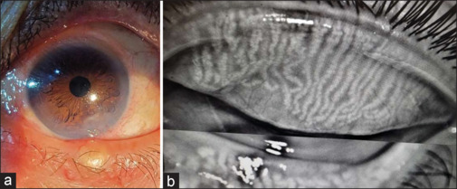
(a) Lower-lid chalazion with inferior bulbar congestion and sterile corneal infiltrate. (b) Blocked meibomian gland in meibography corresponding to the site of chalazion
Eyelid changes have also been noted with chronic inflammation, including lid margin telangiectasia, thickening of the lid margin, ocular rosacea, and even laxity of the eyelid over time. Chronic DB is harmful to the follicles, leading to thinning and loss of eyelashes.
If inflammation persists, ocular surface involvement in the form of blepharoconjunctivitis is a common presentation [Fig. 3b].[70] DB should be considered in the presence of refractory recurrent blepharoconjunctivitis in the pediatric population.[15] In a study of 12 healthy pediatric patients, recalcitrant blepharoconjunctivitis that did not respond to traditional therapy resolved after a short course of tea tree oil (TTO).[74] Staphylococcus epidermidis and Staphylococcus aureus are the main pathogens in pediatric blepharoconjunctivitis, and as has been mentioned, Demodex mites may be a vector for these bacteria in the eye.[74] Demodex infestation may cause various sight-threatening corneal lesions, including superficial corneal vascularization, marginal infiltration [Fig. 3b], a phlyctenule-like lesion, superficial opacity, and nodular scarring of the cornea.[70,75]
Figure 3.
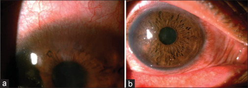
(a) Superficial vascularisation in the superior cornea. (b) Diffuse inflammation with peripheral corneal infiltrates
Lash follicle pouting, a sign that is not often reported, is thought to represent follicular hypertrophy due to inflammation in the underlying skin from Demodex mites impeding the normal growth of the eyelash.[1] This sign is also thought to be specific to ocular demodicosis and may provide an additional surrogate measure alongside the pathognomonic sign of collarettes when diagnosing or assessing the efficacy of treatments for this condition. A potential benefit of assessing lash pouting is that it is an assessable sign in patients who may still have a high Demodex load but have a low number of collarettes due to good lid hygiene.[76]
D. brevis is thought to be the main causative agent in Demodex keratitis as the D. brevis mite is closer to the cornea and more likely to cause corneal inflammation.[15,38,70] In some cases of keratitis, findings can be so severe as to mimic herpes keratitis – in one study, all cases of demodicosis keratitis had been previously misdiagnosed as a viral infection but failed to respond to antiviral therapy.[49] Importantly, mite sampling and response to therapy can be used to distinguish the two entities.[49]
Vascularization of the cornea increases with the chronicity of the disease. Superior [Fig. 3a] and inferior distribution of vascularization can be explained by the fact that the chemical mediators secreted by Demodex spread over the cornea as the eyelids rub them during blinking and cause inflammation in either the superior or inferior part of the cornea [Fig. 4b].[77]
DB can be associated with corneal ectasia, which may potentially develop due to intense rubbing. Comorbidity of lid infestation with eye rubbing may lead to corneal ectasia, even in elder patients with thick cornea.[78] DB-related corneal ectasia may occur first on the frontelevation map, which means the front elevation map could be potentially viewed as a monitoring tool for these patients.[78] It is reversible at an earlier stage with timely instituted proper treatment for the mite.
By altering the ocular surface microbiome, contact lens wear may increase susceptibility to Demodex.[79] Perhaps more importantly for clinical practice, Demodex may be a significant factor in contact lens intolerance and dropout. Approximately half (51%) of contact lens wearers have the mite.[80]
Treatment
Demodex infestations are still underdiagnosed and often misunderstood. This is important because if we see signs of mite infestation and offer a blanket diagnosis of DB and standard treatments, then treatments will not work.
When treating DB, the primary goal is to reduce the number of mites through both chemical and mechanical measures as the entire mite population cannot be completely eliminated.[80,81] Common antiseptic agents, including povidone-iodine 10% and available alcoholic agents, are not effective options.[44] Recent publications have demonstrated that baby shampoo should not be used for lid hygiene as it is ineffective and may harm the tear film; the survival time of DF in 50% baby shampoo was >150 min and showed no change in quantity over 350 days.[81] A wide range of therapeutic agents from sulfur ointment to topical antibiotics, pilocarpine gel, and systemic antihelminthics have been used for ocular demodicosis.[82,83,84]
Although used initially, sulfuric ointment[85] and yellow mercuric ointment[85] have become obsolete. Pilocarpine, a well-known molecule in glaucoma, showed interesting results in gel form.[86] Its antiparasitic effect may be based on parasympathomimetic action resulting in paralysis of mites’ respiration and mobility.[86]
Ivermectin is a broad-spectrum antiparasite drug first derived from Streptomyces avermitilis.
Oral ivermectin is well known for its effect in reducing the number of Demodex organisms and improving tear film stability.[87,88] Antiparasitic and anti-inflammatory activities are the two main mechanisms that are believed to be effective against Demodex infestations. The selective and high-affinity binding of ivermectin with glutamate-gated or g-aminobutyric acid–gated chloride ion channels in the peripheral synapses of neurons leads to an increase in the permeability of chloride ion channels, resulting in the inhibition of nerve or muscle cells, which can cause paralysis and death of the offending parasite.[89] The anti-inflammatory effects of ivermectin are likely due to the inhibition of the production of tumor necrosis factor-α and interleukin-8 and the upregulation of anti-inflammatory cytokines, such as interleukin-10, by downregulating the nuclear transcription factor kappa-B and mitogen-activated protein kinase activation pathways.[90] Choi et al.[91] have shown in their study that the application of topical ivermectin 1% cream on the eyelashes for 15 min once weekly together with a daily eyelid hygiene regimen is effective in improving the subjective symptoms and objective signs of DB.
Martinez-Pulgarin et al.[66] showed that the combination of systemic and topical ivermectin and metronidazole reduces mite counts in DB. Metronidazole has a broad anti-inflammatory effect through neutrophil-mediated reduction of reactive oxygen species and T lymphocytes.[92] However, the effects of these agents may only temporarily alleviate Demodex infestation, and side effects are possible.[61]
A 2018 systematic review including three randomized trials and two meta-analyses indicated that patients in both the ivermectin and metronidazole groups experienced complete relapse within 36 weeks of treatment, at rates of 62.7% and 68.4%, respectively.[93] Furthermore, severe systemic reactions have been observed with oral ivermectin or metronidazole use, such as the Mazzotti reaction, fatal encephalopathy, and increased international normalized ratio with hemorrhage.[23]
Selenium disulfide has previously been studied as an effective treatment for Demodex infestation in combination with other compounds, but research on its individual role and concentration is currently limited.[7]
Based on the current literature, TTO is the most effective treatment.[19] Mechanical elimination may be a complementary measure to chemical regimens, especially in severe DB. Manual lid scrubs, eyelid pads, and DB brushes are commonly used. However, their efficacy in removing eyelash collarettes and mites may be influenced by several factors, including adequate instructions, patients’ compliance with therapy, and severity of DB. To optimize the mechanical removal of Demodex mites, blepharoexfoliation (also known as microblepharoexfoliation) using specific commercial devices has recently been introduced.[41] In-office treatment can be initiated with a microexfoliation procedure to begin the removal of collarettes.
TTO is an essential oil from the Melaleuca alternifolia plant known for its anti-inflammatory and antiseptic properties. It has been effective at reducing the number of mites and associated ocular surface inflammation in patients with DB. Scientists have identified terpinene-4-ol (T4O) as the most active ingredient in TTO and isolated it for the treatment of Demodex.[33] Cliradex (Bio-Tissue), which contains a high concentration of T4O, has been shown to effectively kill adult mites within 40 minutes of exposure.[33,44] TTO, however, is not acaricidal below a concentration of 50%.[94] This concentration of TTO can be very irritating. Commercially available wipes such as Oust™ Demodex® (Richmond, Texas, USA) are well tolerated but contain only 1% TTO. Although T4O is demodicidal, new research indicates that T4O, even at low levels, is toxic to human meibomian gland epithelial cells.[47] Chen et al. recently found a dose- and time-dependent decrease in cell survival of human meibomian gland epithelial cells, with changes noted 15 minutes after exposure to 1% T4O and cell death after 90 minutes.[47] These data confirm the need for evaluation of the meibomian gland structure and function before initiating any TTO-based treatment and continued monitoring for changes over the treatment period. It may also spark us to look for alternative treatments. The primary side effects associated with TTO and T4O use are contact dermatitis, ocular irritation, and allergic reactions.[19,57,58,78]
Lotilaner or TP-03 (Tarsus Pharmaceuticals, Irvine, CA) is a topical ophthalmic medication that targets the Demodex mite and has the potential to be the first prescription topical treatment for DB. In the phase 2a SATURN-1 trial, treatment with TP-03 was effective, achieving collarette cure in 72% of patients and mite eradication in 78% of patients at day 42.[95] A recent multicenter, randomized controlled study has demonstrated that 0.25% lotilaner ophthalmic solution shows significant improvements in mite eradication, collarette elimination, and eyelid erythema reduction compared with vehicle.[56] Lotilaner is an acaricide of the isoxazoline parasiticide class and has completed late-stage clinical trials. It is a veterinary medication approved for use in several countries, including the United States and the European Union, for treatment of ticks and fleas in pets. Lotilaner is lipophilic, making it reach the oily lash follicles where mites reside. Ectoparasites exposed to isoxazolines exhibit spastic paralysis, leading to their starvation and death.[26] The mechanism of action involves the selective inhibition of parasite-specific γ-aminobutyric acid chloride channels, which are essential for the nervous system function of mites. Importantly, lotilaner does not target human γ-aminobutyric acid chloride channels, minimizing the risk of adverse effects on humans.[22,24]
Fishman et al. recently reported the effects of intense pulsed light (IPL) on a Demodex mite extracted from a patient with ocular rosacea.[48] Their results suggest that intense pulsed light application, with settings identical to those used for the treatment of DED, causes destruction of the mite. The heat transfer may be beneficial in killing the mites and for MGD, softening meibum, and reducing inflammatory mediators.[18,74] Some limitations of IPL include cost and the fact that IPL cannot be performed on patients with darker skin tones.
Another recent study suggests that the okra-based topical lid cleanser ZocuFoam (Zocular) may have demodicidal properties.[49] Other natural products proposed for topical use to address DB include Manuka honey, mugwort volatile oil, and castor oil, but robust studies on their effectiveness are lacking.
The comorbidity based on a symbiotic relationship of B. oleronius in Demodex mites also justifies the consideration of a therapeutic strategy directed to killing the symbiotic bacterium via oral antibiotics such as tetracycline and killing and preventing mating/reinfestation of the mites, for example, lid scrub with TTO and general hygiene at the same time.[8]
The wipes and cleansers available in the Indian market and commonly used by practitioners are Oculeaf eye wipes and cleansers, I-dew wipes, Just Tears EN wipes, Eye Cirque lid wipes, Ecotears eye wipes (0.02% TTO), Aquaray eye wipes and cleansers, and Himalaya Herbal Shampoo with 5% TTO. However, the exact percentage of TTO in these wipes is not standardized and has variable efficacy. Some mild generic lid cleansers contain detergents or hypochlorous acid, which are active against bacterial, fungal, and viral pathogens.[96] Hypochlorous acid is effective in controlling biofilms and in wound healing. Studies have shown a reduction in the number of Demodex mites with the management of hypochlorous acid.[96]
Measures of efficacy: Recently, an expert panel concluded that patients with more than 10 collarettes should be treated, irrespective of symptoms, with reductions in collarettes used to evaluate treatment efficacy.[97]
Conclusion
DB has the potential to impair visual quality and ocular comfort and has a substantial impact on quality of life. The symptoms negatively affect patients, prompting many visits to healthcare practitioners and unsuccessful attempts at relief. The chronicity of ocular demodicosis, DED, and/or MGD might trigger compensatory stress-relieving signals both physically and psychologically, similar to what is observed in Sjogren’s syndrome, a representative chronic condition. This mechanism could involve the desensitization or inhibition of polymodal nociceptors due to prolonged or repeated activation[37] caused by long-lasting but not severe inflammatory stimuli induced by demodicosis.[51] The study by Lee et al. showed that ocular demodicosis did not exhibit an association with the severity of MGD and DED in patients aged over 60 years. Thus, the presence of Demodex mites in the eyelids of elderly individuals with MGD or DED may not be the trigger for the same.
Current options for the management of DB are burdensome, inconvenient, and may be ineffective and/or toxic to ocular tissues. The development of a more effective, tolerable, safe, convenient, and regulatory body–approved treatment for DB would benefit both patients and practitioners.
Conflicts of interest
There are no conflicts of interest.
Funding Statement
Nil.
References
- 1.Nicholls SG, Oakley CL, Tan A, Vote BJ. Demodex species in human ocular disease: New clinicopathological aspects. Int Ophthalmol. 2017;37:303–12. doi: 10.1007/s10792-016-0249-9. [DOI] [PubMed] [Google Scholar]
- 2.Damian D, Rogers M. Demodex infestation in a child with leukaemia: Treatment with ivermectin and permethrin. Int J Dermatol. 2003;42:724–6. doi: 10.1046/j.1365-4362.2003.01916.x. [DOI] [PubMed] [Google Scholar]
- 3.Clyti E, Sayavong K, Chanthavisouk K. Demodecidosis in a patient infected by HIV: Successful treatment with ivermectin. Ann Dermatol Venereol. 2005;132:459–61. doi: 10.1016/s0151-9638(05)79308-x. [DOI] [PubMed] [Google Scholar]
- 4.Seyhan ME, Karincaoglu Y, Bayram N, Aycan O, Kuku I. Density of Demodex folliculorum in haematological malignancies. J Int Med Res. 2004;32:4115. doi: 10.1177/147323000403200410. [DOI] [PubMed] [Google Scholar]
- 5.Karincaoglu Y, Esrefoglu Seyhan M, Bayram N, Aycan O, Taskapan H. Incidence of Demodex folliculorum in patients with end stage chronic renal failure. Ren Fail. 2005;27:495–9. doi: 10.1080/08860220500198037. [DOI] [PubMed] [Google Scholar]
- 6.Yeu E, Holdbrook MJ, Baba SN, Gomes PJ. Psychosocial impact of demodex blepharitis. Invest Ophthalmol Vis Sci. 2021;62:1261. doi: 10.2147/OPTH.S374530. [DOI] [PMC free article] [PubMed] [Google Scholar]
- 7.Post CF, Juhlin E. Demodex folliculorum and blepharitis. Arch Dermatol. 1963;88:298302. doi: 10.1001/archderm.1963.01590210056008. [DOI] [PubMed] [Google Scholar]
- 8.Correa Fontt J, Mena Oliva M, Pérez Campos C, Alburquenque Ossandón C. Prevalence of Demodex spp. in eyelashes of a university’s population in Santiago, Chile. Arch Soc Esp Oftalmol (Engl Ed) 2020;95:159–63. doi: 10.1016/j.oftal.2020.01.008. [DOI] [PubMed] [Google Scholar]
- 9.Murphy O, O’Dwyer V, Lloyd-McKernan A. The effect of lid hygiene on the tear film and ocular surface, and the prevalence of Demodex blepharitis in university students. Cont Lens Anterior Eye. 2020;43:159–68. doi: 10.1016/j.clae.2019.09.003. [DOI] [PubMed] [Google Scholar]
- 10.Vargas-ARzola J, Segura-Salvador A, Torres-Aguilar H, Urbina-Mata M, Aguilar-Ruiz S, Díaz-Chiguer DL, et al. Prevalence and risk factors to Demodex folliculorum infection in eyelash follicles from a university population of Mexico. Acta Microbiol Immunol Hung. 2020;67:156–60. doi: 10.1556/030.2020.01067. [DOI] [PubMed] [Google Scholar]
- 11.Biernat MM, Rusiecka-Ziółkowska J, Piątkowska E, Helemejko I, Biernat P, Gościniak G. Occurrence of demodex species in patients with blepharitis and in healthy individuals: A 10-year observational study. Jpn J Ophthalmol. 2018;62:628–33. doi: 10.1007/s10384-018-0624-3. [DOI] [PubMed] [Google Scholar]
- 12.Cheng AM, Hwang J, Dermer H, Galor A. Prevalence of ocular demodicosis in an older population and its association with symptoms and signs of dry eye. Cornea. 2021;40:995–1001. doi: 10.1097/ICO.0000000000002542. [DOI] [PMC free article] [PubMed] [Google Scholar]
- 13.Enginyurt O, Karaman U, Cetin F, Ozer A. The prevalence of Demodex species and its relationship with the metabolic syndrome in women of Malatya province, Turkey. Jundishapur J Microbiol. 2015;8:e24322. doi: 10.5812/jjm.24322. [DOI] [PMC free article] [PubMed] [Google Scholar]
- 14.Palopoli MF, Fergus DJ, Minot S, Pei DT, Simison WB, Fernandez-Silva I, et al. Global divergence of the human follicle mite Demodex folliculorum: Persistent associations between host ancestry and mite lineages. Proc Natl Acad Sci U S A. 2015;112:15958–63. doi: 10.1073/pnas.1512609112. [DOI] [PMC free article] [PubMed] [Google Scholar]
- 15.Liu J, Sheha H, Tseng SC. Pathogenic role of Demodex mites in blepharitis. Curr Opin Allergy Clin Immunol. 2010;10:505–10. doi: 10.1097/ACI.0b013e32833df9f4. [DOI] [PMC free article] [PubMed] [Google Scholar]
- 16.Elston CA, Elston DM. Demodex mites. J Clin Dermatol. 2014;32:739–43. doi: 10.1016/j.clindermatol.2014.02.012. [DOI] [PubMed] [Google Scholar]
- 17.Lee WJ, Kim M, Lee SH, Chun YS, Kim KW. The varied influence of ocular Demodex infestation on dry eye disease and meibomian gland dysfunction across different age groups. Sci Rep. 2023;13:16324. doi: 10.1038/s41598-023-43674-x. [DOI] [PMC free article] [PubMed] [Google Scholar]
- 18.King DF, King LA, Rabson SM. Demodex folliculorum of Simon. J Am Acad Dermatol. 1983;8:907–8. doi: 10.1016/s0190-9622(83)80034-6. [DOI] [PubMed] [Google Scholar]
- 19.Norn MS. CHAPTER I. Historical data and anatomy. Acta Ophthalmol. 1970;48:8–22. [Google Scholar]
- 20.Desch C, Nutting WB. Demodex folliculorum (Simon) and D. brevis Akbulatova of man: Redescription and reevaluation. J Parasitol. 1972;58:169–77. [PubMed] [Google Scholar]
- 21.Coston TO. Demodex folliculorum blepharitis. Trans Am Ophthalmol Soc. 1967;65:361–92. [PMC free article] [PubMed] [Google Scholar]
- 22.Rather PA, Hassan I. Human Demodex mite: The versatile mite of dermatological importance. Indian J Dermatol. 2014;59:60–6. doi: 10.4103/0019-5154.123498. [DOI] [PMC free article] [PubMed] [Google Scholar]
- 23.Navel V, Mulliez A, Benoist d’Azy C, Baker JS, Malecaze J, Chiambaretta F, et al. Efficacy of treatments for Demodex blepharitis: A systematic review and meta-analysis. Ocul Surf. 2019;17:655–69. doi: 10.1016/j.jtos.2019.06.004. [DOI] [PubMed] [Google Scholar]
- 24.Bitton E, Aumond S. Demodex and eye disease: A review. Clin Exp Optom. 2021;104:285–94. doi: 10.1111/cxo.13123. [DOI] [PubMed] [Google Scholar]
- 25.Lam NSK, Long XX, Li X, Yang L, Griffin RC, Doery JC. Comparison of the efficacy of tea tree (melaleuca alternifolia) oil with other current pharmacological management in human demodicosis: A systematic review. Parasitology. 2020;147:1587–613. doi: 10.1017/S003118202000150X. [DOI] [PMC free article] [PubMed] [Google Scholar]
- 26.Fromstein SR, Harthan JS, Patel J, Opitz DL. Demodex blepharitis: Clinical perspectives. Clin Optom (Auckl) 2018;10:57–63. doi: 10.2147/OPTO.S142708. [DOI] [PMC free article] [PubMed] [Google Scholar]
- 27.Gao Y-Y, Di Pascuale MA, Li W, Liu DT-S, Baradaran-Rafii A, Elizondo A, et al. High prevalence of ocular Demodex in lashes with cylindrical dandruffs. Invest Ophthalmol Vis Sci. 2005;46:3089–94. doi: 10.1167/iovs.05-0275. [DOI] [PubMed] [Google Scholar]
- 28.Murphy O, O’Dwyer V, LloydMcKernan A. The efficacy of tea tree face wash, 1, 2- Octanediol and microblepharoexfoliation in treating Demodex folliculorum ble- pharitis, Cont Lens Anterior Eye. 2018;41:77–82. doi: 10.1016/j.clae.2017.10.012. [DOI] [PubMed] [Google Scholar]
- 29.Murphy O, O’Dwyer V, LloydMcKernan A. Ocular Demodex folliculorum: Pre- valence and associated symptoms in an Irish population. Int Ophthalmol. 2019;39:405–17. doi: 10.1007/s10792-018-0826-1. [DOI] [PubMed] [Google Scholar]
- 30.Randon M, Liang H, El Hamdaoui M, Tahiri R, Batellier L, Denoyer A, et al. In vivo confocal microscopy as a novel and reliable tool for the diagnosis of Demodex eyelid infestation. Br J Ophthalmol. 2015;99:336–41. doi: 10.1136/bjophthalmol-2014-305671. [DOI] [PubMed] [Google Scholar]
- 31.Jalbert I, Rejab S. Increased numbers of Demodex in contact lens wearers. Optom Vis Sci. 2015;92:671–8. doi: 10.1097/OPX.0000000000000605. [DOI] [PubMed] [Google Scholar]
- 32.Man Peles I, Zahavi A, Chemodanova E, Vardizer Y. Novel in-office technique for visual confirmation of Demodex infestation in blepharitic patients. Cornea. 2020;39:858–61. doi: 10.1097/ICO.0000000000002254. [DOI] [PubMed] [Google Scholar]
- 33.Kheirkhah A, Blanco G, Casas V, Tseng SC. Fluorescein dye improves microscopic evaluation and counting of Demodex in blepharitis with cylindrical dandruff. Cornea. 2007;26:697–700. doi: 10.1097/ICO.0b013e31805b7eaf. [DOI] [PubMed] [Google Scholar]
- 34.Toutain CE, Seewald W, Jung M. The intravenous and oral pharmacokinetics of lotilaner in dogs. Parasit Vectors. 2017;10:522–9. doi: 10.1186/s13071-017-2475-z. [DOI] [PMC free article] [PubMed] [Google Scholar]
- 35.Muntz A, Purslow C, Wolffsohn JS, Craig JP. Improved Demodex diagnosis in the clinical setting using a novel in situ technique. Cont Lens Anterior Eye. 2020;43:345–9. doi: 10.1016/j.clae.2019.11.009. [DOI] [PubMed] [Google Scholar]
- 36.Mastrota K.M. Method to identify demodex in the eyelash follicle without epilation. Optom Vis Sci. 2013;90:172–4. doi: 10.1097/OPX.0b013e318294c2c0. [DOI] [PubMed] [Google Scholar]
- 37.Ayres BD, Donnenfeld E, Farid M, Gaddie IB, Gupta PK, Holland E, et al. Clinical diagnosis and management of Demodex blepharitis: The Demodex Expert Panel on Treatment and Eyelid Health (DEPTH) Eye (Lond) 2023;7:3249–55. doi: 10.1038/s41433-023-02500-4. [DOI] [PMC free article] [PubMed] [Google Scholar]
- 38.Cheng AMS, Sheha H, Tseng SCG. Recent advances on ocular Demodex infestation. Curr Opin Ophthalmol. 2015;26:295–300. doi: 10.1097/ICU.0000000000000168. [DOI] [PubMed] [Google Scholar]
- 39.Liang L, Ding X, Tseng SC. High prevalence of demodex brevis infestation in chalazia. Am J Ophthalmol. 2014;157:342–8. doi: 10.1016/j.ajo.2013.09.031. [DOI] [PubMed] [Google Scholar]
- 40.Schear MJ, Milman T, Steiner T, Shih C, Udell IJ, Steiner A. The association of demodex with chalazia: A histopathologic study of the eyelid. Ophthalmic Plast Reconstr Surg. 2016;32:275–8. doi: 10.1097/IOP.0000000000000500. [DOI] [PubMed] [Google Scholar]
- 41.Zou D, Lu X, Song F, Zhong X, Chen H, Zhang J, et al. Characteristics of bacterial community in eyelashes of patients with Demodex blepharitis. Parasit Vectors. 2024;17:64. doi: 10.1186/s13071-024-06122-x. [DOI] [PMC free article] [PubMed] [Google Scholar]
- 42.Wesolowska M, Knysz B, Reich A, Blazejewska D, Czarnecki M, Gladysz A, et al. Prevalence of Demodex spp. in eyelash follicles in different populations. Arch Med Sci. 2014;10:319–24. doi: 10.5114/aoms.2014.42585. [DOI] [PMC free article] [PubMed] [Google Scholar]
- 43.English FP, Nutting WB. Demodicosis of ophthalmic concern. Am J Ophthalmol. 1981;91:362–72. doi: 10.1016/0002-9394(81)90291-9. [DOI] [PubMed] [Google Scholar]
- 44.Cheng S, Zhang M, Chen H, Fan W, Huang Y. The correlation between the microstructure of meibomian glands and ocular Demodex infestation: A retrospective case-control study in a Chinese population. Medicine (Baltimore) 2019;98:e15595. doi: 10.1097/MD.0000000000015595. [DOI] [PMC free article] [PubMed] [Google Scholar]
- 45.Zhang AC, Muntz A, Wang MTM, Craig JP, Downie LE. Ocular demodex: A systematic review of the clinical literature. Ophthalmic Physiol Opt. 2020;40:389–432. doi: 10.1111/opo.12691. [DOI] [PubMed] [Google Scholar]
- 46.Hung KH, Lan YH, Lin JY, Kang EY, Tan HY, Chen HC, et al. Potential role and significance of ocular demodicosis in patients with concomitant refractory herpetic keratitis. Clin Ophthalmol. 2020;14:4469–82. doi: 10.2147/OPTH.S282059. [DOI] [PMC free article] [PubMed] [Google Scholar]
- 47.Gao H, Chen H, Xie HT, Xu K-K, Shi B-J, Huang Y-K. Changes in meibum lipid composition with ocular Demodex infestation. Transl Vis Sci Technol. 2021;10:6. doi: 10.1167/tvst.10.14.6. [DOI] [PMC free article] [PubMed] [Google Scholar]
- 48.ADEAC Branch Expert consensus on diagnosis and treatment of Demodex blepharitis in China (2018) Zhonghuayankezazhi. 2018;54:5. [Google Scholar]
- 49.Luo X, Li J, Chen C, Tseng S, Liang L. Ocular demodicosis as a potential cause of ocular surface infammation. Cornea. 2017;36(Suppl 1):S9–14. doi: 10.1097/ICO.0000000000001361. [DOI] [PMC free article] [PubMed] [Google Scholar]
- 50.Rabensteiner DF, Aminfar H, Boldin I, Nitsche-Resch M, Berisha B, Schwantzer G, et al. Demodex mite infestation and its associations with tear film and ocular surface parameters in patients with ocular discomfort. Am J Ophthalmol. 2019;204:7–12. doi: 10.1016/j.ajo.2019.03.007. [DOI] [PubMed] [Google Scholar]
- 51.Zhang XB, Ding YH, He W. The association between demodex infestation and ocular surface manifestations in meibomian gland dysfunction. Int J Ophthalmol. 2018;11:589–92. doi: 10.18240/ijo.2018.04.08. [DOI] [PMC free article] [PubMed] [Google Scholar]
- 52.Gökçe C, Aycan-Kaya O, Yula E, Üstün I, Yengil E, Sefil F, et al. The effect of blood glucose regulation on the presence of opportunistic Demodex folliculorum mites in patients with type 2 diabetes mellitus. J Int Med Res. 2013;41:1752–8. doi: 10.1177/0300060513494730. [DOI] [PubMed] [Google Scholar]
- 53.Yin-Shuo Chang, Yu-Chen Huang. Role of Demodex mite infestation in rosacea: A systematic review and meta-analysis. J Am Acad Dermatol. 2017;77:441–7. doi: 10.1016/j.jaad.2017.03.040. [DOI] [PubMed] [Google Scholar]
- 54.El-Shazly AM, Ghaneum BM, Morsy TA, Aaty HE. The pathogenesis of Demodex folliculorum (hair follicular mites) in females with and without rosacea. J Egypt Soc Parasitol. 2001;31:867–75. [PubMed] [Google Scholar]
- 55.Li J, O’Reilly N, Sheha H, Katz R, Raju VK, Kavanagh K, et al. Correlation between ocular Demodex infestation and serum immunoreactivity to Bacillus proteins in patients with Facial rosacea. Ophthalmology. 2010;117:870–7. doi: 10.1016/j.ophtha.2009.09.057. [DOI] [PMC free article] [PubMed] [Google Scholar]
- 56.Gonzalez-Hinojosa D, Jaime-Villalonga A, Aguilar-Montes G, Lammoglia-Ordiales L. Demodex and rosacea: Is there a relationship? Indian J Ophthalmol. 2018;66:36–8. doi: 10.4103/ijo.IJO_514_17. [DOI] [PMC free article] [PubMed] [Google Scholar]
- 57.Aktas¸ Karabay E, Aksu Çerman A. Demodex folliculorum infestations in common facial dermatoses: Acne vulgaris, rosacea, seborrheic dermatitis. An Bras Dermatol. 2020;95:187–93. doi: 10.1016/j.abd.2019.08.023. [DOI] [PMC free article] [PubMed] [Google Scholar]
- 58.Sánchez España JC, Tenorio Abreu A, Álvarez López A, Naranjo González LE, González Gallego MC, Hidalgo Grass C, et al. PCR Quantification of D. folliculorum in periocular basal cell carcinoma. Ophthalmology. 2016;123:2632–3. doi: 10.1016/j.ophtha.2016.08.005. [DOI] [PubMed] [Google Scholar]
- 59.Tarkowski W, Moneta-Wielgoś J, Młocicki D. Do Demodex mites play a role in pterygium development? Med Hypotheses. 2017;98:6–10. doi: 10.1016/j.mehy.2016.09.003. [DOI] [PubMed] [Google Scholar]
- 60.Zhao Y E, Wu LP, Hu L, Xu JR. Association of blepharitis with Demodex: A meta-analysis. Ophthalmic Epidemiol. 2012;19:95–102. doi: 10.3109/09286586.2011.642052. [DOI] [PubMed] [Google Scholar]
- 61.Shah PP, Stein RL, Perry HD. Update on the management of demodex blepharitis. Cornea. 2022;41:934–9. doi: 10.1097/ICO.0000000000002911. [DOI] [PubMed] [Google Scholar]
- 62.Bhandari V, Reddy JK. Blepharitis: Always remember demodex. Middle East Afr J Ophthalmol. 2014;21:317–20. doi: 10.4103/0974-9233.142268. [DOI] [PMC free article] [PubMed] [Google Scholar]
- 63.O’Dell L, Dierker DS, Devries DK, Garlich J, Whitley WO, Holdbrook M, et al. Psychosocial impact of Demodex blepharitis. Clin Ophthalmol. 2022;16:2979–87. doi: 10.2147/OPTH.S374530. [DOI] [PMC free article] [PubMed] [Google Scholar]
- 64.Tarsus Collarette Prevalence Study. ASCRS. 2021:2020. [Google Scholar]
- 65.Pflugfelder SC, Karpecki PM, Perez VL. Treatment of blepharitis: Recent clinical trials. Ocul Surf. 2014;12:273–84. doi: 10.1016/j.jtos.2014.05.005. [DOI] [PubMed] [Google Scholar]
- 66.Martinez-Pulgarin DF, Avila MY, Rodriguez-Morales AJ. Interventions for demodex blepharitis and their effectiveness: A systematic review and metaanalysis. Cont Lens Anterior Eye. 2021;44:101453. doi: 10.1016/j.clae.2021.101453. [DOI] [PubMed] [Google Scholar]
- 67.Liang L, Liu Y, Ding X, Ke H, Chen C, Tseng HCG. Significant correlation between meibomian gland dysfunction and keratitis in young patients with Demodex brevis infestation. Br J Ophthalmol. 2018;102:1098–102. doi: 10.1136/bjophthalmol-2017-310302. [DOI] [PubMed] [Google Scholar]
- 68.Sun X, Liu Z, Sun S, Zhao S, Zhang X, Huang Y. The correlation between Demodex infestation and meibomian gland dysfunction at diferent ages. BMC Ophthalmol. 2022;22:388. doi: 10.1186/s12886-022-02610-9. [DOI] [PMC free article] [PubMed] [Google Scholar]
- 69.Yam JC, Tang BS, Chan TM, Cheng ACK. Ocular demodicidosis as a risk factor of adult recurrent chalazion. Eur J Ophthalmol. 2014;24:159–63. doi: 10.5301/ejo.5000341. [DOI] [PubMed] [Google Scholar]
- 70.Kheirkhah A, Casas V, Li W, Raju VK, Tseng SC. Corneal manifestations of ocular Demodex infestation. Am J Ophthalmol. 2007;143:743–9. doi: 10.1016/j.ajo.2007.01.054. [DOI] [PubMed] [Google Scholar]
- 71.Kim JT, Lee SH, Chun YS, Kim JC. Tear cytokines and chemokines in patients with Demodex blepharitis. Cytokine. 2011;53:94–9. doi: 10.1016/j.cyto.2010.08.009. [DOI] [PubMed] [Google Scholar]
- 72.Jun SY, Choi YJ, Lee BR, Lee SU, Kim SC. Clinical characteristics of Demodex-associated recurrent hordeola: An observational, comparative study. Sci Rep. 2021;11:21398. doi: 10.1038/s41598-021-00599-7. [DOI] [PMC free article] [PubMed] [Google Scholar]
- 73.Pan S, Chen Y. A clinical study on the correlation between demodex infestation and ocular surface changes in patients with meibomian gland dysfunction. Indian J Ophthalmol. 2021;69:2389–94. doi: 10.4103/ijo.IJO_3641_20. [DOI] [PMC free article] [PubMed] [Google Scholar]
- 74.Liang L, Safran S, Gao Y, Sheha H, Raju VK, Tseng SC. Ocular demodicosis as a potential cause of pediatric blepharoconjunctivitis. Cornea. 2010;29:1386–91. doi: 10.1097/ICO.0b013e3181e2eac5. [DOI] [PubMed] [Google Scholar]
- 75.Gao YY, Di Pascuale MA, Elizondo A, Tseng SC. Clinical treatment of ocular demodecosis by lid scrub with tea tree oil. Cornea. 2007;26:136–43. doi: 10.1097/01.ico.0000244870.62384.79. [DOI] [PubMed] [Google Scholar]
- 76.Smith M, Wolffsohn JS, Chiang JCB. Topical ivermectin 1.0% cream in the treatment of ocular demodicosis. Cont Lens Anterior Eye. 2024;47:102099. doi: 10.1016/j.clae.2023.102099. [DOI] [PubMed] [Google Scholar]
- 77.Patel NV, Mathur U, Gandhi A, Singh M. Demodex blepharokeratoconjunctivitis affecting young patients: A case series. Indian J Ophthalmol. 2020;68:745–9. doi: 10.4103/ijo.IJO_1402_19. [DOI] [PMC free article] [PubMed] [Google Scholar]
- 78.Hung KH, Tan HY, Chen HC, Yeh LK. Clinical characteristics and topographic findings of corneal ectasia in patients with symptomatic Demodex blepharitis. Taiwan J Ophthalmol. 2021;11:146–55. doi: 10.4103/tjo.tjo_45_20. [DOI] [PMC free article] [PubMed] [Google Scholar]
- 79.Ozkan J, Willcox MD. The ocular microbiome: Molecular characterisation of a unique and low microbial environment. Curr Eye Res. 2019;44:685–94. doi: 10.1080/02713683.2019.1570526. [DOI] [PubMed] [Google Scholar]
- 80.Trattler W, Karpecki P, Rapoport Y, Sadri E, Schachter S, Whitley WO, et al. The prevalence of Demodex blepharitis in US eye care clinic patients as determined by collarettes, a pathognomonic sign. Clin Ophthalmol. 2022;16:1153–64. doi: 10.2147/OPTH.S354692. [DOI] [PMC free article] [PubMed] [Google Scholar]
- 81.Gao YY, Di Pascuale MA, Li W, Baradaran-Rafii A, Elizondo A, Kuo CL, et al. In vitro and in vivo killing of ocular Demodex by tea tree oil. Br J Ophthalmol. 2005;89:1468–73. doi: 10.1136/bjo.2005.072363. [DOI] [PMC free article] [PubMed] [Google Scholar]
- 82.Sharma N, Martin E, Pearce EI, Hagan S, Purslow C. Demodex blepharitis: A survey-based approach to investigate knowledge, attitudes, and practices among optometrists in India. Clin Optom (Auckl) 2023;15:55–64. doi: 10.2147/OPTO.S403837. [DOI] [PMC free article] [PubMed] [Google Scholar]
- 83.Farid M, Ayres BD, Donnenfeld E, Gaddie IB, Gupta PK, Holland E, et al. Delphi panel consensus regarding current clinical practice management options for Demodex blepharitis. Clin Ophthalmol. 2023;17:667–79. doi: 10.2147/OPTH.S399989. [DOI] [PMC free article] [PubMed] [Google Scholar]
- 84.Gouin O, L’Herondelle K, Lebonvallet N, Le Gall-Ianotto C, Sakka M, Buhé V, et al. TRPV1 and TRPA1 in cutaneous neurogenic and chronic infammation: Pro-infammatory response induced by their activation and their sensitization. Protein Cell. 2017;8:644–61. doi: 10.1007/s13238-017-0395-5. [DOI] [PMC free article] [PubMed] [Google Scholar]
- 85.Rusiecka-Zió1kowska J, Nokiel M, Fleischer M. Demodex—An old pathogen or a new one? Adv Clin Exp Med. 2014;23:295–8. doi: 10.17219/acem/37081. [DOI] [PubMed] [Google Scholar]
- 86.Fulk GW, Murphy B, Robins MD. Pilocarpine gel for the treatment of demodicosis–a case series. Optom Vis Sci. 1996;73:742–5. doi: 10.1097/00006324-199612000-00004. [DOI] [PubMed] [Google Scholar]
- 87.Filho PA, Hazarbassanov RM, Grisolia AB, Pazos HB, Kaiserman I, Gomes JAP. The efficacy of oral ivermectin for the treatment of chronic blepharitis in patients tested positive for Demodex spp. Br J Ophthalmol. 2011;95:893–5. doi: 10.1136/bjo.2010.201194. [DOI] [PubMed] [Google Scholar]
- 88.Jones L, Downie LE, Korb D, Benitez-Del-Castillo JM, Dana R, Deng SX, Dong PN, et al. TFOS DEWS II management and therapy report. Ocul Surf. 2017;15:575–628. doi: 10.1016/j.jtos.2017.05.006. [DOI] [PubMed] [Google Scholar]
- 89.Dourmishev AL, Dourmishev LA, Schwartz RA. Ivermectin: Pharmacology and application in dermatology. Int J Dermatol. 2005;44:981–8. doi: 10.1111/j.1365-4632.2004.02253.x. [DOI] [PubMed] [Google Scholar]
- 90.Ci X, Li H, Yu Q, Zhang X, Yu L, Chen N, et al. Avermectin exerts anti-inflammatory effect by downregulating the nuclear transcription factor kappa-B and mitogenactivated protein kinase activation pathway. Fundam Clin Pharmacol. 2009;23:449–55. doi: 10.1111/j.1472-8206.2009.00684.x. [DOI] [PubMed] [Google Scholar]
- 91.Choi Y, Eom Y, Yoon EG, Song JS, Kim IH, Kim HM. Efficacy of topical ivermectin 1% in the treatment of demodex blepharitis. Cornea. 2022;41:427–34. doi: 10.1097/ICO.0000000000002802. [DOI] [PMC free article] [PubMed] [Google Scholar]
- 92.Persi A, Rebora A. Metronidazole and demodex folliculorum. Acta Derm Venereol. 1981;61:182–3. [PubMed] [Google Scholar]
- 93.Ebbelaar CCF, Venema AW, Van Dijk MR. Topical ivermectin in the treatment of Papulopustular Rosacea: A systematic review of evidence and clinical guideline recommendations. Dermatol Ther (Heidelb) 2018;8:379–87. doi: 10.1007/s13555-018-0249-y. [DOI] [PMC free article] [PubMed] [Google Scholar]
- 94.Gao YY, Di Pascuale MA, Li W, Elizondo A, Kuo CL, Raju VK, et al. In vitro and in vivo killing of ocular Demodex by tea tree oil. Br J Ophthalmol. 2005;89:1468–73. doi: 10.1136/bjo.2005.072363. [DOI] [PMC free article] [PubMed] [Google Scholar]
- 95.Tarsus releases data from lo and Europa trials for TP-03 to treat Demodex blepharitis and begins enrollment and treatment in phase 2b/3 Saturn-1 trial [press release] PR Newswire. 2020 October 6; [Google Scholar]
- 96.Ngo W, Jones L, Bitton E. Short-term comfort responses associated with the use of eyelid cleansing products to manage Demodex folliculorum. Eye Contact Lens. 2018;44:S87–92. doi: 10.1097/ICL.0000000000000415. [DOI] [PubMed] [Google Scholar]
- 97.Huang J, Guo MX, Xiang DM, Yan LF, Yu Y, Han L, et al. The association of demodex infestation with pediatric chalazia. BMC Ophthalmol. 2022;22:124. doi: 10.1186/s12886-022-02261-w. [DOI] [PMC free article] [PubMed] [Google Scholar]