

Abstract
The green fluorescent protein (GFP) was used as a noninvasive probe to quantify the rheological properties of cell cytoplasm. GFP mutant S65T was purified from recombinant bacteria for solution studies, and expressed in CHO cell cytoplasm. GFP-S65T was brightly fluorescent in solution (lambda ex 492 nm, lambda em 509 nm) with a lifetime of 2.9 ns and a rotational correlation time (tc) of 20 ns. Recovery of GFP fluorescence after photobleaching was complete with a half-time (t1/2) in aqueous saline of 30 +/- 2 ms (5-micron diameter spot), giving a diffusion coefficient of 8.7 x 10(-7) cm2/s. The t1/2 was proportional to solution viscosity and was dependent on spot diameter. In contrast to fluorescein. GFP photobleaching efficiency was not affected by solution O2 content, triplet state quenchers, singlet oxygen scavengers, and general radical quenchers. In solutions of higher viscosity, an additional, rapid GFP recovery process was detected and ascribed to reversible photobleaching. The t1/2 for reversible photobleaching was 1.5-5.5 ms (relative viscosity 5-250), was independent of spot diameter, and was unaffected by O2 or quenchers. In cell cytoplasm, time-resolved microfluorimetry indicated a GFP lifetime of 2.6 ns and a tc of 36 +/- 3 ns, giving a relative viscosity (cytoplasm versus water) of 1.5. Photobleaching recovery of GFP in cytoplasm was 82 +/- 2% complete with a t1/2 of 83 +/- 6 ms, giving a relative viscosity of 3.2. GFP translational diffusion increased 4.7-fold as cells swelled from a relative volume of 0.5 to 2. Taken together with measurements of GFP translation and rotation in aqueous dextran solutions, the data in cytoplasm support the view that the primary barrier to GFP diffusion is collisional interactions between GFP and macromolecular solutes.
Full text
PDF


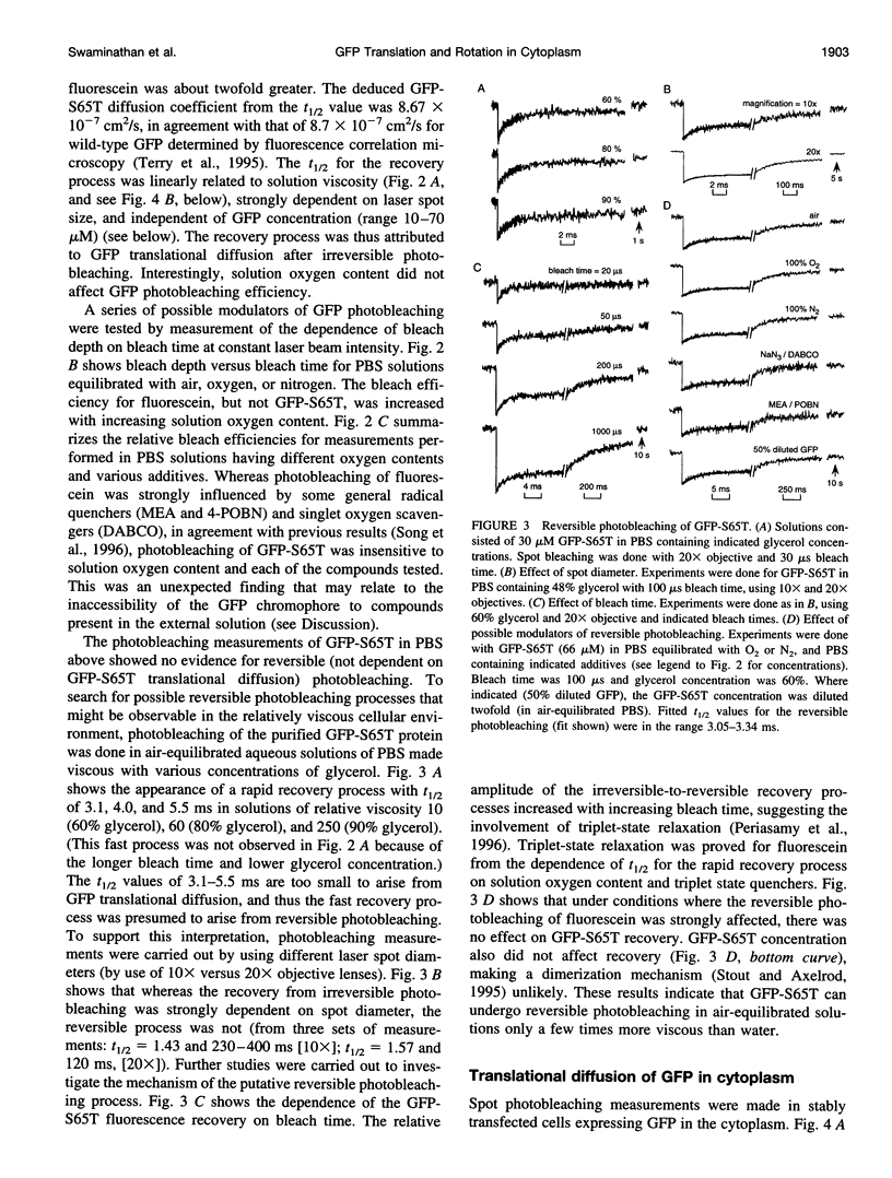


Images in this article
Selected References
These references are in PubMed. This may not be the complete list of references from this article.
- Bicknese S., Periasamy N., Shohet S. B., Verkman A. S. Cytoplasmic viscosity near the cell plasma membrane: measurement by evanescent field frequency-domain microfluorimetry. Biophys J. 1993 Sep;65(3):1272–1282. doi: 10.1016/S0006-3495(93)81179-2. [DOI] [PMC free article] [PubMed] [Google Scholar]
- Chalfie M. Green fluorescent protein. Photochem Photobiol. 1995 Oct;62(4):651–656. doi: 10.1111/j.1751-1097.1995.tb08712.x. [DOI] [PubMed] [Google Scholar]
- Chalfie M., Tu Y., Euskirchen G., Ward W. W., Prasher D. C. Green fluorescent protein as a marker for gene expression. Science. 1994 Feb 11;263(5148):802–805. doi: 10.1126/science.8303295. [DOI] [PubMed] [Google Scholar]
- Cole N. B., Smith C. L., Sciaky N., Terasaki M., Edidin M., Lippincott-Schwartz J. Diffusional mobility of Golgi proteins in membranes of living cells. Science. 1996 Aug 9;273(5276):797–801. doi: 10.1126/science.273.5276.797. [DOI] [PubMed] [Google Scholar]
- Cubitt A. B., Heim R., Adams S. R., Boyd A. E., Gross L. A., Tsien R. Y. Understanding, improving and using green fluorescent proteins. Trends Biochem Sci. 1995 Nov;20(11):448–455. doi: 10.1016/s0968-0004(00)89099-4. [DOI] [PubMed] [Google Scholar]
- De Giorgi F., Brini M., Bastianutto C., Marsault R., Montero M., Pizzo P., Rossi R., Rizzuto R. Targeting aequorin and green fluorescent protein to intracellular organelles. Gene. 1996;173(1 Spec No):113–117. doi: 10.1016/0378-1119(95)00687-7. [DOI] [PubMed] [Google Scholar]
- Fushimi K., Verkman A. S. Low viscosity in the aqueous domain of cell cytoplasm measured by picosecond polarization microfluorimetry. J Cell Biol. 1991 Feb;112(4):719–725. doi: 10.1083/jcb.112.4.719. [DOI] [PMC free article] [PubMed] [Google Scholar]
- Gerdes H. H., Kaether C. Green fluorescent protein: applications in cell biology. FEBS Lett. 1996 Jun 24;389(1):44–47. doi: 10.1016/0014-5793(96)00586-8. [DOI] [PubMed] [Google Scholar]
- Heim R., Cubitt A. B., Tsien R. Y. Improved green fluorescence. Nature. 1995 Feb 23;373(6516):663–664. doi: 10.1038/373663b0. [DOI] [PubMed] [Google Scholar]
- Heim R., Tsien R. Y. Engineering green fluorescent protein for improved brightness, longer wavelengths and fluorescence resonance energy transfer. Curr Biol. 1996 Feb 1;6(2):178–182. doi: 10.1016/s0960-9822(02)00450-5. [DOI] [PubMed] [Google Scholar]
- Kao H. P., Abney J. R., Verkman A. S. Determinants of the translational mobility of a small solute in cell cytoplasm. J Cell Biol. 1993 Jan;120(1):175–184. doi: 10.1083/jcb.120.1.175. [DOI] [PMC free article] [PubMed] [Google Scholar]
- Kao H. P., Verkman A. S. Construction and performance of a photobleaching recovery apparatus with microsecond time resolution. Biophys Chem. 1996 Mar 7;59(1-2):203–210. doi: 10.1016/0301-4622(95)00139-5. [DOI] [PubMed] [Google Scholar]
- Luby-Phelps K., Castle P. E., Taylor D. L., Lanni F. Hindered diffusion of inert tracer particles in the cytoplasm of mouse 3T3 cells. Proc Natl Acad Sci U S A. 1987 Jul;84(14):4910–4913. doi: 10.1073/pnas.84.14.4910. [DOI] [PMC free article] [PubMed] [Google Scholar]
- Luby-Phelps K., Hori M., Phelps J. M., Won D. Ca(2+)-regulated dynamic compartmentalization of calmodulin in living smooth muscle cells. J Biol Chem. 1995 Sep 15;270(37):21532–21538. doi: 10.1074/jbc.270.37.21532. [DOI] [PubMed] [Google Scholar]
- Luby-Phelps K. Physical properties of cytoplasm. Curr Opin Cell Biol. 1994 Feb;6(1):3–9. doi: 10.1016/0955-0674(94)90109-0. [DOI] [PubMed] [Google Scholar]
- Luby-Phelps K., Taylor D. L., Lanni F. Probing the structure of cytoplasm. J Cell Biol. 1986 Jun;102(6):2015–2022. doi: 10.1083/jcb.102.6.2015. [DOI] [PMC free article] [PubMed] [Google Scholar]
- Ormö M., Cubitt A. B., Kallio K., Gross L. A., Tsien R. Y., Remington S. J. Crystal structure of the Aequorea victoria green fluorescent protein. Science. 1996 Sep 6;273(5280):1392–1395. doi: 10.1126/science.273.5280.1392. [DOI] [PubMed] [Google Scholar]
- Periasamy N., Bicknese S., Verkman A. S. Reversible photobleaching of fluorescein conjugates in air-saturated viscous solutions: singlet and triplet state quenching by tryptophan. Photochem Photobiol. 1996 Mar;63(3):265–271. doi: 10.1111/j.1751-1097.1996.tb03023.x. [DOI] [PubMed] [Google Scholar]
- Rizzuto R., Brini M., De Giorgi F., Rossi R., Heim R., Tsien R. Y., Pozzan T. Double labelling of subcellular structures with organelle-targeted GFP mutants in vivo. Curr Biol. 1996 Feb 1;6(2):183–188. doi: 10.1016/s0960-9822(02)00451-7. [DOI] [PubMed] [Google Scholar]
- Rizzuto R., Brini M., Pizzo P., Murgia M., Pozzan T. Chimeric green fluorescent protein as a tool for visualizing subcellular organelles in living cells. Curr Biol. 1995 Jun 1;5(6):635–642. doi: 10.1016/s0960-9822(95)00128-x. [DOI] [PubMed] [Google Scholar]
- Song L., Varma C. A., Verhoeven J. W., Tanke H. J. Influence of the triplet excited state on the photobleaching kinetics of fluorescein in microscopy. Biophys J. 1996 Jun;70(6):2959–2968. doi: 10.1016/S0006-3495(96)79866-1. [DOI] [PMC free article] [PubMed] [Google Scholar]
- Stout A. L., Axelrod D. Spontaneous recovery of fluorescence by photobleached surface-adsorbed proteins. Photochem Photobiol. 1995 Aug;62(2):239–244. doi: 10.1111/j.1751-1097.1995.tb05264.x. [DOI] [PubMed] [Google Scholar]
- Swaminathan R., Bicknese S., Periasamy N., Verkman A. S. Cytoplasmic viscosity near the cell plasma membrane: translational diffusion of a small fluorescent solute measured by total internal reflection-fluorescence photobleaching recovery. Biophys J. 1996 Aug;71(2):1140–1151. doi: 10.1016/S0006-3495(96)79316-5. [DOI] [PMC free article] [PubMed] [Google Scholar]
- Terry B. R., Matthews E. K., Haseloff J. Molecular characterisation of recombinant green fluorescent protein by fluorescence correlation microscopy. Biochem Biophys Res Commun. 1995 Dec 5;217(1):21–27. doi: 10.1006/bbrc.1995.2740. [DOI] [PubMed] [Google Scholar]
- Thevenin B. J., Periasamy N., Shohet S. B., Verkman A. S. Segmental dynamics of the cytoplasmic domain of erythrocyte band 3 determined by time-resolved fluorescence anisotropy: sensitivity to pH and ligand binding. Proc Natl Acad Sci U S A. 1994 Mar 1;91(5):1741–1745. doi: 10.1073/pnas.91.5.1741. [DOI] [PMC free article] [PubMed] [Google Scholar]
- Velez M., Axelrod D. Polarized fluorescence photobleaching recovery for measuring rotational diffusion in solutions and membranes. Biophys J. 1988 Apr;53(4):575–591. doi: 10.1016/S0006-3495(88)83137-0. [DOI] [PMC free article] [PubMed] [Google Scholar]
- Verkman A. S., Armijo M., Fushimi K. Construction and evaluation of a frequency-domain epifluorescence microscope for lifetime and anisotropy decay measurements in subcellular domains. Biophys Chem. 1991 Apr;40(1):117–125. doi: 10.1016/0301-4622(91)85036-p. [DOI] [PubMed] [Google Scholar]
- Welch G. R., Easterby J. S. Metabolic channeling versus free diffusion: transition-time analysis. Trends Biochem Sci. 1994 May;19(5):193–197. doi: 10.1016/0968-0004(94)90019-1. [DOI] [PubMed] [Google Scholar]
- Yang B., Brown D., Verkman A. S. The mercurial insensitive water channel (AQP-4) forms orthogonal arrays in stably transfected Chinese hamster ovary cells. J Biol Chem. 1996 Mar 1;271(9):4577–4580. [PubMed] [Google Scholar]