

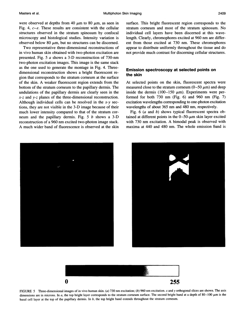
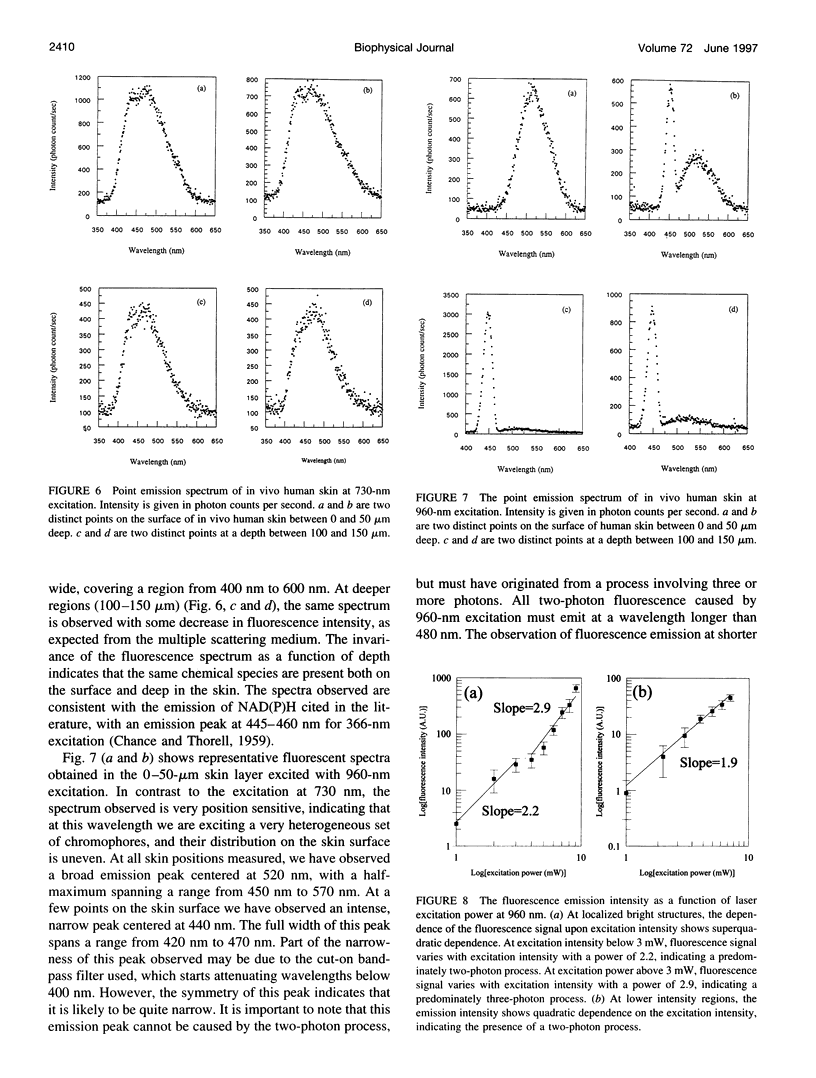
Abstract
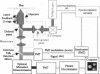
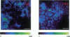
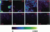
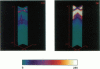
Multiphoton excitation microscopy at 730 nm and 960 nm was used to image in vivo human skin autofluorescence from the surface to a depth of approximately 200 microm. The emission spectra and fluorescence lifetime images were obtained at selected locations near the surface (0-50 microm) and at deeper depths (100-150 microm) for both excitation wavelengths. Cell borders and cell nuclei were the prominent structures observed. The spectroscopic data suggest that reduced pyridine nucleotides, NAD(P)H, are the primary source of the skin autofluorescence at 730 nm excitation. With 960 nm excitation, a two-photon fluorescence emission at 520 nm indicates the presence of a variable, position-dependent intensity component of flavoprotein. A second fluorescence emission component, which starts at 425 nm, is observed with 960-nm excitation. Such fluorescence emission at wavelengths less than half the excitation wavelength suggests an excitation process involving three or more photons. This conjecture is further confirmed by the observation of the super-quadratic dependence of the fluorescence intensity on the excitation power. Further work is required to spectroscopically identify these emitting species. This study demonstrates the use of multiphoton excitation microscopy for functional imaging of the metabolic states of in vivo human skin cells.
Full text
PDF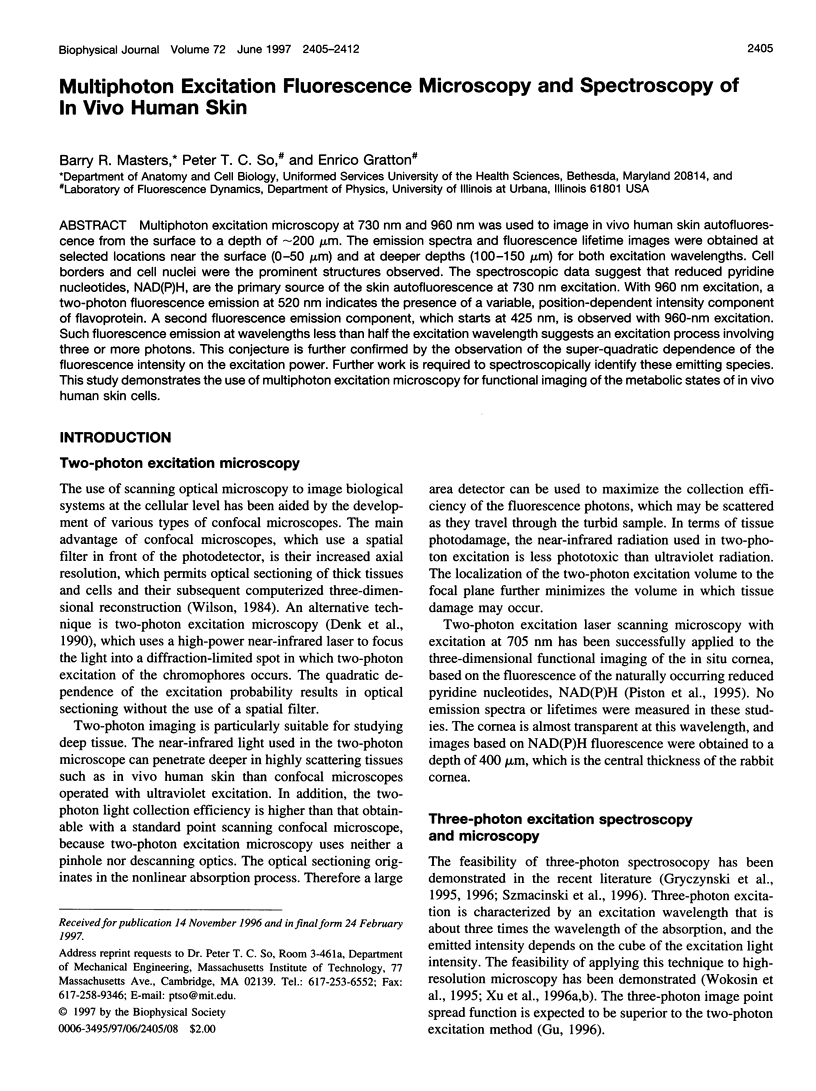
Images in this article
Selected References
These references are in PubMed. This may not be the complete list of references from this article.
- Bertrand C., Corcuff P. In vivo spatio-temporal visualization of the human skin by real-time confocal microscopy. Scanning. 1994 May-Jun;16(3):150–154. doi: 10.1002/sca.4950160301. [DOI] [PubMed] [Google Scholar]
- CHANCE B., THORELL B. Localization and kinetics of reduced pyridine nucleotide in living cells by microfluorometry. J Biol Chem. 1959 Nov;234:3044–3050. [PubMed] [Google Scholar]
- Chance B., Lieberman M. Intrinsic fluorescence emission from the cornea at low temperatures: evidence of mitochondrial signals and their differing redox states in epithelial and endothelial sides. Exp Eye Res. 1978 Jan;26(1):111–117. doi: 10.1016/0014-4835(78)90159-8. [DOI] [PubMed] [Google Scholar]
- Chance B., Schoener B., Oshino R., Itshak F., Nakase Y. Oxidation-reduction ratio studies of mitochondria in freeze-trapped samples. NADH and flavoprotein fluorescence signals. J Biol Chem. 1979 Jun 10;254(11):4764–4771. [PubMed] [Google Scholar]
- Corcuff P., Bertrand C., Leveque J. L. Morphometry of human epidermis in vivo by real-time confocal microscopy. Arch Dermatol Res. 1993;285(8):475–481. doi: 10.1007/BF00376820. [DOI] [PubMed] [Google Scholar]
- Corcuff P., Lévêque J. L. In vivo vision of the human skin with the tandem scanning microscope. Dermatology. 1993;186(1):50–54. doi: 10.1159/000247302. [DOI] [PubMed] [Google Scholar]
- Denk W., Strickler J. H., Webb W. W. Two-photon laser scanning fluorescence microscopy. Science. 1990 Apr 6;248(4951):73–76. doi: 10.1126/science.2321027. [DOI] [PubMed] [Google Scholar]
- Dong C. Y., So P. T., French T., Gratton E. Fluorescence lifetime imaging by asynchronous pump-probe microscopy. Biophys J. 1995 Dec;69(6):2234–2242. doi: 10.1016/S0006-3495(95)80148-7. [DOI] [PMC free article] [PubMed] [Google Scholar]
- Eng J., Lynch R. M., Balaban R. S. Nicotinamide adenine dinucleotide fluorescence spectroscopy and imaging of isolated cardiac myocytes. Biophys J. 1989 Apr;55(4):621–630. doi: 10.1016/S0006-3495(89)82859-0. [DOI] [PMC free article] [PubMed] [Google Scholar]
- Gryczynski I., Szmacinski H., Lakowicz J. R. On the possibility of calcium imaging using Indo-1 with three-photon excitation. Photochem Photobiol. 1995 Oct;62(4):804–808. doi: 10.1111/j.1751-1097.1995.tb08733.x. [DOI] [PubMed] [Google Scholar]
- König K., So P. T., Mantulin W. W., Tromberg B. J., Gratton E. Two-photon excited lifetime imaging of autofluorescence in cells during UVA and NIR photostress. J Microsc. 1996 Sep;183(Pt 3):197–204. [PubMed] [Google Scholar]
- Piston D. W., Masters B. R., Webb W. W. Three-dimensionally resolved NAD(P)H cellular metabolic redox imaging of the in situ cornea with two-photon excitation laser scanning microscopy. J Microsc. 1995 Apr;178(Pt 1):20–27. doi: 10.1111/j.1365-2818.1995.tb03576.x. [DOI] [PubMed] [Google Scholar]
- Piérard G. E. In vivo confocal microscopy: a new paradigm in dermatology. Dermatology. 1993;186(1):4–5. doi: 10.1159/000247294. [DOI] [PubMed] [Google Scholar]
- Rajadhyaksha M., Grossman M., Esterowitz D., Webb R. H., Anderson R. R. In vivo confocal scanning laser microscopy of human skin: melanin provides strong contrast. J Invest Dermatol. 1995 Jun;104(6):946–952. doi: 10.1111/1523-1747.ep12606215. [DOI] [PubMed] [Google Scholar]
- Szmacinski H., Gryczynski I., Lakowicz J. R. Three-photon induced fluorescence of the calcium probe Indo-1. Biophys J. 1996 Jan;70(1):547–555. doi: 10.1016/S0006-3495(96)79601-7. [DOI] [PMC free article] [PubMed] [Google Scholar]
- Xu C., Zipfel W., Shear J. B., Williams R. M., Webb W. W. Multiphoton fluorescence excitation: new spectral windows for biological nonlinear microscopy. Proc Natl Acad Sci U S A. 1996 Oct 1;93(20):10763–10768. doi: 10.1073/pnas.93.20.10763. [DOI] [PMC free article] [PubMed] [Google Scholar]