

Abstract
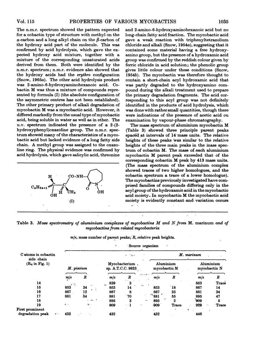
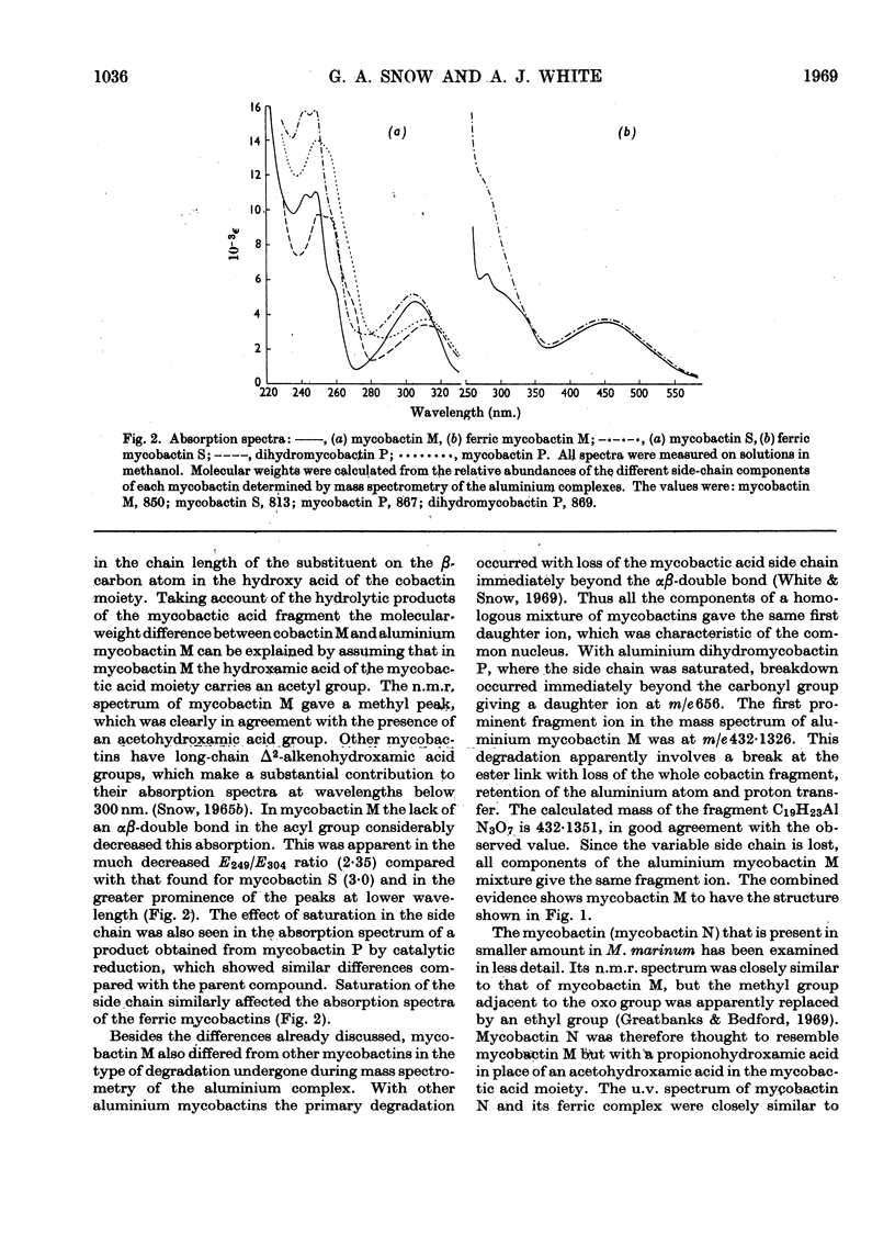
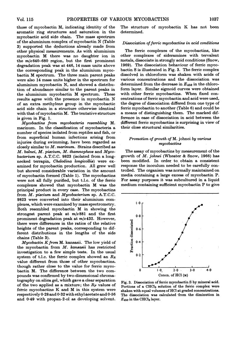
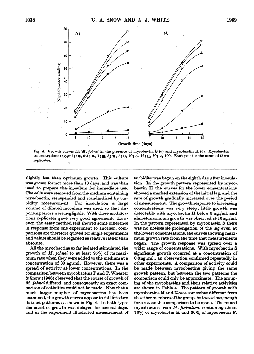
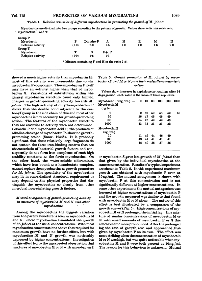
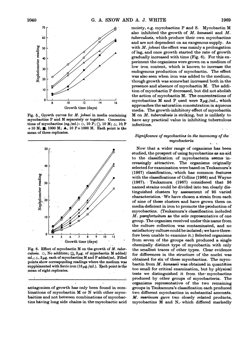
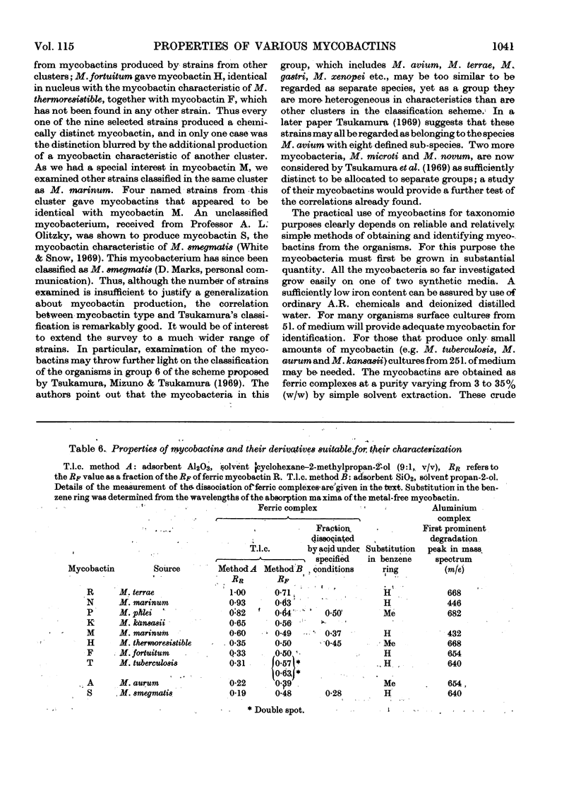
Nine different strains of mycobacteria grown on media deficient in iron all produced mycobactins. Most strains produced one mycobactin in great preponderance. Mycobacteria from clearly distinct taxonomic groups gave mycobactins differing in the structure of their nuclei. One group of taxonomically related mycobacteria produced mycobactins having the same nucleus but with different distributions of side chains within the homologous mixtures. Simple methods are described for identifying mycobactins on a small scale; these may be of value in classifying mycobacteria. Structures are proposed for mycobactin A from Mycobacterium aurum, mycobactin R from M. terrae, mycobactin F, produced together with mycobactin H by M. fortuitum, and mycobactins M and N from M. marinum. The first three of these differ from known mycobactins in details of substitution and configuration of asymmetric centres in the nucleus. Mycobactins M and N are substantially different, having only small acyl groups (acetyl and propionyl respectively) at the hydroxamic acid centre of the mycobactic acid moiety. Both are homologous mixtures having long-chain saturated 3-hydroxy-2-methyl acid fragments in the cobactin moiety. All mycobactins so far isolated promote almost maximal growth of M. johnei at 30ng./ml. in liquid medium. The activity of some mycobactins extends to much lower concentrations, mycobactin S showing significant growth promotion at 0·3ng./ml. Mycobactin M or N in combination with mycobactins having a long side chain in the mycobactic acid moiety exerts a mutually antagonistic effect on the growth of M. johnei, the mixture giving less growth than either mycobactin separately. Mycobactin M also decreases the growth of M. kansasii and M. tuberculosis on liquid media. These antagonistic effects are probably caused by a lengthening of the lag phase.
Full text
PDF
Selected References
These references are in PubMed. This may not be the complete list of references from this article.
- Collins C. H. Revised classification of anonymous mycobacteria. J Clin Pathol. 1966 Sep;19(5):433–437. doi: 10.1136/jcp.19.5.433. [DOI] [PMC free article] [PubMed] [Google Scholar]
- Greatbanks D., Bedford G. R. Identification of mycobactins by nuclear-magnetic-resonance spectroscopy. Biochem J. 1969 Dec;115(5):1047–1050. doi: 10.1042/bj1151047. [DOI] [PMC free article] [PubMed] [Google Scholar]
- Nakayama Y., Takeya K. Esterase zymogram method for classifying mycobacteria. Nature. 1967 Feb 4;213(5075):504–504. doi: 10.1038/213504a0. [DOI] [PubMed] [Google Scholar]
- Reiner E., Beam R. E., Kubica G. P. A rapid chemotaxonomic method for distinguishing mycobacterial strains. J Chromatogr. 1967 Apr;27(2):495–496. doi: 10.1016/s0021-9673(01)85910-0. [DOI] [PubMed] [Google Scholar]
- SNOW G. A. THE STRUCTURE OF MYCOBACTIN P, A GROWTH FACTOR FOR MYCOBACTERIUM JOHNEI, AND THE SIGNIFICANCE OF ITS IRON COMPLEX. Biochem J. 1965 Jan;94:160–165. doi: 10.1042/bj0940160. [DOI] [PMC free article] [PubMed] [Google Scholar]
- Snow G. A. Isolation and structure of mycobactin T, a growth factor from Mycobacterium tuberculosis. Biochem J. 1965 Oct;97(1):166–175. doi: 10.1042/bj0970166. [DOI] [PMC free article] [PubMed] [Google Scholar]
- Snow G. A. Metal complexes of mycobactin P and of desferrisideramines. Biochem J. 1969 Nov;115(2):199–205. doi: 10.1042/bj1150199. [DOI] [PMC free article] [PubMed] [Google Scholar]
- Stanford J. L., Beck A. An antigenic analysis of the mycobacteria, Mycobacterium fortuitum, Myco. kansasii, Myco. phlei, Myco. smegmatis and Myco. tuberculosis. J Pathol Bacteriol. 1968 Jan;95(1):131–139. doi: 10.1002/path.1700950116. [DOI] [PubMed] [Google Scholar]
- Szulga T., Jenkins P. A., Marks J. Thin-layer chromatography of mycobacterial lipids as an aid to classification; Mycobacterium kansasii; and Mycobacterium marinum (balnei). Tubercle. 1966 Mar;47(1):130–136. doi: 10.1016/s0041-3879(66)80055-7. [DOI] [PubMed] [Google Scholar]
- Tsukamura M. Identification of mycobacteria. Tubercle. 1967 Dec;48(4):311–338. doi: 10.1016/s0041-3879(67)80040-0. [DOI] [PubMed] [Google Scholar]
- Tsukamura M., Mizuno S., Tsukamura S. Numerical classification of slowly growing mycobacteria. Am Rev Respir Dis. 1969 Feb;99(2):299–303. doi: 10.1164/arrd.1969.99.2.299. [DOI] [PubMed] [Google Scholar]
- Tuboly S. Studies on the antigenic structure of mycobacteria. I. Comparison of the antigenic structure of pathogenic and saprophytic mycobacteria. Acta Microbiol Acad Sci Hung. 1965;12(3):233–240. [PubMed] [Google Scholar]
- Wayne L. G. Selection of characters for an Adansonian analysis of mycobacterial taxonomy. J Bacteriol. 1967 Apr;93(4):1382–1391. doi: 10.1128/jb.93.4.1382-1391.1967. [DOI] [PMC free article] [PubMed] [Google Scholar]
- Wheather D. W., Snow G. A. Assay of the mycobactins by measurement of the growth of Mycobacterium johnei. Biochem J. 1966 Jul;100(1):47–49. doi: 10.1042/bj1000047. [DOI] [PMC free article] [PubMed] [Google Scholar]
- White A. J., Snow G. A. Isolation of mycobactinss from various mycobacteria. The properties of mycobactin S and H. Biochem J. 1969 Mar;111(5):785–792. doi: 10.1042/bj1110785. [DOI] [PMC free article] [PubMed] [Google Scholar]
- White A. J., Snow G. A. Methods for the separation and identification of mycobactins from various species of mycobacteria. Biochem J. 1968 Jul;108(4):593–597. doi: 10.1042/bj1080593. [DOI] [PMC free article] [PubMed] [Google Scholar]