Abstract
The medical records of equine gastrointestinal colic cases presented to the Western College of Veterinary Medicine between 1992 and 2002 are reviewed. There was no breed predisposition to colic. Geldings were more prone to colic than females and stallions. Overall, the 3 most common causes of colic were large colon impaction (20.8%), large colon displacement (16.5%), and spasmodic colic (11.7%), after excluding the 13% of cases in which the diagnosis was undetermined. Of the medical cases, large colon impaction (38.4%) and spasmodic colic (22.5%) were the most common. Of the surgical cases, large colon displacement (24.5%), large colon torsion (14.3%), and strangulating lipoma (13.5%) were the most common. Recovery rate for the medical cases was 93.6%. Recovery rate for surgical cases was 73.5%. In conclusion, most of the equine colic cases were medical, and the recovery rates for both surgical and medical cases were comparable with those of other studies.
Résumé
Causes de coliques gastro-intestinales chez des chevaux de l’Ouest du Canada : 604 cas (1992 à 2002). Les dossiers médicaux des cas de coliques gastro-intestinales présentés au Western College of Veterinary Medicne entre 1992 et 2002 ont été passés en revue. Il n’y avait pas de prédisposition raciales aux coliques. Les hongres étaient plus sujets aux coliques que les femelles ou les étalons. Globalement, les 3 principales causes de coliques étaient l’impaction du gros colon (20,8 %), le déplacement du gros côlon (16,5 %) et les coliques spasmodiques (11,7 %), après exclusion de 13 % de cas où le diagnostic était indéterminé. De tous les cas médicaux, l’impaction du gros côlon (38,4 %) et la colique spasmodique (22,5 %) étaient les plus fréquents. Des cas chirurgicaux, le déplacement du gros côlon (24,5 %), la torsion du côlon (14,3 %) et l’étranglement par lipomes (13,5 %) étaient les plus fréquents. Le taux de rétablissement des cas médicaux était de 93,6 % et celui des cas chirurgicaux de 73,5 %. Il a été conclu que la majorité des coliques équines étaient d’origine médicale et que les taux de rétablissement des cas chirurgicaux et médicaux étaient comparables à ceux d’autres études.
(Traduit par Docteur André Blouin)
Introduction
Colic is one of the most common problems in equine practice. It has a significant economic impact on the racehorse industry and is a major concern for owners (1–3). Equine colic can be divided into 2 major categories; gastrointestinal and nongastrointestinal (2). Nongastrointestinal colic cases can usually be excluded based on physical examination findings; these include signs of abdominal discomfort due to urinary urolithiasis and disorders of reproductive, nervous, respiratory, or musculoskeletal systems (2). Causes of gastrointestinal colic (GC) are gut distension, tension on the root of mesentery, ischemia, deep ulcers in the stomach or bowel, and peritoneal pain (2). The decision whether a colic case should be managed medically or surgically depends on 5 main points; severity of pain (responsive vs. nonresponsive to analgesia), cardiovascular and systemic status, findings on transrectal palpation, the presence of nasogastric reflux, and results of abdominocentesis (3). Most causes of GC can be managed medically, only a small percentage (4% to 10%) require surgery (3–7).
Gastrointestinal colic can be caused by different conditions, ranging from a harmless spasmodic colic to a life-threatening strangulating obstruction (8). Commonly, it is difficult to reach a final diagnosis without an exploratory laparotomy, but the clinician should attempt to provide the owner with all possible outcomes and a prognosis (8).
The purpose of this study was to explore the different causes of equine GC in western Canada, with a view to providing equine practitioners with a better understanding of the most likely causes and a short-term prognosis for survival.
Materials and methods
Criteria for selection of cases
The medical records of horses presented with signs of abdominal discomfort (colic) at the Western College of Veterinary Medicine between December 1992 and December 2002 were reviewed. Signs included were pawing, sweating, flank watching, bruxism, stretching, kicking at the abdomen, and lying down or rolling. Cases where the signs were attributed to urinary, reproductive, respiratory, or musculoskeletal system disorders were not included (nongastrointestinal colic). Diagnosis of GC was achieved by physical examination, transrectal examination, abdominocentesis, the presence of nasogastric reflux, surgical exploration or at necropsy.
Methods of analysis
Age, breed, sex, duration of clinical signs, referral case or not, type of analgesia (α2 agonists, with or without opiates), heart rate (beats/min), the presence of nasogastric reflux (> 2 L net), transrectal examination findings, diagnosis, hospitalization days, and the outcome were recorded. Data were analyzed by using descriptive statistics, and nonparametric (Kruskal-Wallis One Way ANOVA) and chi-square tests with the help of a computerized statistical package (Student Statistix 7; Analytical Software, Tallahassee, Florida, USA).
Horses were considered a “surgical colic” when, in the opinion of the attending clinicians, the horse exhibited evidence of having a surgical lesion or could no longer be managed medically. Spasmodic colic, in this study, was when a horse showed brief attacks of pain associated with hypermotility and loud intestinal sounds of the intestine on auscultation. It can affect both small and large intestines; however, in this study, it was considered as a large intestinal cause of colic.
Results
Six hundred and four cases met the inclusion criteria. A diagnosis was not reached in 79 (13%) of these cases, which were treated medically and observed for 1 d before being discharged; they were not included in the Tables.
Median age at presentation was 7 y (< 1 mo to 36 y). Median duration of clinical signs was 12 h (range 0 to 504 h). Median number of hospitalized days was 2 (0 to 75). Median heart rate (HR) was 52 beats/min (range 24 to 160). Quarter horses and Thoroughbreds represented 32.3% and 20.6% of the cases, respectively. Geldings accounted for 50% of the cases; mares and stallions for 30% and 20%, respectively. When compared with the hospital horse population during the same period of time, there was no breed predisposition. However, geldings were more prone to colic (P < 0.0001), females and stallions less prone (P < 0.0001, P = 0.0114, respectively).
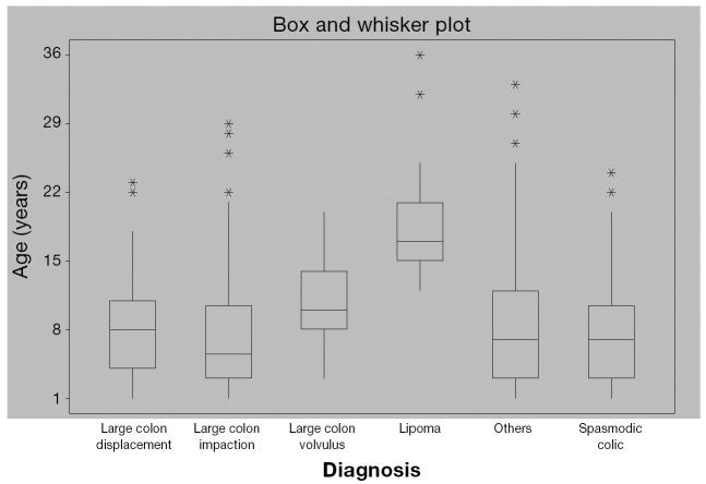
The 9 most common causes of colic are tabulated in Table 1. The median ages of horses in the 5 most common causes of colic differed significantly (P < 0.0001), (Table 2, Figure 1).
Table 1.
The 9 most common causes of colic presented to the Western College of Veterinary Medicine during the period from 1992 to 2002
| Diagnosis | Percentage |
|---|---|
| Large colon impaction | 20.8 |
| Large colon displacement | 16.5 |
| Spasmodic colic | 11.7 |
| Large colon volvulus | 7.3 |
| Lipoma | 6.9 |
| Strangulating SI lesion (other) | 4.2 |
| Enteritis | 3.4 |
| Peritonitis | 2.7 |
| Verminous arteritis | 2.1 |
SI — small intestine
Table 2.
Comparison of mean ranks of age for types of gastrointestinal colic diagnosed at the Western College of Veterinary Medicine
| Diagnosis | Mean rank | Median (range) |
|---|---|---|
| Lipoma | 401.8a | 17 (12–36) |
| Large colon volvulus | 288.0b | 10 (3–20) |
| Large colon displacement | 219.8b | 8 (1–23) |
| Impaction | 194.8b | 5.5 (1–29) |
| Others | 219.6b | 7 (1–33) |
| Spasmodic colic | 205.0b,c | 7 (1–24) |
Values with different superscripts within columns are different (P < 0.05)
Figure 1.
The distribution of the 6 most common diagnoses of colic, by age, presented to the Western College of Veterinary Medicine during the period of 1992 to 2002.
There were 327 medical and 277 surgical colic cases in the 10 y. Animals with surgical colic were significantly older (median 8 y, < 1 mo to 33 y) compared with those with medical colic (median 6 y, < 1 mo to 36 y, P = 0.01). Surgical cases that were not euthanized remained in the hospital longer (median 6 d, range 1 to 75 d) than did medical cases (median 2 d, range 0 to 36 d; P = 0.028). Surgical cases received significantly higher doses of analgesics (P < 0.0001) than did medical cases for all categories of pain medication (α2 agonists, opiates, and nonsteroidal anti-inflammatories). Significantly more animals with surgical colic were euthanized than were animals with medical colic (P < 0.0001). Overall, 306/327 (93.6%) medical cases survived to discharge, whereas only 90/277 (32.5%) surgical cases survived, or 90/151 (59.6%) when the cases euthanized prior to surgery are excluded. The duration of clinical signs negatively affected overall survival (P = 0.039), with survivors showing clinical signs for a mean of 18 h (s = 36.8 h) and these euthanized showing signs for a mean of 22.2 h (s = 41.1 h). Large intestinal cases were not more likely to survive a prolonged duration of clinical signs than were small intestinal cases (P = 0.12).
The distribution between large and small intestinal causes of colic defined at physical examination, exploratory laparotomy, or necropsy can be found in Tables 3 and 4. Small intestinal strangulating lesions were divided to 2 groups; strangulating lipoma and others. Those horses having a strangulating lipoma were significantly older than those with other small intestinal lesions (18.6 s = 5.1 and 8.4 s = 7.3 y, respectively, P < 0.0001). Horses with a strangulating lipoma were also significantly more likely to require euthanasia than were those horses with other small intestinal lesions (χ2 = 6.2, P = 0.012).
Table 3.
The 10 most common causes of gastric and small intestinal colic presented to the Western College of Veterinary Medicine from 1992 to 2002
| Diagnosis | Number | Percentage |
|---|---|---|
| Lipoma | 36 | 22.2 |
| Other small intestinal strangulating lesion | 22 | 13.6 |
| Enteritis | 14 | 8.6 |
| Verminous arteritis | 11 | 6.8 |
| Anterior enteritis | 9 | 5.5 |
| Mesenteric volvulus | 9 | 5.5 |
| Gastric (impaction, dilatation, ulcer, rupture, and others) | 8 | 4.9 |
| Small intestinal obstruction | 4 | 2.5 |
| Heal impaction | 4 | 2.5 |
| Intussusception | 3 | 1.9 |
Table 4.
The 10 most common causes of large intestinal colic presented to the Western College of Veterinary Medicine from 1992 to 2002
| Diagnosis | Number | Percentage |
|---|---|---|
| Large colon impaction | 109 | 30.1 |
| Large colon displacement | 87 | 24 |
| Spasmodic colic | 61 | 16.9 |
| Large colon volvulus | 38 | 10.5 |
| Cecal impaction | 11 | 3 |
| Small colon impaction | 10 | 2.8 |
| Typhlitis/colitis | 6 | 1.7 |
| Meconium impaction | 5 | 1.4 |
| Sand colic | 3 | 0.8 |
| Small colon strangulation | 3 | 0.8 |
Referred cases accounted for 384 animals and represented 63.6% of cases; these included the cases referred from WCVM-Field Service. Of these referred horses, 50.3% represented medical cases, whereas 49.7% were surgical cases. Of horses referred for medical reasons, 39.6% had a large colon impaction and 20.8% were suspected to have spasmodic colic. Of horses defined as having a surgical lesion, 28.7% had large colon displacement, 14.1% had large colon volvulus, and 12.5% were diagnosed as having lipoma. Referred horses were more likely to remain in the clinic longer than nonreferred horses. Despite the fact that referred horses had been exhibiting colic symptoms longer than nonreferred horses by the time they were presented to the WCVM (P = 0.016), this did not affect survival. Surgery was performed in significantly more referred than nonreferred horses (P = 0.0006). More surgical nonreferred cases were euthanized than referred cases (34% and 50%, respectively, P = 0.01).
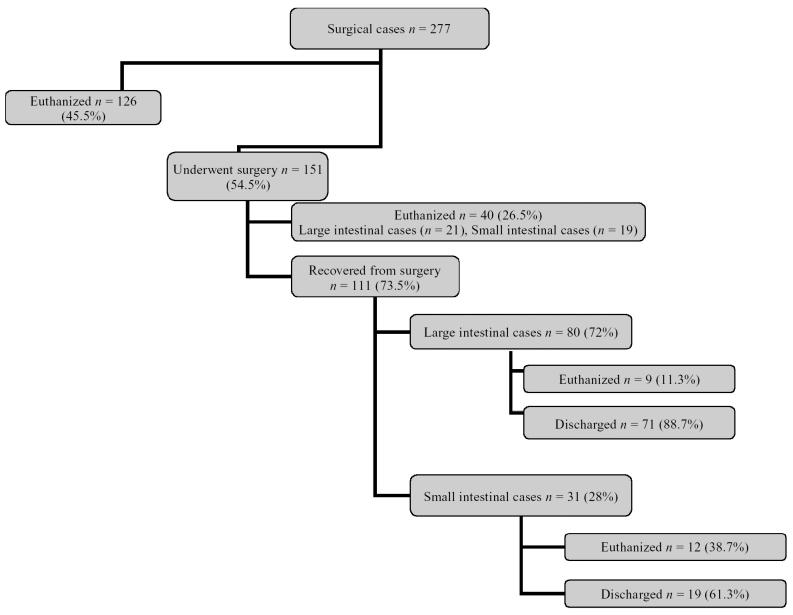
Of 277 surgical colic cases, the overall rate of survival was 32.5%. However, surgery was performed in only 151 cases (101 large intestinal colic cases and 50 small intestinal ones), the other 126 cases were euthanized for various reasons including poor prognosis and financial constraints. Of the 151 cases, 111 (73.5%) recovered from surgery (80 large intestinal and 31 small intestinal). However, 9/80 large intestinal and 12/31 small intestinal cases were subsequently euthanized prior to discharge. Thus 71/101 (69.3%) large intestine surgical colic cases and 19/50 (38%) small intestinal colic cases survived to discharge (Figure 2).
Figure 2.
Surgical colic cases presented to the Western College of Veterinary Medicine during the period 1992 to 2002.
Age was available as a variable in 574 cases. Distribution was examined by using descriptive statistics, and based on this, several age categories were delineated. These were < 1 y (n = 75), 1 to 7 y (n = 231), 8 to 15 y (n = 169), and > 15 y (n = 99). Causes of colic were then examined within these categories and the top 5 causes were listed (Table 5).
Table 5.
The 5 most common causes of colic presented to the Western College of Veterinary Medicine from 1992 to 2002, sorted by age
| Age (years) | Cause 1 | Cause 2 | Cause 3 | Cause 4 | Cause 5 |
|---|---|---|---|---|---|
| ≤ 1 | Large colon impaction (17.9%) | Spasmodic colic (16.4%) | Large colon displacement (10.4%i) | Enteritis (9%) | Meconium impaction (7.5%) |
| > 1 to 7 | Large colon impaction (27.7%) | Large colon displacement (20.5%) | Spasmodic colic (12.6%) | Large colon volvulus (4.2%) | Other small intestinal strangulation and peritonitis (3.1% each) |
| > 7 to 15 | Large colon displacement (17.9%)) | Large colon impaction (16%)) | Large colon volvulus (14.1%) | Spasmodic colic (11. 5%) | Lipoma (6.4%) |
| > 15 | Lipoma (28.6%) | Large colon impaction (14.3%>) | Large colon displacement (10.8%)) | Other small intestinal strangulation (8.3%>) | Large colon volvulus (7.1%i) |
To make the information presented in Table 5 more usable by practitioners, a cut point of 7 y old was chosen, so that groups 1 and 2 and groups 3 and 4 were pooled together, and the prevalence of the most encountered diagnoses were compared between horses aged ≤ 7 y and > 7 y old, using the chi-square test. Horses > 7 y were significantly more prone to large colon volvulus and lipoma, but less prone to large colon impaction (P < 0.0001 each). The prevalence of large colon displacement was not different between horses ≤ 7 y and > 7 y.
Discussion
Evaluation of equine patients for colic by veterinarians continues to be a common procedure in western Canada. The population of horses examined for colic at the WCVM, mainly the American quarter horse and the Thoroughbred, is consistent with the rest of the equine population presented to the WCVM. However, geldings were more prone to colic than stallions and mares. To the authors’ knowledge, this has never been reported before and the exact reason for this difference is unknown. Perhaps, mares and stallions, as valuable breeding animals, are better cared for.
As a teaching institution, the WCVM examines and treats both nonreferred and referred equine colic cases. Services are provided mainly to the 4 western provinces but occasionally to the northern American states. Numerous clients have to transport their animals substantial distances for veterinary care, and commonly state that they decided to bring the animal to the WCVM because they were already hauling the horse and that the extra distance was relatively inconsequential. Another reason for the relatively high primary case load is the large local horse population with few private practices in the immediate area.
The fact that large colon impactions and spasmodic colic comprised 39.6% and 20.8% of medical referrals, respectively, indicates that practitioners are not set up to hospitalize these animals.
Most veterinary practices in western Canada cannot afford to put the resources towards managing medical colic cases without compromising other aspects of their practice. Private practitioners manage the colic cases that can be dealt with effectively on-farm, which are the vast majority, but some refractory cases have to be referred to the WCVM. Another reason for referral of medical colic cases could be to provide the horse owner with a 2nd opinion, since it is not uncommon for horse owners to request a 2nd opinion, even if they are very confident in their veterinarian’s diagnostic abilities. If a 2nd opinion is not readily available locally, owners are required to transport their horses, which may lead them to a referral institution such as the WCVM. A 3rd reason for the seemingly large medical referral caseload is related to the vast area serviced by the WCVM; it is not uncommon for horses to be referred from as far away as 8 h by road. In these instances, medical colic cases may be referred, not because of the inability of a practice to manage the case, but because the clinical course of the colic may progress, necessitating surgical intervention in the future, and it is safer to refer an “acute abdomen” rather than wait until it becomes an emergency. Our results suggest that while duration of colic symptoms impacted negatively on survival overall, the subset of horses that were referred to the WCVM did not follow this trend, perhaps because more referred horses underwent surgery and survived, which was statistically significant and may have been influenced by the fact that owners of referred horses were more likely to consent to pursue an exploratory laparotomy.
The substantially higher percentage of equine colic cases requiring surgery that were euthanized compared with the percentage of those receiving medical treatment that were euthanized can be explained by the following factors: Horses with surgical conditions are more likely to carry a poorer prognosis compared with medical cases. The financial investment required for surgical treatment of a colic case is drastically higher, in our institution, than that for medical ailments. The exceptional distance travelled to the WCVM would have an effect on the severity of the lesions and degree of damage, reducing the survival rate. The combination of these factors commonly influences owners toward euthanasia of surgical cases. Overall the survival rate of surgical colic was 32.5% (90/277) (Figure 2). Of the horses undergoing exploratory laparotomy, 73.5% of cases that underwent surgery recovered. This indicates the selective nature of the cases taken to surgery, owners being more likely to request surgery if the preoperative diagnosis and subsequent prognosis are promising. Our overall survival rate was comparable with those of other studies where the rate ranged from 20% to 78.5% (9–12).
The percentage of large colon impaction cases presented to the WCVM are comparable with those of other referral centers; these have ranged from 13.4% (13) to 30% (14,15) of all “acute abdomens” examined. Some of the more common risk factors significantly associated with large colon obstructions (displacements and impactions) have included crib-biting, increased time stabled, changes in regular exercise regimes, no administration of ivermectin or moxidectin anthelmintics in the previous 12 mo, and a history of travel in the previous 24 h (16). The vast majority of simple obstructions of the large colon will respond to medical management, which includes intravenous or oral fluids, mineral oil, dioctyl sodium succinate, and analgesics (3). It is recommended that if an impaction persists and has not softened with 3 d of appropriate medical therapy or if the horses deteriorate clinically, surgery is indicated (3). However, horses undergoing surgical treatment for large colon impactions have a significantly higher fatality rate compared with those managed medically (13).
Large colon volvulus has been reported to occur in 11% to 17% of horses requiring surgery for colic (17). Our records indicate a similar trend in which 14.3% of colic cases requiring surgery were diagnosed as large colon volvulus. Spasmodic colic has been estimated to be as high as 72% of all colic cases examined in general practice in the UK (6). The large difference in our numbers (16.9%) can be explained at least in part by the fact that a substantial number of our cases were referrals.
In our retrospective study, the median age of horses with a strangulating lipoma was 17 y (range 12 to 36 y), which is similar to that in other recent reports (18). Therefore, clinicians in western Canada should have strangulating lipoma high on a differential diagnoses list when examining an aged horse with a suspected small intestinal disease.
Small intestinal strangulating lesions included volvulus of a portion of the jejunum or ileum, epiploic foramen entrapment, intussusceptions, inguinal hernias, vitelloumbilical bands and strangulated umbilical hernias, and others. These diseases were much more sporadic in occurrence and were less prevalent (13.6%) than strangulating lipomas in our hospital population. Epiploic foramen entrapments have long been considered a disease of aged horses (19). Recent literature suggests that this is not the case; there was no significant difference between the mean age of horses with epiploic foramen entrapment (9.6 y) and horses with miscellaneous small intestinal lesions (7.7 y) (18). Verminous arteritis can involve any part of the intestines supplied by the cranial mesenteric artery and, therefore, can affect either large or small intestine. However, in this study, it was classified as a small intestinal cause of colic. Verminous arteritis was a common diagnosis prior to the widespread introduction of anthelmintics drugs. No colic cases requiring surgical intervention in the past 5 y at the WCVM have been diagnosed with verminous arteritis. Duodenitis-proximal jejunitis was infrequently diagnosed at the WCVM, which is not surprising as the disease is more prevalent in the southwestern and southeastern portions of the United States (20).
In our population, horses > 7 y are significantly more prone to large colon volvulus, and less prone to large colon impaction than those < 7 y. The exact reason for that is unknown. CVJ
Footnotes
Reprints will not be available from the authors.
References
- 1.Parry BW. Prognostic evaluation of equine colic cases. Compend Contin Educ Pract Vet. 1986;8:S98–S104. [Google Scholar]
- 2.Smith BP. Large animal internal medicine. 3rd ed. St Louis: Mosby, 2002:108–111.
- 3.Singer ER, Smith MA. Examination of the horse with colic: is it medical or surgical? Equine Vet Educ. 2002;34:87–96. [Google Scholar]
- 4.Proudman CJ, Smith JE, Edwards GB, French NP. Long-term survival of equine surgical cases. Part 1: patterns of mortality and morbidity. Equine Vet J. 2002;34:432–437. doi: 10.2746/042516402776117845. [DOI] [PubMed] [Google Scholar]
- 5.Tinker MK, White NA, Lessard P, et al. Prospective study of equine colic incidence and mortality. Equine Vet J. 1997;29:448–453. doi: 10.1111/j.2042-3306.1997.tb03157.x. [DOI] [PubMed] [Google Scholar]
- 6.Proudman CJ. A two-year, prospective survey of equine colic in general practice. Equine Vet J. 1992;24:90–93. doi: 10.1111/j.2042-3306.1992.tb02789.x. [DOI] [PubMed] [Google Scholar]
- 7.Hillyer MH, Taylor FG, French NP. A cross-sectional study of colic in horses on thoroughbred training premises in the British Isles in 1997. Equine Vet J. 2001;33:380–385. doi: 10.2746/042516401776249499. [DOI] [PubMed] [Google Scholar]
- 8.van der Linden MA, Laffont CM, van Oldruitenborgh-Oosterbaan MMS. Prognosis in equine medical and surgical colic. J Vet Intern Med. 2003;17:343–348. doi: 10.1111/j.1939-1676.2003.tb02459.x. [DOI] [PubMed] [Google Scholar]
- 9.Pascoe PJ, McDonell WN, Trim CM, Gorder JV. Mortality rates and associated factors in equine colic operations-a retrospective study of 341 operations. Can Vet J. 1983;24:76–85. [PMC free article] [PubMed] [Google Scholar]
- 10.Parker JE, Fubini SL, Todhunter RJ. Retrospective evaluation of repeat celiotomy in 53 horses with acute gastrointestinal disease. Vet Surg. 1989;18:424–431. doi: 10.1111/j.1532-950x.1990.tb01118.x. [DOI] [PubMed] [Google Scholar]
- 11.Ducharme NG, Hackett RP, Ducharme GR, Long S. Surgical treatment of colic results in 181 horses. Vet Surg. 1983;12:206–209. [Google Scholar]
- 12.Phillips TJ, Walmsley JP. Retrospective analysis of the results of 151 exploratory laparotomies in horses with gastrointestinal disease. Equine Vet J. 1993;25:427–431. doi: 10.1111/j.2042-3306.1993.tb02985.x. [DOI] [PubMed] [Google Scholar]
- 13.Dabareiner RM, White NA. Large colon impaction in horses: 147 cases (1985–1991) J Am Vet Med Assoc. 1995;206:679–685. [PubMed] [Google Scholar]
- 14.White NA. Epidemiology and etiology of colic. In: White NA, ed. The Equine Acute Abdomen. Philadelphia: Lea & Febiger, 1990: 53–56.
- 15.Tennant BC, Wheat JD, Meagher DM. Observation on the cause and incidence of acute intestinal obstruction in the horse. Proc 18th Annu Meet Am Assoc Equine Pract. 1972:251–257. [Google Scholar]
- 16.Hillyer MH, Taylor FGR, Proudman CJ, Edwards GB, Smith JE, French NP. Case control study to identify risk factors for simple colonic obstruction and distension in horses. Equine Vet J. 2002;34:455–463. doi: 10.2746/042516402776117746. [DOI] [PubMed] [Google Scholar]
- 17.Fischer AT, Meagher DM. Strangulating torsion of the equine large colon. Compend Contin Educ Pract Vet. 1986;8:S25–S30. [Google Scholar]
- 18.Freeman DE, Schaeffer DJ. Age distribution of horses with strangulation of the small intestine by a lipoma or in the epiploic foramen: 46 cases (1994–2000) J Am Vet Med Assoc. 2001;219:87–89. doi: 10.2460/javma.2001.219.87. [DOI] [PubMed] [Google Scholar]
- 19.Wheat JD. Diseases of the small intestine: Diagnosis and treatment. Proc 18th Annu Meet Am Assoc Equine Pract. 1972:265. [Google Scholar]
- 20.Edwards GB. Duodenitis-proximal jejunitis (anterior enteritis) as a surgical problem. Equine Vet Educ. 2000;12:411–414. [Google Scholar]


