Correction to: Virology Journal (2023) 20:158 10.1186/s12985-023-02126-8
In this article [1], Fig(s) 4 and 5 appeared incorrectly and have now been corrected in the original publication. For completeness and transparency, both the incorrect and correct versions are displayed below.
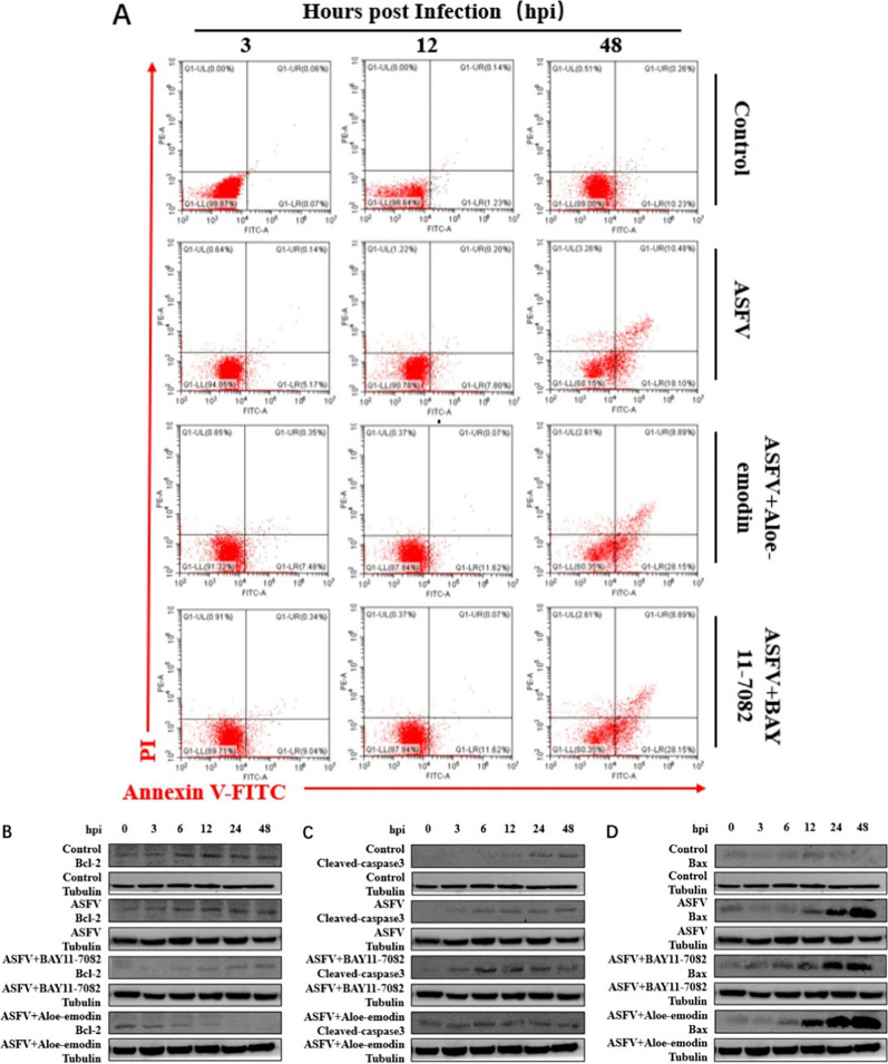
Fig. 4.
Ae inhibits the NF-κB signaling pathway activated by ASFV infection. The expression level of (A) MyD88 protein, (B) phospho-NF-κB p65 protein, and (C) pIκB protein in the ASFV-infected group, ASFV-infected group treated with BAY11-7082 or Ae was detected by western blot. The expression of tubulin was used as a positive control. qPCR was used to detect the changes of (D) IL-1β mRNA levels and (E) IL-8 mRNA levels in the ASFV-infected group, ASFV-infected group treated with BAY11-7082, or Ae at different time points. The mRNA level of GAPDH was used as a positive control. All control cells were normal cultured PAMs. The results of three independent experiments (mean ± SD) were represented by one data. Significant differences that were compared to the control group were indicated by * (P < 0.05), ** (P < 0.01) and *** (P < 0.001)
Fig. 5.
Ae promotes apoptosis by inhibiting the NF-κB signaling pathway. (A) The apoptosis of PAMs in the ASFV infection group, BAY11-7082 or Ae-treated ASFV infection group was detected by flow cytometry, and the apoptosis of induced cells was detected at 3, 12 and 48 h after treatment. Untreated cells served as negative controls. The expression level of (B) Bcl-2 protein, (C) cleaved-caspase3 protein, and (D) Bax protein in the ASFV infection group, BAY11-7082 or Ae-treated ASFV infection group was detected by western blot. All control cells were normal cultured PAMs. The tubulin expression was used as a positive control
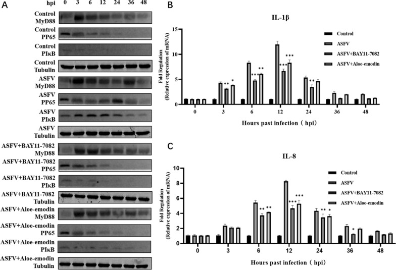
Fig. 4.
Ae inhibits the NF-κB signaling pathway activated by ASFV infection. A The expression level ofMyD88 protein, phospho-NF-κB p65 protein, and pIκB protein in the ASFV-infected group, ASFV-infected group treated with BAY11-7082 or Ae was detected by western blot. The expression of tubulin was used as a positive control. qPCR was used to detect the changes of B IL-1β mRNA levels and C IL-8 mRNA levels in the ASFV-infected group, ASFV-infected group treated with BAY11-7082, or Ae at different time points. The mRNA level of GAPDH was used as a positive control. All control cells were normal cultured PAMs. The results of three independent experiments (mean ± SD) were represented by one data. Significant differences that were compared to the control group were indicated by * (P < 0.05), ** (P < 0.01) and *** (P < 0.001)
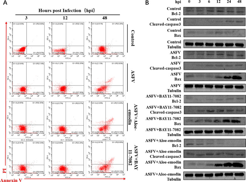
Fig. 5.
Ae promotes apoptosis by inhibiting the NF-κB signaling pathway. A The apoptosis of PAMs in the ASFV infection group, BAY11-7082 or Ae-treated ASFV infection group was detected by flow cytometry, and the apoptosis of induced cells was detected at 3, 12 and 48 h after treatment. Untreated cells served as negative controls. The expression level of B Bcl-2 protein, cleaved-caspase3 protein, and Bax protein in the ASFV infection group, BAY11-7082 or Ae-treated ASFV infection group was detected by western blot. All control cells were normal cultured PAMs. The tubulin expression was used as a positive control
Footnotes
Publisher's Note
Springer Nature remains neutral with regard to jurisdictional claims in published maps and institutional affiliations.
Reference
- 1.Luo Y, Yang Y, Wang W, et al. Aloe-emodin inhibits African swine fever virus replication by promoting apoptosis via regulating NF-κB signaling pathway. Virol J. 2023;20:158. 10.1186/s12985-023-02126-8. [DOI] [PMC free article] [PubMed] [Google Scholar]


