Abstract
Acetyl-CoA carboxylases (ACC1 and ACC2) catalyze the carboxylation of acetyl-CoA to form malonyl-CoA, an intermediate metabolite that plays a pivotal role in the regulation of fatty acid metabolism. We previously reported that ACC2 null mice are viable, and that ACC2 plays an important role in the regulation of fatty acid oxidation through the inhibition of carnitine palmitoyltransferase I, a mitochondrial component of the fatty-acyl shuttle system. Herein, we used gene targeting to knock out the ACC1 gene. The heterozygous mutant mice (Acc1+/–) had normal fertility and lifespans and maintained a similar body weight to that of their wild-type cohorts. The mRNA level of ACC1 in the tissues of Acc1+/– mice was half that of the wild type; however, the protein level of ACC1 and the total malonyl-CoA level were similar. In addition, there was no difference in the acetate incorporation into fatty acids nor in the fatty acid oxidation between the hepatocytes of Acc1+/– mice and those of the wild type. In contrast to Acc2–/– mice, Acc1–/– mice were not detected after mating. Timed pregnancies of heterozygotes revealed that Acc–/– embryos are already undeveloped at embryonic day (E)7.5, they die by E8.5, and are completely resorbed at E11.5. Our previous results of the ACC2 knockout mice and current studies of ACC1 knockout mice further confirm our hypotheses that malonyl-CoA exists in two independent pools, and that ACC1 and ACC2 have distinct roles in fatty acid metabolism.
Keywords: malonyl-CoA
Acetyl-CoA carboxylases (ACCs) are biotin-containing enzymes that catalyze the carboxylation of acetyl-CoA to form malonyl-CoA, an intermediate metabolite that plays a key role in the regulation of fatty acid metabolism. In addition, malonyl-CoA, as a precursor of the synthesis of long-chain fatty acids, has been implicated as a signal molecule for insulin secretion from the pancreatic β-islets (1, 2). The role that ACC plays in energy metabolism in lipogenic tissues (liver and adipose) and in oxidative tissues (liver, heart, and skeletal muscle) have become the focus of many studies (3–8). In lipogenic tissues, malonyl-CoA is the C2-unit donor for de novo synthesis of long-chain fatty acids catalyzed by fatty acid synthase (FAS) and for the chain elongation of fatty acid to very long-chain fatty acids. Moreover, increasing evidence suggests that malonyl-CoA is a regulator of fatty acid oxidation through the inhibition of carnitine palmitoyltransferase I, an enzyme that controls the entry of long-chain fatty acids into the mitochondria for β-oxidation (3). In animals, including humans, ACC1 (Mr = 265,000) and ACC2 (Mr = 280,000) are the two isoforms of acetyl-CoA carboxylase that are encoded by two separate genes, ACC1 and ACC2, respectively, and they display distinct tissue distribution (9–12). ACC1 is abundant in lipogenic tissues, such as liver and adipose tissue, whereas ACC2 is highly expressed in heart, skeletal muscle, and liver (9, 10). Recently, Loftus et al. (13) proposed that malonyl-CoA plays a role in the central nervous system as a component of appetite control. This suggestion was based on their finding that C75, a FAS inhibitor, may act as an anorectic signal in mice because of the increase in malonyl-CoA level. They also reported that the effect of C75 was offset by administering tetradecycloxyfluroic acid, an inhibitor of ACC that decreased the malonyl-CoA levels.
ACC1 is subject to long-term control at the transcriptional and translational levels, short-term regulation by phosphorylation/dephosphorylation of targeted serine residues, and allosteric transformation by citrate or palmitoyl-CoA (14–20). The amino acid sequences of ACC1 of yeast (21), rat (22), chicken (23), and human (12) are very similar, with ≈90% identity among the animal ACC1 carboxylases and ≈35% similarity between the animal and yeast ACC1 enzymes. In the yeast, Saccharomyces cerevisiae, acetyl-CoA carboxylase (ACC1/FAS3), the ACC homolog of animal ACC1, is essential for the growth and viability of the yeast cells, and supplementation of fatty acids failed to rescue the acc1 null mutants (24, 25).
To dissect the roles of the two animal ACC isoforms, we perused knockouts of the ACC1 and ACC2 genes in mice. Our studies with Acc2 knockout mice have shown that Acc2–/– mice live and breed well, continuously oxidize fatty acids, eat more food, and gain less weight than their wild-type cohorts (4). More importantly, Acc2–/– mice are resistant to obesity and diabetes when fed a high-fat/high-carbohydrate diet and retain insulin sensitivity and normal glucose levels compared to wild-type cohorts that become obese and type 2 diabetic (26). In addition, the adipose tissue of Acc2–/– mice has higher levels of lipolysis, fatty acid, and glucose oxidations when fed normal chow or a high-fat/high-carbohydrate diet (27).
In this communication, we provide further evidence about the different physiological roles of each ACC isoform in mice. In contrast to the Acc2 knockouts, Acc1 homozygous mutant mice are embryonically lethal, and the mutant embryos are already undeveloped at embryonic day (E)7.5. These results suggest that malonyl-CoA, produced by ACC1, plays an important role in the development of mouse embryos through the de novo synthesis of long-chain fatty acids. In addition, and based on the knockout studies of both ACC2 and ACC1, we conclude that the two enzymes work independently and that there is little, if any, overlap of function between them.
Materials and Methods
Materials. All restriction enzymes, T4 DNA ligase, and Klenow DNA polymerase were purchased from New England Biolabs. The sources of the following are as follows: TaqDNA polymerase (PerkinElmer), dideoxy sequencing kits (United States Biochemical), 32P-labeled nucleotides (Amersham Pharmacia), Quick-Clone mouse heart cDNA (Clontech), random primer labeling kits (Stratagene), and Western blot kits (Novagen). The immunoblot analysis was carried out by using anti-phospho-ACC (Ser-79, Upstate Biotechnology) as the primary antibody and horseradish peroxidase goat anti-rabbit as the secondary antibody. All chemicals used were of the highest quality commercially available.
Generation of an Acc1 Mutant Allele in Mice. ACC1 isoform is highly conserved among animal species. We designed a forward primer (5′-GGATATCGCATCACAATTGGC-3′) based on the human ACC1 cDNA and a reverse primer (5′-CCTCGGAGTGCCGTGCTCTGGATC-3′) that contained the biotin-binding site and used them to amplify a 335-bp cDNA probe by using mouse cDNA as a template. A 129/SvEv mouse genomic library was screened with the PCR fragment. A 14-kbp genomic fragment was isolated, mapped with restriction enzymes, and analyzed by Southern blotting. Several exonic sequences, including the one that contains the Met-Lys-Met motif, were identified and confirmed by DNA sequencing. A 1.5-kb 3′ NotI-XbaI fragment and a 6-kb 5′ KpnI-SpeI fragment were isolated and used to construct a replacement gene-targeting vector, and hypoxanthine phosphoribosyltransferase cassette was inserted in opposite orientation on the blunt end of the SpeI site. The linearized targeting vector (25 μg) was electroporated into 107 AB2.2 ES cells. DNA samples from 96 hypoxanthine/aminopterin/thymidine/1-(2′-deoxy-2′-f luoro-β-d-arabinofuranosyl)-5-iodouracil-resistant ES clones were digested in duplicate with the SphI endonuclease and subjected to Southern blot analysis. The blots were hybridized with either a 5′ probe or 3′ probe. A correctly targeted clone, ACC1-103-B12, was microinjected into C57BL/6J mouse blastocysts, which were then implanted into the uterine horns of pseudopregnant female mice as described in ref. 28. The male chimeras thus generated were bred with C57BL/6J mates, and the Acc1 heterozygous offsprings were intercrossed. To obtain embryos from different stages, heterozygotes were intercrossed to produce timed pregnancies. At 1 p.m., we checked the vaginal plug and considered it as E0.5. Embryos were isolated at different days of the pregnancy to be analyzed and genotyped.
Northern Blot Analysis. Total RNA was isolated from different mouse tissues or hepatocytes derived from perfused livers of the wild typed and Acc1 heterozygous by using the Tri reagent (Sigma). Ten to fifty micrograms of RNA was subjected to 1% agarose electrophoresis in the presence of formalin. The fractionated RNA was transferred to Hybond N (Amersham Pharmacia Biotech). The filters were hybridized with 32P-labeled cDNA probes: 362 bp mouse cDNA PCR fragment. To show equal loading, the gel was stained with ethidium bromide to visualize the 28S and 18S rRNA.
Liver Perfusion and Studies with Hepatocytes. Hepatocytes from the livers of 3- to 4-month-old wild-type and Acc1 heterozygous mice were isolated by using the collagenase perfusion method (29). The cells were plated at a density of ≈105 cells per bottle in medium containing 10% FCS in 24 well plates at 37°C under humidified 95% O2/5% CO2. For studies with hormones (insulin and T3), the hepatocytes were allowed to grow for 24 h after perfusion, and cultures without T3 and insulin were allowed to grow for an additional 24–48 h. At the end of the indicated time, the cells were washed with PBS, and the RNA was isolated and subjected to Northern blot analysis.
Fatty Acid Oxidation. For the determination of fatty acid oxidation, hepatocytes were seeded in polylysine coated bottles (105 cells per bottle) in DMEM with 10% FBS for 24 h. On the second day, the media of hepatocytes were replaced with 1 ml of Krebs-Ringer bicarbonate buffer containing 1.0 mM [14C]-palmitic acid (1 μCi; 1 Ci = 37 GBq) and gassed for 30 sec under humidified 95% O2/5% CO2. The bottles were capped with a rubber stopper with a centered well containing 0.2 ml of benzethonium solution. After incubating for 1 h at 37°C, the reaction was stopped by injecting 0.3 ml of H2SO4 (4 M), and the radioactivity trapped in the center wells was determined (30).
Acetate Incorporation into Fatty Acids. Hepatocytes (105 cells per well) were incubated overnight with DMEM at 37°C. On the second day, 1 μCi of [1-14C]acetate (59.5 mCi/mmol, ICN) was added to the cells for 2–3 h. The medium was removed, and the cells were washed three times with Krebs buffer. Total cellular lipids were extracted with methanol:chloroform (1:2 vol/vol), aliquots from each well were added to scintillation mixture, and the radioactivity was measured.
Western Blot Analysis. Samples of mice liver (0.5–1.0 g) were minced in ice-cold 10 mM Tris·HCI, pH 7.5 containing 225 mM mannitol, 75 mM sucrose, 0.05 mM EDTA, 2.5 mM MnC12, 5 mM potassium citrate, and 5 μg/μl each of leupeptin, aprotinin, and antitrypsin and were homogenized (3–4 min) by using a Brinkmann-Polytron. The homogenate volumes were adjusted to 20 ml per gram of liver tissues and centrifuged at 20,000 × g for 15 min. The supernatant fluids were isolated, and the proteins were estimated by using Bio-Rad Protein Assay. Samples of 30 μg were subjected to 6.5% SDS/PAGE. The proteins were transferred onto a nitrocellulose membrane and probed with avidin peroxidase for the detection of biotin-containing proteins. To evaluate the phosphorylation status of ACC1, we probed the transferred proteins with anti-phospho-ACC (Ser-79) antibodies.
Determination of Malonyl-CoA Levels in Mice Livers. The livers of both wild-type and Acc1+/– mice (matched by age and sex) were isolated, and their malonyl-CoA contents were determined and compared. Protein-free extracts of these tissues were prepared by using 5% HClO4 (wt/vol) and were neutralized and analyzed for their malonyl-CoA contents, as measured by the incorporation of 14C-acetyl-CoA into palmitate in the presence of NADPH and highly purified fatty acid synthase (7, 31).
Results
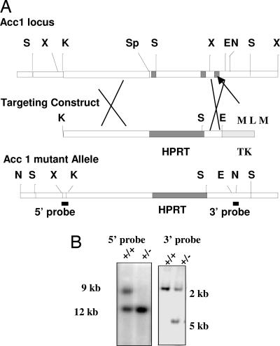
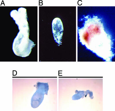
Absence of ACC1 in Mice Results in Early Embryonic Lethality. A mouse ACC1 14-kb genomic clone was isolated by using an Acc1 cDNA probe, and the clone was mapped with restriction enzymes and analyzed by Southern blot as depicted in Fig. 1. Several exonic sequences, including the one containing the Met-Lys-Met motif, were identified and confirmed by DNA sequencing. A 1.5-kb 3′ NotI-XbaI fragment and a 6-kb 5′ KpnI-SpeI fragment were isolated and used to construct a replacement gene-targeting vector (Fig. 1) for gene targeting in ES cells. A correctly targeted clone (Fig. 1B) was microinjected into C57BL/6J mouse blastocysts, which were then implanted into the uterine horns of pseudopregnant female mice. A high percentage in male chimeras, thus generated, was bred with C57BL/6J females to produce Acc1tm1AL heterozygote (herein called Acc1+/–) offspring that were interbred. As shown in Fig. 1, and as was expected, the mRNA level of Acc1 in the white adipose tissue of Acc1+/– mice was half that of the wild type. After analyzing the genomic tail DNA of >300 progenies by Southern blot with both the 5′ and 3′ probes, we were unable to obtain Acc1–/– (homozygous mutant) offspring. The litter sizes were less than average, being six or seven; 35% of the progeny were wild type, and 65% were heterozygous (Table 1). These results demonstrate that the Acc1 mutation causes embryonic lethality in the homozygous state. To characterize the embryonic lethality, we timed the mating of the heterozygotes and genotyped the resulting embryos. By genotyping DNA from embryos at gestation days E11.5 and E12.5, we found that the viable embryos were 35% wild type and 65% heterozygous, indicating that lethality had occurred earlier. At E11.5, we found that, of the 26 embryos, 14 were heterozygotes, 7 were wild type, and 5 were resorbed and disappeared in their degenerating deciduas (Table 1), an indication that the homozygotes had died several days earlier. At E9.5, we were able to recover remains of the homozygotes, and at E8.5, we recovered degenerating embryos from inside the ectoplacental cone (Fig. 2). At E7.5, approximately one-fourth of the embryos were less than approximately one-third the size of their wild-type or heterozygous littermates and did not develop beyond the egg cylinder stage (Fig. 2).
Fig. 1.
Targeted mutation of the Acc1 locus. (A) Strategy used to create the targeted mutation. The exon (dark box) that contains two of the exons upstream the biotin-binding motif (Met-Lys-Met) was replaced with a hypoxanthine phosphoribosyltransferase (HPRT) expression cassette. The 3′ and 5′ probes used for Southern blot analysis are indicated. (B) A typical pattern observed in genotyping by Southern blot analyses of genomic DNA extracted from mouse tails. The DNA was digested with SphI in duplicate. The blots were probed with the 5′ and 3′ probes indicated in A. The presence of only wild-type (+/+) and heterozygous (+/–) genotypes indicated that no homozygous (–/–) mice were born.
TABLE 1. Genotypes of offspring from intercrosses of heterozygous Acc1+/- mice.
| Genotype
|
|||||
|---|---|---|---|---|---|
| Age | +/+ | +/- | -/- | Resorbed | Total |
| 3-4 weeks | 124 | 218 | 0 | NA | 342 |
| E11.5 | 6 | 14 | NA | 5 | 25 |
| E9.5 | NA | NA | NA | 9* | 42 |
NA, not applicable.
Remains of embryos are observed.
Fig. 2.
Developmental abnormalities in E9.5 and E7.5 of ACC1 mutant embryos. Whole embryos at the late head-fold stage (E8.5) and E6.5 were extracted from the uterus of a heterozygous (+/–) female mouse after crossbreeding with a heterozygous male (+/–) and were mounted for morphological study. (A) A wild-type or heterozygous embryo at E8.5. (B) A degenerating Acc1 mutant embryo recovered by dissecting the ectoplacental cone shown in C. (D) A normal, developed embryo at E7.5 compared to a smaller and unorganized embryo (E). The embryos in B and E account for ≈25% of all of the offspring of heterozygous female mice crossbred with heterozygous males.
Regulation of Acc1 mRNA and Protein Synthesis in Acc1+/– Mice. As expected, the Acc1 mRNA level in the white adipose tissue was 2-fold higher in the wild type compared to Acc1+/– mice, confirming that there is only one gene copy in the Acc1+/– mice (Fig. 3A). We prepared hepatocytes from the livers of both wild-type and Acc1+/– mice and examined the level of mRNA in response to hormones. In the hepatocytes of both wild-type and Acc1+/– mice, the level of mRNA was induced after 24 h of treatment with insulin and T3; however, the mRNA level in the wild type continued to be 2-fold higher than in the Acc1+/– mice (Fig. 3B). As for the protein level, as Fig. 3C shows, the phosphorylated ACC was similar in Acc1+/– mice and in the wild type. These results suggest that there is no transcriptional compensation as a result of deleting one copy of the gene but that there was translational and/or proterlytion compensation.
Fig. 3.
Northern and Western blot analyses of total RNA from white adipose tissue and perfused hepatocytes of wild-type and heterozygous mutant Acc1+/–mice. Total RNA from white adipose (A) and hepatocytes (B) of wild-type and mutant Acc+/– mice were subjected to 1% agarose gel in the presence of formalin. The fractionated RNA was transferred to Hybond N (Amersham Pharmacia Biotech). The filters were hybridized with 32P-labeled cDNA of a 362-bp mouse cDNA PCR fragment. To show equal loading, the gel were stained with ethedium bromide to visualize the 28S and 18S rRNA. (C and D) Western blot of liver crude extracts (25 μg) probed with avidin peroxidase and antiphospho-ACC (Ser-79) respectively; lanes: 1, wild type; 2, heterozygous (+/–); M, myosin.
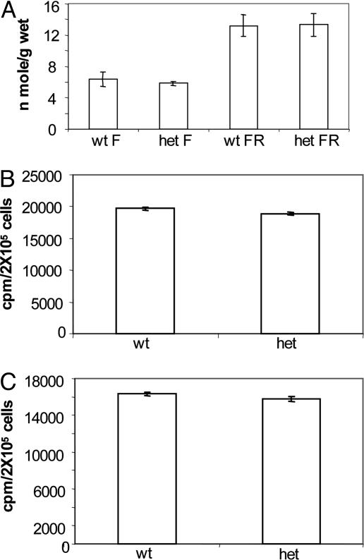
We measured the level of malonyl-CoA in the liver extracts of Acc1+/– mutant mice and compared it to that of their wild-type littermates. In the liver and tissues where ACC1 is highly expressed, the level of malonyl-CoA was similar in both the wild-type and Acc1+/– mutant mice when the mice were fed normal chow (6.35 ± 0.6 and 5.4 ± 0.15 nmol/g wet, for wild-type and heterozygote, respectively, Fig. 4). ACC1 in the liver is known to be down-regulated at the mRNA and protein levels after starvation and up-regulated after refeeding a high-carbohydrate, fat-free diet. We measured the malonyl-CoA level and ACC activity in the livers of the control and Acc1 heterozygote mice after fasting and refeeding with a high-carbohydrate diet. As expected, the level of malonyl-CoA increased by 2-fold in the liver when the mice were starved for 48 h, followed by 48 h of refeeding a high-carbohydrate, fat-free diet (13.2 ± 1.34 compared to 12.48 ± 0.23 nmol/g wet tissue for wild-type and Acc1+/–, respectively). These results may suggest that despite the 2-fold difference in mRNA levels of the wild-type and Acc1+/– mice, there was no obvious difference in the level of the ACC1 protein as determined by Western blot analysis, further suggesting that in the heterozygotes, the translation level is twice that of the wild type. We measured ACC activity and phosphorylation status in the livers under normal feeding conditions. The ACC1 protein was equally expressed and phosphorylated suggesting that, in the Acc1+/– mice, there is compensation at the level of translation. Because malonyl-CoA plays an important role in fatty acid synthesis and oxidation, we measured the levels of acetate incorporation into fatty acids and palmitate oxidation in hepatocyets from wild-type and Acc1+/– mice. The results showed that there were no significant differences in either acetate incorporation into fatty acids or palmitate oxidation between the heterozygote Acc1+/– mice and their wild-type cohorts (Fig. 4).
Fig. 4.
Malonyl-CoA levels and fatty acid oxidation synthesis in hepatocytes of Acc+/– and wild-type mice. (A) The levels of malonyl-CoA in liver extracts of wild-type (wt) and heterozygous (het) mice were determined by the incorporation of [3H]acetyl-CoA into palmitate in the presence of NADPH and highly purified chicken fatty acid synthase. The [3H]palmitic acid synthesized was extracted with petroleum ether, and the radioactivity was measured. The mice were either fed a normal chow (F) or were fasted for 48 h, followed by feeding (RF) a fat-free/high-carbohydrate diet for another 48 h before they were killed. (B) Hepatocytes prepared from perfused livers (2 × 10⁵ cells) were cultured in polylysine-coated bottles with shaking in Krebs-Ringer bicarbonate buffer. Fatty acid oxidation was determined by measuring the 14CO2 generated from the oxidation of [1-14C] palmitate and trapped with benzethonium solution in the center wells. (C) [3H]acetyl-CoA incorporation into total lipids in perfused hepatocytes.
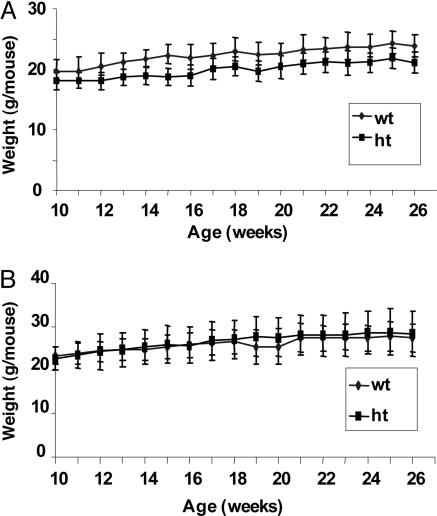
Survival and Body Weight Analysis of Wild-Type and ACC1+/– Mice. Because we found that Acc–/– mice died in utero, we wanted to determine whether deletion of one copy of the Acc1 gene altered survival or body weight. However, in all parameters analyzed, there were no obvious differences between Acc1+/– and their wild-type littermates. Both had similar lifespans and bred normally. We therefore carried out feeding experiments on both male and female wild-type and Acc1+/– mice. Approximately 10-week-old male and female mice were fed normal chow for 27 weeks, and their body weight and food consumption were determined weekly. As shown in Fig. 5A, the average body weight of the males was very similar up to 27 weeks. In females, body weight was slightly lower among heterozygotes than among their wild-type littermates; however, this difference is not statistically significant (Fig. 5B).
Fig. 5.
Body weight of male and female Acc+/– heterozygous and wild-type mice. Ten- to 12-week-old female (A) and male (B) mice were fed normal diets, and their weights were determined weekly for 24 weeks. The data are shown as means ± SD (n = 5).
Discussion
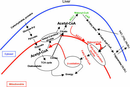
The principal finding of this study is that Acc1 null mice die embryonically, and at E7.5, ≈25% of the embryos did not develop normally. These findings suggest that malonyl-CoA, generated by ACC1, is essential for the early development of the embryos. The presence of intact copies of the ACC2 wild gene did not complement ACC1 function and did not save the mice embryos from failure to develop to the full term. On the other hand, Acc2–/– mutants have interesting phenotypes in their livers and adipose tissues where ACC1 is the dominant isoform that contributes most of the malonyl-CoA. Based on our previous report about the Acc2 knockout studies (4) and our current ACC1 studies, we further confirm our hypothesis that malonyl-CoA, the product of ACC1 and ACC2, exists in two separate compartments in the cell: cytosol and mitochondria (Fig. 6).
Fig. 6.
A schematic model shows that ACC1 and ACC2 are compartmentalized and produce two independent malonyl-CoA Pools. Shown are the biochemical pathways for generating acetyl-CoA and malonyl-CoA, the precursors for de novo fatty acid synthesis and regulators for fatty acid oxidation.
We propose that acetyl-CoA is the key intermediate in the metabolism of glucose, fatty acids, and amino acids. It is generated in the cytosol, mitochondria, and combination thereof (Fig. 6). The pyruvate produced through glycolysis is transported to the mitochondria, where it is converted to acetyl-CoA by the action of pyruvate dehydrogenase. The acetyl-CoA may either be transported to the cytosol through the acetyl-carnitine shuttle pathway or condensed with oxaloacetate to form citrate. The latter is either oxidized through the tricarboxylic acid cycle and the electron transport system to generate energy and CO2 or is transported to the cytosol, where it is cleaved to acetyl-CoA and oxaloacetate through the action of citrate lyase. The acetyl-CoA, thus generated, is the substrate for many synthetic reactions, including the synthesis of malonyl-CoA by the carboxylases, ACC1 and ACC2. Because ACC1 and ACC2 are located respectively at the cytosol and mitochondrial membrane, hence, the malonyl-CoA they generate exists in two independent pools that do not mix: the cytosolic pool, used in fatty acid synthesis, and the mitochondrial pool that regulates carnitine palmitoyltransferase, hence, fatty acid oxidation (Fig. 6). Consequently, lipogenesis and fatty acid oxidation do not occur simultaneously. The newly synthesized fatty acids are spared from oxidation and used in the synthesis of triglycerides (TG) and transported from the liver as very low density lipoproteins (VLDL) to other parts of the body (Fig. 6).
Based on the high homology between ACC1 and ACC2 regarding the phosphorylation sites on Ser 80 and 81 for ACC1, and Ser 219 and 220 for ACC2, we believe that both ACC isoforms are regulated simultaneously by the same insulin-regulated kinases and phosphatases. This tight regulation would prevent a futile cycle of fatty acid synthesis and oxidation under normal physiological conditions. Our studies on both Acc1 and Acc2 knockouts provides convincing evidence that malonyl-CoA, made by ACC2, regulates fatty acid oxidation by regulating the activity of CPT1. Previously, we showed that fatty acid oxidation in hepatocytes increased and triglyceride levels in the livers of Acc2–/– mice decreased despite the fact that there was little change in the overall amount of malonyl-CoA in the livers of these mice compared to wild-type mice (4, 26). We speculated that malonyl-CoA in the liver, which is mostly produced by ACC1 that resides in the cytoplasm (11), is not accessible to CPT1 in the mitochondria and, therefore, does not regulate CPT1 directly. This conclusion is further supported by the recent work of An et al. (32) who reported that despite a 7-fold increase in malonyl-CoA decarboxylase in the cytosol of hepatocytes that significantly decreased the levels of malonyl-CoA and triglyceride, there was a modest change in fatty acid oxidation (32).
In lower eukaryotes, cytosolic ACC, which is the homolog to animal ACC1, is essential to the viability and development of yeast and the embryos of Arabidopsis (24, 33). A null mutation in the acc1 gene of Arabidopsis resulted in embryo lethality, and it was suggested that this lethality is caused by the lack of synthesis of very long-chain fatty acids (33). Long-chain fatty acids, such as palmitate, are abundant in the food we consume; however, very long-chain fatty acids are the products of in vivo elongation where malonyl-CoA is needed to provide the C2 unit for the synthesis of very long-chain fatty acids. Interestingly, in our studies of the knockout of FAS, we found that Fasn–/– mice also die in utero, secondary to embryonic lethality at a much earlier stage of the development of the mouse embryos (34). These results suggested that the transport of fatty acids through the placenta is limited and that there is a dire need for de novo synthesis of long- and very long-chain fatty acids in the embryo. Long-chain fatty acids (C14, C16, and C18), products of the FAS reaction that rely on cytosolic malonyl-CoA-generated ACC1, are required for embryonic development. Here, we expect that Acc1–/– null mutant embryos would die at the same stage or even earlier than the Fasn–/– embryos. However, our current mouse knockout study shows that we could recover embryos at a much later stage than the Fasn knockout. For this reason, we cannot rule out the possibility that the absence of Fasn caused the accumulation of cytosolic malonyl-CoA that can lead to a cytotoxic effect and, ultimately, the death of the embryo at a very early stage.
ACC1 in lipogenic tissues, such as the liver and white adipose, is known to be regulated by diet and hormones. As has always been the case, animals fed a fat-free diet after 48 h of starvation actively synthesize long-chain fatty acids, partially due to an abundance of the substrates, acetyl-CoA and NADPH, generated through glycolysis and activation of ACC1 and ACC2 through their dephosphorylation and citrate activation. Interestingly, the levels of malonyl-CoA were very similar in the livers of both heterozygote and wild-type littermates after the starving/refeeding experiments, which may be due to regulation of ACC1 at the translational level, where the amount of ACC1 protein in Acc1+/– mutants become similar to that of the wild type, or it may result from increased enzymatic activity in the Acc1+/– mutants. In both situations, the activity of ACC1 will be similar. Western blot analysis of proteins from liver protein extracts showed that the amount and phosphorylation status of ACC1 were similar in Acc1+/– mutants and wild type cohorts, confirming and supporting this conclusion (Fig. 3C). In animals, ACC1 is regulated by a variety of complex control mechanisms that include transcription, differential splicing, multiple promoters that respond differently to hormones and nutrients, and mRNA stability, in addition to covalent modifications (35, 36). The similar-fold increases in mRNA level in hepatocytes of wild-type and Acc1+/– mutants, in response to insulin and T3 while maintaining a 2:1 ratio, suggest that under these conditions, there was no compensation at the mRNA level. These results clearly suggest that there is compensation at the translational level of ACC 1 in the Acc1+/– mice that result in equal levels of protein, phosphorylation state, and activity of the enzyme as we demonstrated in liver tissues.
Unlike the Fasn+/– mice, which exhibited haploid insufficiency, Acc+/– mice did not have any obvious phenotype, a fact that was reflected in the similar body weight of both male and female mice and similar malonyl-CoA levels in their livers. Unlike ACC1, FAS is not known to be acutely regulated by posttranslational modification such as phosphorylation/dephosphorylation. Thus, the level of fatty acid synthesis carried out by the FAS enzyme correlates with its abundance. Finally, our previous studies of the ACC2 knockout and our current report support the hypothesis of the distinct roles of ACC1 and ACC2 in animals. ACC1 is essential for the development of mice embryo, and ACC2, through its product, is a regulator of fatty acid oxidation and obesity.
Acknowledgments
We thank Dr. Subrahmanyam S. Chirala, and Jianqiang Mao for discussions and comments. This work was supported in part by the Clayton Foundation for Research and National Institutes of Health Grant GM-63115 (to S.J.W.) and National Institutes of Health Grant HD-42500 (M.M.M.).
Author contributions: L.A.-E., M.M.M., P.K., W.O., T.S., and Z.G. performed research; L.A.-E. and S.J.W. analyzed data; and L.A.-E., M.M.M., and S.J.W. wrote the paper.
Abbreviations: ACC, acetyl-CoA carboxylase; En, embryonic day n; FAS, fatty acid synthase.
References
- 1.Prentki, M., Joly, E., El-Assaad, W. & Roduit, R. (2002) Diabetes 51, S405–S413. [DOI] [PubMed] [Google Scholar]
- 2.Roduit, R., Nolan, C., Alarcon, C., Moore, P., Barbeau, A., Delghingaro-Augusto, V., Przybykowski, E., Morin, J., Masse, F., Massie, B., et al. (2004) Diabetes 53, 1007–1019. [DOI] [PubMed] [Google Scholar]
- 3.McGarry, J. D. & Brown, N. F. (1997) Eur. J. Biochem. 244, 1–14. [DOI] [PubMed] [Google Scholar]
- 4.Abu-Elheiga, L., Matzuk, M. M., Abo-Hashema, K. A. H. & Wakil, S. J. (2001) Science 291, 2613–2616. [DOI] [PubMed] [Google Scholar]
- 5.Lopaschuk, G. & Gamble, J. (1994) Can. J. Physiol. Pharmacol. 72, 1101–1109. [DOI] [PubMed] [Google Scholar]
- 6.Vavvas, D., Apazidis, A., Saha, A. K., Gamble, J., Patel, A., Kemp, B. E., Witters, L. A. & Ruderman, W.B. (1997) J. Biol. Chem. 272, 13255–13261. [DOI] [PubMed] [Google Scholar]
- 7.Alam, N. & Saggerson, E. D. (1998) Biochem. J. 334, 233–241. [DOI] [PMC free article] [PubMed] [Google Scholar]
- 8.Ruderman, N. B., Saha, A. K., Vavvas, D. & Witters, L. A. (1999) Am. J. Physiol. 276, E1–E18. [DOI] [PubMed] [Google Scholar]
- 9.Abu-Elheiga, L., Almarza-Ortega, D. B., Baldini, A. & Wakil, S. J. (1997) J. Biol. Chem. 272, 10669–10677. [DOI] [PubMed] [Google Scholar]
- 10.Ha, J., Lee, J.-K., Kim, K.-S., Witters, L. A. & Kim, K.-H. (1996) Proc. Natl. Acad. Sci. USA 93, 11466–11470. [DOI] [PMC free article] [PubMed] [Google Scholar]
- 11.Abu-Elheiga, L., Brinkley, W. R., Zhong, L., Chirala, S. S., Woldegiorgis, G. & Wakil, S. J. (2000) Proc. Natl. Acad. Sci. USA 97, 1444–1449. [DOI] [PMC free article] [PubMed] [Google Scholar]
- 12.Abu-Elheiga, L., Jayakumar, A., Baldini, A., Chirala, S. S. & Wakil, S. J. (1995) Proc. Natl. Acad. Sci. USA 92, 4011–4015. [DOI] [PMC free article] [PubMed] [Google Scholar]
- 13.Loftus, T. M., Jaworsky, D. E., Frehywot, G. L., Townsend, C. A., Ronnett, G. V., Lane, M. D. & Kuhajda, F. P. (2000) Science 288, 2379–2381. [DOI] [PubMed] [Google Scholar]
- 14.Thampy, K. G. & Wakil, S. J. (1988) J. Biol. Chem. 263, 6447–6453. [PubMed] [Google Scholar]
- 15.Thampy, K. G. & Wakil, S. J. (1988) J. Biol. Chem. 263, 6454–6458. [PubMed] [Google Scholar]
- 16.Kim, K. H., Lopez-Casillas, F., Bai, D. H., Luo, X. & Pape, M. E. (1989) FASEB J. 3, 2250–2256. [DOI] [PubMed] [Google Scholar]
- 17.Mabrouk, G. M., Helmy, I. M., Thampy, K. G. & Wakil, S. J. (1990) J. Biol. Chem. 265, 6330–6338. [PubMed] [Google Scholar]
- 18.Quayle, K. A., Denton, R. M. & Brownsey, R. W. (1993) Biochem. J. 292, 75–84. [DOI] [PMC free article] [PubMed] [Google Scholar]
- 19.Mohamed, A. H., Huang, W. Y., Huang, W., Venkatachalam, K. V. & Wakil, S. J. (1994) J. Biol. Chem. 269, 6859–6865. [PubMed] [Google Scholar]
- 20.Thampy, K. G. (1989) J. Biol. Chem. 264, 17631–17634. [PubMed] [Google Scholar]
- 21.Al-Feel, W., Chirala, S. S. & Wakil, S. J. (1992) Proc. Natl. Acad. Sci. USA 89, 4534–4538. [DOI] [PMC free article] [PubMed] [Google Scholar]
- 22.Lopez-Casillas, F., Bai, D.-H., Luo, X., Kong, I.-S., Hermodson, M. A. & Kim, K.-H. (1988) Proc. Natl. Acad. Sci. USA 85, 5784–5788. [DOI] [PMC free article] [PubMed] [Google Scholar]
- 23.Takai, T., Yokoyama, C., Wada, K. & Tanabe, T. (1988) J. Biol. Chem. 263, 2651–2657. [PubMed] [Google Scholar]
- 24.Hasslacher, M., Ivessa, A., Paltauf, F. & Kohlwein, S. (1993) J. Biol. Chem. 268, 10946–10952. [PubMed] [Google Scholar]
- 25.Al-Feel, W., DeMar, J. C. & Wakil, S. J. (2003) Proc. Natl. Acad. Sci. USA 100, 3095–3100. [DOI] [PMC free article] [PubMed] [Google Scholar]
- 26.Abu-Elheiga, L., Oh, W., Kordari, P. & Wakil, S. J. (2003) Proc. Natl. Acad. Sci. USA 100, 10207–19212. [DOI] [PMC free article] [PubMed] [Google Scholar]
- 27.Oh, W., Abu-Elheiga, L., Kordari, P., Gu, Z., Shaikenov, T., Chirala, S. S. & Wakil, S. J. (2005) Proc. Natl. Acad. Sci. USA 102, 1384–1389. [DOI] [PMC free article] [PubMed] [Google Scholar]
- 28.Matzuk, M. M., Finegold, M., Su, J. G., Hsueh, A. J. & Bradley, A. (1992) Nature 360, 313–319. [DOI] [PubMed] [Google Scholar]
- 29.Neufeld, D.S. (1997) Methods Mol. Biol. 75, 145–151. [DOI] [PubMed] [Google Scholar]
- 30.Hamel, F. G., Bennett, R. G., Upward, J. L. & Duckworth, W. C. (2001) Endocrinology 142, 2702–2706. [DOI] [PubMed] [Google Scholar]
- 31.McGarry, J. D., Stark, M. J. & Foster, D. W. (1978) J. Biol. Chem. 253, 8291–8293. [PubMed] [Google Scholar]
- 32.An, J., Muoio, D. M., Shiota, M., Fujimoto, Y., Cline, G. W., Shulman, G. I., Koves, T. R., Stevens, R., Millington, D. & Newgard, C. B. (2004) Nat. Med. 10, 268–274. [DOI] [PubMed] [Google Scholar]
- 33.Baud, S., Guyon, V., Kronenberger, J., Wuilleme, S., Miquel, M., Caboche, M., Lepiniec, L. & Rochat, C. (2003) Plant J. 33, 75–86. [DOI] [PubMed] [Google Scholar]
- 34.Chirala, S. S., Chang, H., Matzuk, M., Abu-Elheiga, L., Mao, J., Mahon, K., Finegold, M. & Wakil, S. J. (2003) Proc. Natl. Acad. Sci. USA 100, 6358–6363. [DOI] [PMC free article] [PubMed] [Google Scholar]
- 35.Kim, K.-H. (1997) Annu. Rev. Nutr. 17, 77–99. [DOI] [PubMed] [Google Scholar]
- 36.Mao, J., Chirala, S. S. & Wakil, S. J. (2003) Proc. Natl. Acad. Sci. USA 100, 7515–7520. [DOI] [PMC free article] [PubMed] [Google Scholar]


