



ABSTRACT
Background
Cerebral autosomal dominant arteriopathy with subcortical infarcts and leukoencephalopathy (CADASIL) is the most common hereditary cerebral small vessel disease (CSVD). Intracerebral hemorrhage (ICH) is reported to be increasing in CADASIL patients from areas where the p.R544C mutation is prevalent (e.g., Jeju and Chinese Taiwan) but is rare in Caucasians. We attempted to determine potentially genetic, clinical, and/or neuroimaging risk factors for ICH in Chinese CADASIL patients.
Methods
This retrospective observational study included 190 patients with CADASIL and 179 patients with sporadic CSVD. NOTCH3 genotypes as well as clinical and neuroimaging manifestations were compared between ICH and non‐ICH patients, and both logistic regression and a subgroup analysis were used to adjust for confounding factors.
Results
Of 190 CADASIL patients in the present study, 43 patients (22.6%) had ICH lesions. A total of 62 ICH lesions were recorded. Thalamic ICH lesions were the most common (40.3%), followed by basal ganglia (32.3%) and temporal lobe (8.1%). In subgroup analysis, the ICH group had a higher prevalence of CMB than the non‐ICH group, including in the basal ganglia region (58.3% vs. 23.3%, p = 0.037) and thalamus (75.0% vs. 38.3%, p = 0.020). The p.R544C mutation (aOR 6.390; 95% CI, 1.308–31.225; p = 0.022) and total SVD score (aOR 1.731; 95% CI, 1.003–2.990; p = 0.049) were independently associated with ICH.
Conclusions
ICH is a common clinical manifestation of CADASIL patients in southeast coastal China. Hypertension, total SVD score, and the p.R544C mutation are associated with CADASIL ICH.
Trial Registration
ClinicalTrials.gov identifier: (NCT04318119)
Keywords: CADASIL, hypertension, intracerebral hemorrhage, p.R544C mutation, risk factors
Abbreviations
- CADASIL
Cerebral autosomal dominant arteriopathy with subcortical infarcts and leukoencephalopathy
- CMBs
cerebral microbleeds
- CSVD
cerebral small vessel disease
- CT
computed Tomography
- DPWM
deep periventricular white matter
- EPVS
enlarged perivascular space
- GOM
granular osmiophilic material
- ICH
intracerebral hemorrhage
- MRI
magnetic resonance imaging
- N3ECD
Notch3 extracellular domain
- SWI
susceptibility‐weighted imaging
- T2*‐GRE
T2*‐weighted gradient‐recalled echo
- WMH
white matter hyperintensity
1. Introduction
Cerebral autosomal dominant arteriopathy with subcortical infarcts and leukoencephalopathy (CADASIL) is the most common hereditary cerebral small vessel disease (CSVD), caused by mutations in the NOTCH3 gene [1]. The classical clinical manifestations of CADASIL are recurrent ischemic stroke, cognitive impairment, mood disorder, and migraine [2]. Intracerebral hemorrhage (ICH) was previously considered a rare clinical manifestation of CADASIL. In Caucasian patients with CADASIL, intracerebral hemorrhage (ICH) has primarily been reported in case reports [3, 4, 5, 6], with a prevalence of 2%–3.7% [7, 8]. Whereas the prevalence of ICH in East Asian patients with CADASIL has been reported at 17%–42% [9, 10, 11].
In East Asia, the p.R544C mutation accounts for 70.5%–94.4% of CADASIL cases [10, 12, 13]. However, the p.R544C mutation is unfound in several Caucasian CADASIL studies, totaling more than 800 patients [14, 15, 16]. Recently, more studies have focused on hemorrhagic phenotypes in CADASIL. The cysteine‐sparing NOTCH3 p.R75P mutation is associated with Pro‐Hemorrhagic CADASIL [17]. The difference in the prevalence of ICH between Caucasian and East Asian patients with CADASIL may be attributable to genotype differences. The association of the p.R544C mutation and ICH is yet unclear. ICH in CADASIL mostly occurs in patients accompanied by hypertension, and it is more likely to occur in common sites of hypertensive ICH [18]. It is currently believed that ICH in CADASIL is generally attributed to hypertension [9, 10]. Nonetheless, the prevalence of ICH in non‐hypertensive CADASIL patients still remains unclear.
The present observational study was based on the Cerebral Small Vessel Disease Registry Study (NCT04318119). We aimed to assess potential risk factors for ICH in Chinese CADASIL patients and reveal the imaging characteristics of ICH in CADASIL.
2. Method
2.1. Participants, Standard Protocol Approvals, and Consent
This study was based on the prospective Cerebral Small Vessel Disease Registry Study (NCT04318119) cohort that enrolled participants with neuroimaging features of CSVD in the First Affiliated Hospital of Fujian Medical University. The inclusion criteria are as follows: CSVD burden score > 2 points, with at least one of the following manifestations: family history of stroke or cognitive impairment, young stroke (< 50 years old), no risk factors for cerebrovascular disease, or special imaging signs such as temporal pole lesions. A total of 455 participants underwent NOTCH3 gene testing. CADASIL was diagnosed by identifying a cysteine‐altering mutation in the NOTCH3 gene (n = 190). Participants with suspected sporadic CSVD were enrolled if they had at least two MRI markers of CSVD and underwent NOTCH3 gene testing to exclude the most common hereditary forms of CSVD. We then excluded patients with hereditary cerebral small vessel disease as much as possible based on clinical and neuroradiological features. They were confirmed to have no family history of ICH through interviews and medical record reviews to rule out potential hereditary ICH. Sporadic CSVD patients were matched with CADASIL patients based on the severity of WMH (Figure 1). Informed consent was obtained from all the patients and/or their relatives.
FIGURE 1.
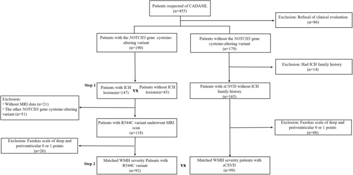
Flowchart of participant recruitment: CSVD, cerebral small vessel disease; ICH, Intracerebral hemorrhage; MRI, magnetic resonance imaging; sCSVD, sporadic cerebral small vessel disease; WMH, white matter hyperintensity.
The study was approved by the Ethics Committee of the First Affiliated Hospital of Fujian Medical University. The study was conducted in accordance with the principles of the Declaration of Helsinki.
2.2. Clinical Assessment
The following clinical information was collected for patients with CADASIL: sex; age at examination; symptoms (migraine, migraine with aura, psychiatric disorders, ICH and cognitive impairment); cerebrovascular risk factors, including hypertension, diabetes, dyslipidemia, and smoking. The diagnostic criteria for hypertension were a systolic blood pressure ≥ 140 mmHg and/or a diastolic blood pressure ≥ 90 mmHg measured three times independently and/or the use of antihypertensive medication. Regarding the diagnosis of ICH patients without hypertension, we reviewed old medical records of 13 patients with ICH without hypertension, and reviewed their blood pressure values. The blood pressure of 10 patients had normal values, while the blood pressure values of 3 patients were unavailable. Details about the definition of the clinical symptoms and cardiovascular risk factors can be found in the Table S1.
2.3. Brain MRI Acquisition and Analysis
Among the 190 patients with CADASIL, 128 patients had undergone a 3.0 Tesla scanner. T1‐weighted imaging (T1WI), T2‐weighted imaging (T2WI), fluid attenuation inversion recovery (FLAIR), and susceptibility‐weighted imaging (SWI) were obtained. One experienced neuroradiologist, blinded to clinical data, reviewed all the images. WMH, Lacunes of presumed vascular origin (abbreviated as lacunes in the following), CMBs, and perivascular space (PVS) were identified according to the neuroimaging standards for research into small vessel disease [19]. The severity of WMH in the periventricular and deep white matter was evaluated by the Fazekas scale [20]. The number of lacunes in the cerebral lobes (including frontal, parietal, occipital, and temporal lobes), infratentorial, and basal ganglia was counted by using FLAIR images [21]. PVS at the basal ganglia and centrum semiovale was assessed on axial T2WI with a 4‐point rating scale (grade 0 = no PVS, grade 1 = < 11 PVSs, grade 2 = 11–20 PVSs, grade 3 = 21–40 PVSs, grade 4 = more than 40 PVSs) [22]. CMBs were evaluated by the Microbleed Anatomical Rating Scale (MARS) [23]. We assess the global burden of SVD by counting the presence of each of the four MRI features [24].
2.4. Genetic Analysis
DNA was extracted from patient peripheral blood samples. PCR and Sanger sequencing were performed on exons 3–6, 11–12, and 18–19 of the NOTCH3 gene. Patients who tested negative for NOTCH3 gene hotspot mutations but had a CADASIL score ≥ 15 [25] received testing of all 33 exons of the NOTCH3 gene using Sanger sequencing or targeted next‐generation sequencing. Missense mutations that lead to the gain or loss of a cysteine residue in one of the 34 epidermal growth factor‐like repeat (EGFR) domains of the notch3 protein were identified (Table S2). Additionally, genotyping of ε2/ε3/ε4 variants in the APOE gene was conducted.
2.5. Statistical Analysis
Variables conforming to a normal distribution were expressed as mean ± SD, and independent sample Student's t‐tests were used. Variables that did not conform to a normal distribution were expressed using the median (interquartile range), and the Mann–Whitney U test was used. Count data were expressed as percentages, and comparisons between two groups were performed using the chi‐square test or a continuously corrected chi‐square test. We applied logistic regression models to estimate the impact of the p.R544C mutation on ICH and calculated the odds ratio (OR) and 95% confidence interval (CI). Age, sex, hypertension, lacunes, and total SVD score were adjusted in the multivariate analysis [9, 26, 27]. All statistical analyses were performed in SPSS (version 25.0). Two‐sided p < 0.05 was considered statistically significant.
3. Results
3.1. Comparison of Demographic and Cerebrovascular Risk Factors Between ICH Group and Non‐ICH Group
Patients in the ICH group were older than those in the non‐ICH group (59.4 ± 8.4 vs. 55.2 ± 10.9, p = 0.020). The ICH group had a significantly higher prevalence of hypertension (69.8% vs. 30.6%, p < 0.001) and diabetes (25.6% vs. 8.2%, p = 0.002) compared to the non‐ICH group. Additionally, the proportion of patients with the p.R544C mutation was higher in the ICH group than in the non‐ICH group (93.0% vs. 61.9%, p < 0.001). There were no significant differences between the two groups in antithrombotic therapy (35.9% vs. 35.7%, p = 0.901) and APOE genotype. There was no significant difference in the prevalence of ischemic stroke (39.5% vs. 52.4%, p = 0.138), cognitive dysfunction (81.4% vs. 76.2%, p = 0.473), psychiatric disorders (16.3% vs. 17.0%, p = 0.911), and migraine (7.0% vs. 10.2%, p = 0.734) between the ICH group and the non‐ICH group (Table 1).
TABLE 1.
Comparison of demographic and cerebrovascular risk factors in CADASIL patients with and without ICH lesions.
| ICH (N = 43) | Non‐ICH (N = 147) | p | |
|---|---|---|---|
| Male, n (%) | 26 (60.5) | 79 (53.7) | 0.435 |
| Age at examination, mean (SD), y a | 59.4 (8.4) | 55.2 (10.9) | 0.020 |
| Proband, n (%) | 39 (90.7) | 127 (86.4) | 0.455 |
| p.R544C mutation, n (%) | 40 (93.0) | 91 (61.9) | < 0.001 |
| Vascular risk factors, n (%) | |||
| Hypertension | 30 (69.8) | 45 (30.6) | < 0.001 |
| Diabetes | 11 (25.6) | 12 (8.2) | 0.002 |
| Dyslipidemia | 27 (62.8) | 72 (49.0) | 0.111 |
| Ever‐smoking | 22 (51.2) | 54 (36.7) | 0.089 |
| Alcohol consumption | 14 (32.6) | 40 (27.2) | 0.494 |
| Family history, n (%) | 35 (81.4) | 99 (67.3) | 0.076 |
| Clinical manifestation | |||
| Ischemic stroke | 17 (39.5) | 77 (52.4) | 0.138 |
| Cognitive dysfunction | 35 (81.4) | 112 (76.2) | 0.473 |
| Psychiatric disorders | 7 (16.3) | 25 (17.0) | 0.911 |
| Migraine | 3 (7.0) | 15 (10.2) | 0.734 b |
| APOE genotyping, n (%) | |||
| APOE ε2/ε3 | 4 (9.3) | 16 (10.9) | 0.804 c |
| APOE ε2/ε4 | 0 (0) | 1 (0.7) | |
| APOE ε3/ε3 | 33 (76.7) | 110 (74.8) | |
| APOE ε3/ε4 | 5 (11.6) | 19 (12.9) | |
| APOE ε4/ε4 | 1 (2.3) | 1 (0.7) | |
| Antithrombotic drug, n (%) | 15 (35.7) | 46 (35.9) d | 0.901 |
Note: Data are presented as mean (standard deviation) or number (percent).
Abbreviations: APOE ε2/ε3, apolipoprotein E ε2/ε3 allele; APOE ε2/ε4, apolipoprotein E ε2/ε4 allele; APOE ε3/ε3, apolipoprotein E ε3/ε3 allele; APOE ε3/ε4, apolipoprotein E ε3/ε4 allele; APOE ε4/ε4, apolipoprotein E ε4/ε4 allele; ICH, intracerebral hemorrhage.
Age at examination was defined by the time of data collection.
Using Chi‐squared test with Yates' continuity correction.
Using Fisher's exact test.
128 patients in the non‐ICH group provided information on the use of antithrombotic medications.
In subgroup analysis, we excluded patients with hypertension. The ICH group had a significantly higher prevalence of diabetes than the non‐ICH group (30.8% vs. 4.9%, p = 0.006). The ICH group also had a higher carriage rate of the p.R544C mutation than the non‐ICH group (84.6% vs. 51.0%, p = 0.022) (Table S3).
3.2. Comparison of Neuroimaging Characteristics Between ICH Group and Non‐ICH Group
A total of 128 participants had heme‐sensitive MRI sequences. The ICH group had a higher incidence of CMBs in brain lobes (78.9% vs. 45.6%, p < 0.001), brainstem (68.4% vs. 36.7%, p = 0.001), cerebellum (52.6% vs. 20.0%, p < 0.001), basal ganglia region (78.9% vs. 27.8%, p < 0.001), thalamus (86.8% vs. 46.7%, p < 0.001), and deep periventricular white matter (DPWM) (26.3% vs. 11.1%, p = 0.030) than the non‐ICH group. The ICH group had a higher prevalence of lacunes than the non‐ICH group (97.4% vs. 80.0%, p = 0.012). Compared to the non‐ICH group, the ICH group had a higher total SVD score (median [IQR]; 4 [3–4] vs. 3 [2–4] p = 0.002) (Table 2).
TABLE 2.
Comparison of CSVD MRI Markers in CADASIL patients with and without ICH Lesions.
| ICH (n = 38) | Non‐ICH (n = 90) | p | |
|---|---|---|---|
| Presence of CMBs, n (%) | |||
| Lobar | 30 (78.9) | 41 (45.6) | < 0.001 |
| Brain stem | 26 (68.4) | 33 (36.7) | 0.001 |
| Cerebellum | 20 (52.6) | 18 (20.0) | < 0.001 |
| Basal ganglia region | 30 (78.9) | 25 (27.8) | < 0.001 |
| Thalamus | 33 (86.8) | 42 (46.7) | < 0.001 |
| Callosum | 1 (2.6) | 0 (0) | 0.297 b |
| DPWM | 10 (26.3) | 10 (11.1) | 0.030 |
| Fazekas scale of WHM, median (IQR) | |||
| Periventricular | 2.5 (2–3) | 2 (2–3) | 0.545 |
| Deep | 3 (2–3) | 2 (2–3) | 0.229 |
| Total score | 5 (4–6) | 4.5 (3.75–6) | 0.336 |
| Lacunes | |||
| Number of lacunes, median (IQR) | 7 (3–12.25) | 6.5 (1–16) | 0.712 |
| Moderate‐to‐severe lacunes, n (%) a | 22 (57.9) | 53 (58.9) | 0.917 |
| Presence of lacunes, n (%) | |||
| Whole Brain | 37 (97.4) | 72 (80.0) | 0.012 |
| Lobar | 33 (86.8) | 65 (72.2) | 0.074 |
| Subtentorial | 8 (23.7) | 20 (22.2) | 0.857 |
| Basal ganglia | 30 (78.9) | 55 (61.1) | 0.051 |
| Moderate‐to‐severe EPVS, n (%) | |||
| Basal ganglia | 29 (76.3) | 59 (65.6) | 0.230 |
| Centrum semiovale | 21 (55.3) | 60 (66.7) | 0.221 |
| Total SVD score, median (IQR) | 4 (3–4) | 3 (2–4) | 0.002 |
Note: Data are presented as number (percent) or median (interquartile range).
Abbreviations: CMBs, cerebral microbleeds; CSVD, cerebral small vessel disease; DPWM, deep periventricular white matter; EPVS, enlarged perivascular space; EPVS, enlarged perivascular space; ICH, intracerebral hemorrhage; MRI, magnetic resonance imaging; NA, not applicable; WHM, white matter hyperintensity.
The grades of the lacunes were grouped as being absent, mild (1–3), moderate (4–10), or severe (> 10). Moderate‐to‐severe lacunes refer to the number of lacunes more than 4.
Using Fisher's exact test.
In subgroup analysis, we excluded patients with hypertension. The ICH group had a higher incidence of CMBs in the basal ganglia region (58.3% vs. 23.3%, p = 0.037) and thalamus (75.0% vs. 38.3%, p = 0.020) than the non‐ICH group (Table S4).
3.3. Factors Associated With ICH in Patients With CADASIL
In model 1, after adjusting for age, hypertension, and diabetes, the p.R544C mutation (adjusted odds ratio [aOR] 6.660; 95% CI, 1.383–32.080; p = 0.018) and hypertension (aOR 2.485; 95% CI, 1.023–6.038; p = 0.045) were independently associated with ICH. In model 2, after adjusting for age, hypertension, diabetes, and Presence of lacunes in the whole brain, the p.R544C mutation (aOR 7.240; 95% CI, 1.496–35.033; p = 0.014) was independently associated with ICH. In model 3, after adjusting for age, hypertension, diabetes, and total SVD score, the p.R544C mutation (aOR 6.390; 95% CI, 1.308–31.225; p = 0.022) and total SVD score (aOR 1.731; 95% CI, 1.003–2.990; p = 0.049) were independently associated with ICH (Table 3).
TABLE 3.
Factors Associated with ICH in Patients with Cerebral Autosomal Dominant Arteriopathy with Subcortical Infarcts and Leukoencephalopathy.
| All patients (N = 128) | p | |
|---|---|---|
| OR (95% CI) | ||
| Model 1 | ||
| Age at examination, y | 1.026 (0.982–1.072) | 0.259 |
| p.R544C mutation | 6.660 (1.383–32.080) | 0.018 |
| Hypertension | 2.485 (1.023–6.038) | 0.045 |
| Diabetes | 2.701 (0.945–7.721) | 0.064 |
| Model 2 | ||
| Age at examination, y | 1.022 (0.976–1.070) | 0.358 |
| p.R544C mutation | 7.240 (1.496–35.033) | 0.014 |
| Hypertension | 2.345 (0.948–5.801) | 0.065 |
| Diabetes | 2.456 (0.838–7.197) | 0.101 |
| Presence of lacunes in whole brain | 6.935 (0.827–58.169) | 0.074 |
| Model 3 | ||
| Age at examination, y | 1.013 (0.968–1.061) | 0.576 |
| p.R544C mutation | 6.390 (1.308–31.225) | 0.022 |
| Hypertension | 2.164 (0.861–5.439) | 0.101 |
| Diabetes | 2.464 (0.833–7.289) | 0.103 |
| Total SVD score | 1.731 (1.003–2.990) | 0.049 |
Abbreviations: CI, confidence interval; CSVD, cerebral small vessel disease; ICH, intracerebral hemorrhage; MRI, magnetic resonance imaging; OR, odds ratio.
3.4. ICH Characteristics of Patients With CADASIL
Of the 43 patients with CADASIL who had ICH lesions, 40 (93.0%) patients harbored the p.R544C mutation, and 3 (7.0%) patients harbored the other mutations (p.R182C, p.C457S, p.R607C). 10 of the 43 patients had two or more ICH lesions, and all of them harbored the p.R544C mutation. Among the 190 CADASIL patients in the cohort, 21 had ICH as their first clinical manifestation (Table S5). A total of 62 ICH lesions were recorded, of which 27 were asymptomatic. The most common asymptomatic ICH lesions were in the basal ganglia (37.0%), followed by the thalamus (29.6%), temporal lobe (14.8%), frontal lobe I (7.4%), occipital lobe (3.7%), brainstem (3.7%), and periventricular (3.7%). According to brain regions, deep ICH lesions were the most common (74.2%), followed by the cerebral lobe (16.1%), and the least common was infratentorial (9.7%). According to the specific location, thalamic ICH lesions were the most common (40.3%), followed by basal ganglia (32.3%), temporal lobe (8.1%), cerebellum (4.8%), brainstem (4.8%), parietal lobe (3.2%), frontal lobe (3.2%), occipital lobe (1.6%), and paraventricular (1.6%) (Table S6). For patients without hypertension, thalamic ICH lesions were the most common (35.3%), followed by basal ganglia (23.5%), temporal lobe (5.9%), cerebellum (5.9%), brainstem (5.9%), parietal lobe (5.9%), frontal lobe (11.8%), occipital lobe (5.9%), and paraventricular (0%) (Table S7).
We enrolled patients with sporadic CSVD who had similar cerebrovascular disease prevalence and white matter hyperintensity severity. Patients with the p.R544C mutation had a higher prevalence of ICH than patients with sporadic CSVD (35.9% vs. 18.2%, p = 0.006) (Table 4).
TABLE 4.
Comparison of ICH prevalence between CADASIL patients with p.R544C mutation and sCSVD patients.
| Variable | p.R544C mutation (N = 92) | sCSVD (N = 99) | p |
|---|---|---|---|
| Male, n (%) | 50 (54.3) | 64 (64.6) | 0.147 |
| Age of examination, mean (SD), y a | 60.3 (9.6) | 63.6 (10.4) | 0.025 |
| Vascular risk factors, n (%) | |||
| Hypertension | 56 (60.9) | 69 (69.7) | 0.200 |
| Diabetes mellitus | 16 (17.4) | 19 (19.2) | 0.748 |
| Dyslipidemia | 56 (60.9) | 53 (55.2) b | 0.432 |
| Smoking | 39 (42.4) | 40 (40.4) | 0.781 |
| Alcohol consumption | 26 (28.3) | 27 (27.6) c | 0.913 |
| Intracerebral hemorrhage, n (%) | 33 (35.9) | 18 (18.2) | 0.006 |
| Family history of ICH, n (%) | 17 (18.5) | 0 (0) | < 0.001 |
| Fezkas score, median (IQR) | 5 (4–6) | 5 (4–6) | 0.191 |
| Deep Fazekas scale, n (%) | |||
| 2 score | 40 (43.5) | 52 (52.5) | 0.211 |
| 3 score | 52 (56.5) | 47 (47.5) | 0.211 |
| Periventricular Fazekas scale, n (%) | |||
| 2 score | 35 (38.0) | 46 (46.5) | 0.239 |
| 3 score | 57 (62.0) | 53 (53.5) | 0.239 |
Note: Data are presented as mean (standard deviation), median (interquartile range) or number (percent).
Abbreviations: ICH, intracerebral hemorrhage; sCSVD, sporadic cerebral small vessel disease.
Age at examination was defined by the time of data collection.
3 patients with sCSVD missing blood lipid data.
1 patients with CSVD are missing alcohol consumption data.
4. Discussion
The present study demonstrated that up to 22.6% (43 of the 190) CADASIL patients had ICH. There were three major findings from our study. First, in southeastern coastal China, ICH is a common clinical presentation in CADASIL patients. Second, we mainly revealed the association between the p.R544C mutation and ICH. This novel cysteine‐altering NOTCH3 genotype–phenotype association enriches the concept of pro‐hemorrhagic CADASIL. Third, hypertension and total SVD score were associated with ICH lesions.
We found that 22.6% of patients with CADASIL had ICH lesions in southeastern coastal China. Similarly, ICH was identified in 21.3% of the Chinese Taiwan patients with CADASIL [10]. In a study from Korea, there were 17% of patients with CADASIL who had ICH [9]. ICH is a common clinical manifestation of CADASIL in East Asia. Our study suggested that physicians should consider the possibility of CADASIL even in patients with ICH. Interestingly, all these studies came from areas where the p.R544C variant was prevalent. However, a study of 214 Chinese patients with CADASIL found that the prevalence of ICH was only 3%, which may be related to the fact that the p.R544C mutation accounted for only 15.48% in the study [28]. The p.R544C mutation, an East Asian‐specific genotype [29] was previously considered a potential risk mutation for ICH. However, a clear association has not been established, possibly due to the insufficient number of non‐p.R544C mutation patients in Chinese Taiwan and Jeju Island, which makes it difficult to observe this association.
Studies have revealed that the phenotype of CADASIL is associated with the NOTCH3 gene genotype [30, 31, 32]. The R544C mutation is located at the boundary of EGFR 13 and EGFR 14, distinguishing it from other NOTCH3 mutations [17]. CADASIL mutations result in the loss or gain of a cysteine residue, causing an odd number of cysteine residues within an EGF domain [33]. Previous research suggested that the deposition of NOTCH3 extracellular domain (N3ECD) was due to unpaired sulfhydryl groups [34]. The NOTCH3 p.R544C mutation of cysteine residue between EGFR 13 and EGFR 14 could have a partially free cysteine thiol leading to the acquisition of the aggregation property, which was lower than that of the conventional mutation with a completely free cysteine thiol [17]. In Alzheimer's disease and cerebral amyloid angiopathy, soluble amyloid‐β oligomers inflict greater damage on cerebral vascular walls compared to insoluble amyloid‐β aggregates [35]. Given the lower aggregation property in patients with the p.R544C mutation, Soluble or non‐aggregated forms of N3ECD in the extracellular matrix may exert greater toxicity on vascular cells than aggregated forms, potentially resulting in microvascular rupture. We speculate that the hemorrhagic phenotype of patients with the p.R544C mutation is related to the unique EGFR domain where this mutation is located.
Consistent with previous study [10], we found that hypertension is a risk factor for ICH in CADASIL. Hypertension as a risk factor for spontaneous ICH [36, 37] is an important modifier for the disease severity of CADASIL patients [38]. A key pathological hallmark of hypertension is structural and functional changes in small arteries [39]. The NOTCH3 mutation typically causes damage to small arterial smooth muscle cells [40]. Several studies of patients hospitalized with a diagnosis of ICH have reported that antihypertensive therapy reduces the recurrence rate of ICH [41, 42, 43]. We found that CADASIL ICH lesions are mainly located in the thalamus, followed by the basal ganglia. Consistent with our study, ICH in CADASIL mostly occurred in deep brain regions including the thalamus and basal ganglia [10]. Our study showed that the thalamus was also the most common location of ICH, followed by the basal ganglia for CADASIL patients without hypertension. The common sites of CADASIL ICH are similar to those of hypertensive ICH, both of which involve deep perforating arterioles, suggesting that their downstream pathogenic mechanisms may be similar. We speculated that CADASIL mutations may damage deep perforating arterioles similarly to hypertension. Given that both hypertension and the NOTCH3 mutation damage deep perforating arterioles, blood pressure control may need to be more intensive in CADASIL patients with ICH.
The present study found a higher total SVD score in the ICH group than in the non‐ICH group. Consistent with our study, a study from Chinese Taiwan also found it [10]. Patients with a higher SVD score have a greater risk of ICH and should be more cautious in taking antithrombotic or anticoagulation measures. A study revealed that CADASIL patients with ICH had worse outcomes than those with ischemic stroke [11]. The Rotterdam study found that participants with a higher SVD score had a much greater risk for stroke, dementia, and mortality during follow‐up [44]. CADASIL patients with ICH probably had a greater burden of vasculopathy.
Our study has limitations that should be considered when interpreting the findings. First, the estimation of the prevalence of hypertension and ICH in CADASIL is suboptimal, owing to the retrospective study design. Prospective studies would be needed to establish causal relationships between the p.R544C mutation and ICH. Second, the two‐stage gene sequencing used in the present study could have missed the patients in the first step, although it should be noted that we examined all NOTCH3 exons for those patients whose CADASIL scale score was over 15. In addition, 96.8% of the CADASIL patients in our study were diagnosed by the first step, a finding consistent with previous studies [28]. Finally, not all CADASIL patients underwent the heme‐sensitive sequences, which could have introduced bias.
In conclusion, ICH is a common clinical manifestation of CADASIL patients in southeast coastal China. Hypertension, total SVD score, and the p.R544C mutation are associated with CADASIL ICH.
Author Contributions
Fangwei Hu: writing – original draft, data curation, visualization, methodology, investigation. Weijie Xie: conceptualization, data curation, writing – review and editing. Mengting Fan: data curation, investigation. Yuanrong Wang: data curation, investigation. Shuyan Xu: investigation, data curation. Wenxin Qiu: investigation, data curation. Tao Yang: investigation, data curation. Huimin Lv: data curation. Huiqing Huang: investigation. Yijia Wu: investigation. Ying Fu: conceptualization, methodology. Bin Cai: conceptualization, methodology, software, resources, supervision, writing – review and editing.
Conflicts of Interest
The authors declare no conflicts of interest.
Supporting information
Data S1.
Acknowledgments
We thank the study participants and their families for their Cooperation.
Funding: This study was supported by grant No. 82471299 (B.C.) and 82071277 (B.C.) from the Natural Science Foundation of China, grant No. 2023Y9039 (B.C.) and 2018Y9084 (B.C.) Joint Funds for the Innovation of Science and Technology, Fujian Province.
Fangwei Hu, Weijie Xie and Mengting Fan are joint first authors.
Bin Cai and Ying Fu are the co‐corresponding authors.
Data Availability Statement
The data that support the findings of this study are available from the corresponding author upon reasonable request. The data are not publicly available due to privacy or ethical restrictions.
References
- 1. Chabriat H., Joutel A., Dichgans M., Tournier‐Lasserve E., and Bousser M. G., “Cadasil,” Lancet Neurology 8 (2009): 643–653. [DOI] [PubMed] [Google Scholar]
- 2. Chabriat H., Joutel A., Tournier‐Lasserve E., and Bousser M. G., “CADASIL: Yesterday, Today, Tomorrow,” European Journal of Neurology 27 (2020): 1588–1595. [DOI] [PubMed] [Google Scholar]
- 3. Maclean A. V., Woods R., Alderson L. M., et al., “Spontaneous Lobar Haemorrhage in CADASIL,” Journal of Neurology, Neurosurgery, and Psychiatry 76 (2005): 456–457. [DOI] [PMC free article] [PubMed] [Google Scholar]
- 4. Koutroulou I., Karapanayiotides T., Grigoriadis N., and Karacostas D., “CADASIL Presenting With Spontaneous Intracerebral Hemorrhage: Report of a Case and Description of the First Family in Northern Greece,” Hippokratia 20 (2016): 76–79. [PMC free article] [PubMed] [Google Scholar]
- 5. Pradotto L., Orsi L., Daniele D., et al., “A New NOTCH3 Mutation Presenting as Primary Intracerebral Haemorrhage,” Journal of the Neurological Sciences 315 (2012): 143–145. [DOI] [PubMed] [Google Scholar]
- 6. Mehta S., Mehndiratta P., and Sila C. A., “Spontaneous Cerebellar Hemorrhage Associated With a Novel Notch3 Mutation,” Journal of Clinical Neuroscience 20 (2013): 1034–1036. [DOI] [PubMed] [Google Scholar]
- 7. Nannucci S., Rinnoci V., Pracucci G., et al., “Location, Number and Factors Associated With Cerebral Microbleeds in an Italian‐British Cohort of CADASIL Patients,” PLoS One 13 (2018): e0190878. [DOI] [PMC free article] [PubMed] [Google Scholar]
- 8. Mönkäre S., Kuuluvainen L., Schleutker J., Myllykangas L., and Pöyhönen M., “Clinical Features and Spectrum of NOTCH3 Variants in Finnish Patients With Cerebral Autosomal Dominant Arteriopathy With Subcortical Infarcts and Leukoencephalopathy (CADASIL),” Acta Neurologica Scandinavica 146 (2022): 643–651. [DOI] [PMC free article] [PubMed] [Google Scholar]
- 9. Lee J. S., Ko K., Oh J. H., et al., “Cerebral Microbleeds, Hypertension, and Intracerebral Hemorrhage in Cerebral Autosomal‐Dominant Arteriopathy With Subcortical Infarcts and Leukoencephalopathy,” Frontiers in Neurology 8 (2017): 203. [DOI] [PMC free article] [PubMed] [Google Scholar]
- 10. Liao Y. C., Hu Y. C., Chung C. P., et al., “Intracerebral Hemorrhage in Cerebral Autosomal Dominant Arteriopathy With Subcortical Infarcts and Leukoencephalopathy: Prevalence, Clinical and Neuroimaging Features and Risk Factors,” Stroke 52, no. 3 (2021): 985–993, 10.1161/STROKEAHA.120.030664. [DOI] [PubMed] [Google Scholar]
- 11. Chen C. H., Tang S. C., Cheng Y. W., et al., “Detrimental Effects of Intracerebral Haemorrhage on Patients With CADASIL Harbouring NOTCH3 R544C Mutation,” Journal of Neurology, Neurosurgery, and Psychiatry 90 (2019): 841–843. [DOI] [PMC free article] [PubMed] [Google Scholar]
- 12. Liao Y. C., Hsiao C. T., Fuh J. L., et al., “Characterization of CADASIL Among the Han Chinese in Taiwan: Distinct Genotypic and Phenotypic Profiles,” PLoS One 10 (2015): e0136501. [DOI] [PMC free article] [PubMed] [Google Scholar]
- 13. Choi J. C., Lee J. S., and Kim K., “Importance of CADASIL Research in Jeju: A Review and Update on Epidemiology, Diagnosis, and Clinical Spectrum,” Journal of Medicine and Life Science 17 (2020): 65–73. [Google Scholar]
- 14. Opherk C., Peters N., Herzog J., Luedtke R., and Dichgans M., “Long‐Term Prognosis and Causes of Death in CADASIL: A Retrospective Study in 411 Patients,” Brain 127 (2004): 2533–2539. [DOI] [PubMed] [Google Scholar]
- 15. Adib‐Samii P., Brice G., Martin R. J., and Markus H. S., “Clinical Spectrum of CADASIL and the Effect of Cardiovascular Risk Factors on Phenotype: Study in 200 Consecutively Recruited Individuals,” Stroke 41 (2010): 630–634. [DOI] [PubMed] [Google Scholar]
- 16. Bianchi S., Zicari E., Carluccio A., et al., “CADASIL in Central Italy: A Retrospective Clinical and Genetic Study in 229 Patients,” Journal of Neurology 262 (2015): 134–141. [DOI] [PubMed] [Google Scholar]
- 17. Ishiyama H., Kim H., Saito S., et al., “Pro‐Hemorrhagic Cerebral Autosomal Dominant Arteriopathy With Subcortical Infarcts and Leukoencephalopathy Associated With NOTCH3 p.R75P Mutation With Low Vascular NOTCH3 Aggregation Property,” Annals of Neurology 95 (2024): 1040–1054. [DOI] [PubMed] [Google Scholar]
- 18. Sukhonpanich N. and Markus H. S., “Prevalence, Clinical Characteristics, and Risk Factors of Intracerebral Haemorrhage in CADASIL: A Case Series and Systematic Review,” Journal of Neurology 271 (2024): 2423–2433. [DOI] [PMC free article] [PubMed] [Google Scholar]
- 19. Wardlaw J. M., Smith E. E., Biessels G. J., et al., “Neuroimaging Standards for Research Into Small Vessel Disease and Its Contribution to Ageing and Neurodegeneration,” Lancet Neurology 12 (2013): 822–838. [DOI] [PMC free article] [PubMed] [Google Scholar]
- 20. Fazekas F., Chawluk J. B., Alavi A., Hurtig H. I., and Zimmerman R. A., “MR Signal Abnormalities at 1.5 T in Alzheimer's Dementia and Normal Aging,” American Journal of Roentgenology 149 (1987): 351–356. [DOI] [PubMed] [Google Scholar]
- 21. Lee S. H., Bae H. J., Ko S. B., Kim H., Yoon B. W., and Roh J. K., “Comparative Analysis of the Spatial Distribution and Severity of Cerebral Microbleeds and Old Lacunes,” Journal of Neurology, Neurosurgery, and Psychiatry 75 (2004): 423–427. [DOI] [PMC free article] [PubMed] [Google Scholar]
- 22. Doubal F. N., MacLullich A. M., Ferguson K. J., Dennis M. S., and Wardlaw J. M., “Enlarged Perivascular Spaces on MRI Are a Feature of Cerebral Small Vessel Disease,” Stroke 41 (2010): 450–454. [DOI] [PubMed] [Google Scholar]
- 23. Gregoire S. M., Chaudhary U. J., Brown M. M., et al., “The Microbleed Anatomical Rating Scale (MARS): Reliability of a Tool to Map Brain Microbleeds,” Neurology 73 (2009): 1759–1766. [DOI] [PubMed] [Google Scholar]
- 24. Staals J., Makin S. D., Doubal F. N., Dennis M. S., and Wardlaw J. M., “Stroke Subtype, Vascular Risk Factors, and Total MRI Brain Small‐Vessel Disease Burden,” Neurology 83 (2014): 1228–1234. [DOI] [PMC free article] [PubMed] [Google Scholar]
- 25. Pescini F., Nannucci S., Bertaccini B., et al., “The Cerebral Autosomal‐Dominant Arteriopathy With Subcortical Infarcts and Leukoencephalopathy (CADASIL) Scale: A Screening Tool to Select Patients for NOTCH3 Gene Analysis,” Stroke 43, no. 11 (2012): 2871–2876, 10.1161/STROKEAHA.112.665927. [DOI] [PubMed] [Google Scholar]
- 26. Brown R., Low A., and Markus H. S., “Rate of, and Risk Factors for, White Matter Hyperintensity Growth: A Systematic Review and Meta‐Analysis With Implications for Clinical Trial Design,” Journal of Neurology, Neurosurgery, and Psychiatry 92 (2021): 1271–1277. [DOI] [PubMed] [Google Scholar]
- 27. Lesnik Oberstein S. A., van den Boom R., van Buchem M. A., et al., “Cerebral Microbleeds in CADASIL,” Neurology 57 (2001): 1066–1070. [DOI] [PubMed] [Google Scholar]
- 28. Chen S., Ni W., Yin X. Z., et al., “Clinical Features and Mutation Spectrum in Chinese Patients With CADASIL: A Multicenter Retrospective Study,” CNS Neuroscience & Therapeutics 23 (2017): 707–716. [DOI] [PMC free article] [PubMed] [Google Scholar]
- 29. Ni W., Zhang Y., Zhang L., Xie J. J., Li H. F., and Wu Z. Y., “Genetic Spectrum of NOTCH3 and Clinical Phenotype of CADASIL Patients in Different Populations,” CNS Neuroscience & Therapeutics 28 (2022): 1779–1789. [DOI] [PMC free article] [PubMed] [Google Scholar]
- 30. Rutten J. W., Van Eijsden B. J., Duering M., et al., “The Effect of NOTCH3 Pathogenic Variant Position on CADASIL Disease Severity: NOTCH3 EGFr 1–6 Pathogenic Variant Are Associated With a More Severe Phenotype and Lower Survival Compared With EGFr 7–34 Pathogenic Variant,” Genetics in Medicine 21 (2019): 676–682. [DOI] [PMC free article] [PubMed] [Google Scholar]
- 31. Cho B. P. H., Jolly A. A., Nannoni S., Tozer D., Bell S., and Markus H. S., “Association of NOTCH3 Variant Position With Stroke Onset and Other Clinical Features Among Patients With CADASIL,” Neurology 99 (2022): e430–e439. [DOI] [PMC free article] [PubMed] [Google Scholar]
- 32. Monet‐Leprêtre M., Bardot B., Lemaire B., et al., “Distinct Phenotypic and Functional Features of CADASIL Mutations in the Notch3 Ligand Binding Domain,” Brain 132 (2009): 1601–1612. [DOI] [PMC free article] [PubMed] [Google Scholar]
- 33. Joutel A., Vahedi K., Corpechot C., et al., “Strong Clustering and Stereotyped Nature of Notch3 Mutations in CADASIL Patients,” Lancet 350, no. 9090 (1997): 1511–1515, 10.1016/S0140-6736(97)08083-5. [DOI] [PubMed] [Google Scholar]
- 34. Opherk C., Duering M., Peters N., et al., “CADASIL Mutations Enhance Spontaneous Multimerization of NOTCH3,” Human Molecular Genetics 18 (2009): 2761–2767. [DOI] [PubMed] [Google Scholar]
- 35. Dietrich H. H., Xiang C., Han B. H., Zipfel G. J., and Holtzman D. M., “Soluble Amyloid‐Beta, Effect on Cerebral Arteriolar Regulation and Vascular Cells,” Molecular Neurodegeneration 5 (2010): 15. [DOI] [PMC free article] [PubMed] [Google Scholar]
- 36. Qureshi A. I., Tuhrim S., Broderick J. P., Batjer H. H., Hondo H., and Hanley D. F., “Spontaneous Intracerebral Hemorrhage,” New England Journal of Medicine 344 (2001): 1450–1460. [DOI] [PubMed] [Google Scholar]
- 37. Saliba W., Barnett‐Griness O., Gronich N., et al., “Association of Diabetes and Glycated Hemoglobin With the Risk of Intracerebral Hemorrhage: A Population‐Based Cohort Study,” Diabetes Care 42 (2019): 682–688. [DOI] [PubMed] [Google Scholar]
- 38. Hack R. J., Cerfontaine M. N., Gravesteijn G., et al., “Effect of NOTCH3 EGFr Group, Sex, and Cardiovascular Risk Factors on CADASIL Clinical and Neuroimaging Outcomes,” Stroke 53 (2022): 3133–3144. [DOI] [PMC free article] [PubMed] [Google Scholar]
- 39. Brown I. A. M., Diederich L., Good M. E., et al., “Vascular Smooth Muscle Remodeling in Conductive and Resistance Arteries in Hypertension,” Arteriosclerosis, Thrombosis, and Vascular Biology 38 (2018): 1969–1985. [DOI] [PMC free article] [PubMed] [Google Scholar]
- 40. Monet‐Leprêtre M., Haddad I., Baron‐Menguy C., et al., “Abnormal Recruitment of Extracellular Matrix Proteins by Excess Notch3 ECD: A New Pathomechanism in CADASIL,” Brain 136 (2013): 1830–1845. [DOI] [PMC free article] [PubMed] [Google Scholar]
- 41. Schmidt L. B., Goertz S., Wohlfahrt J., Melbye M., and Munch T. N., “Recurrent Intracerebral Hemorrhage: Associations With Comorbidities and Medicine With Antithrombotic Effects,” PLoS One 11 (2016): e0166223. [DOI] [PMC free article] [PubMed] [Google Scholar]
- 42. Kim J., Bushnell C. D., Lee H. S., and Han S. W., “Effect of Adherence to Antihypertensive Medication on the Long‐Term Outcome After Hemorrhagic Stroke in Korea,” Hypertension 72 (2018): 391–398. [DOI] [PubMed] [Google Scholar]
- 43. Biffi A., Anderson C. D., Battey T. W., et al., “Association Between Blood Pressure Control and Risk of Recurrent Intracerebral Hemorrhage,” Journal of the American Medical Association 314 (2015): 904–912. [DOI] [PMC free article] [PubMed] [Google Scholar]
- 44. Yilmaz P., Ikram M. K., Niessen W. J., Ikram M. A., and Vernooij M. W., “Practical Small Vessel Disease Score Relates to Stroke, Dementia, and Death,” Stroke 49 (2018): 2857–2865. [DOI] [PubMed] [Google Scholar]
Associated Data
This section collects any data citations, data availability statements, or supplementary materials included in this article.
Supplementary Materials
Data S1.
Data Availability Statement
The data that support the findings of this study are available from the corresponding author upon reasonable request. The data are not publicly available due to privacy or ethical restrictions.