Abstract
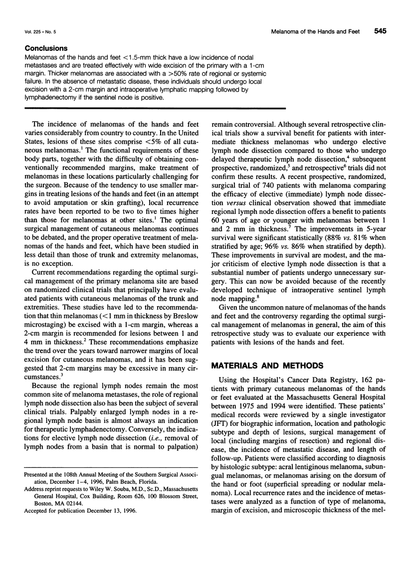
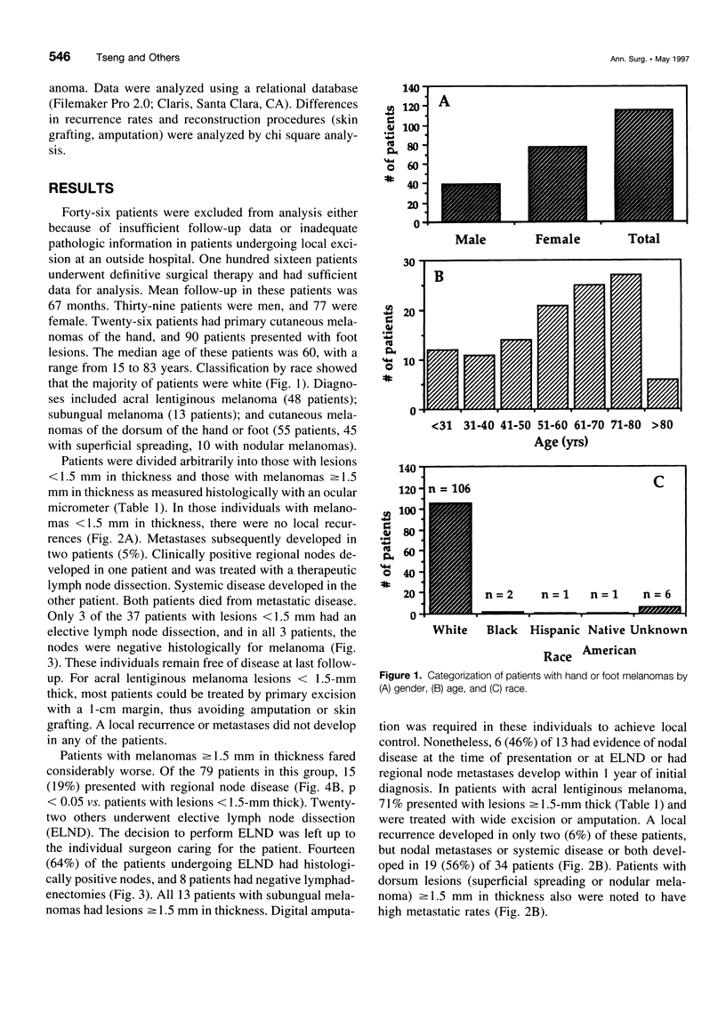
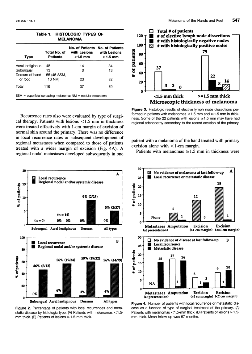
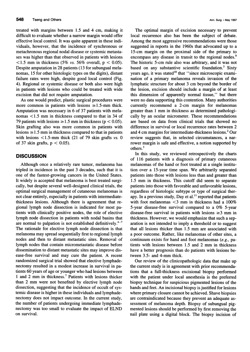
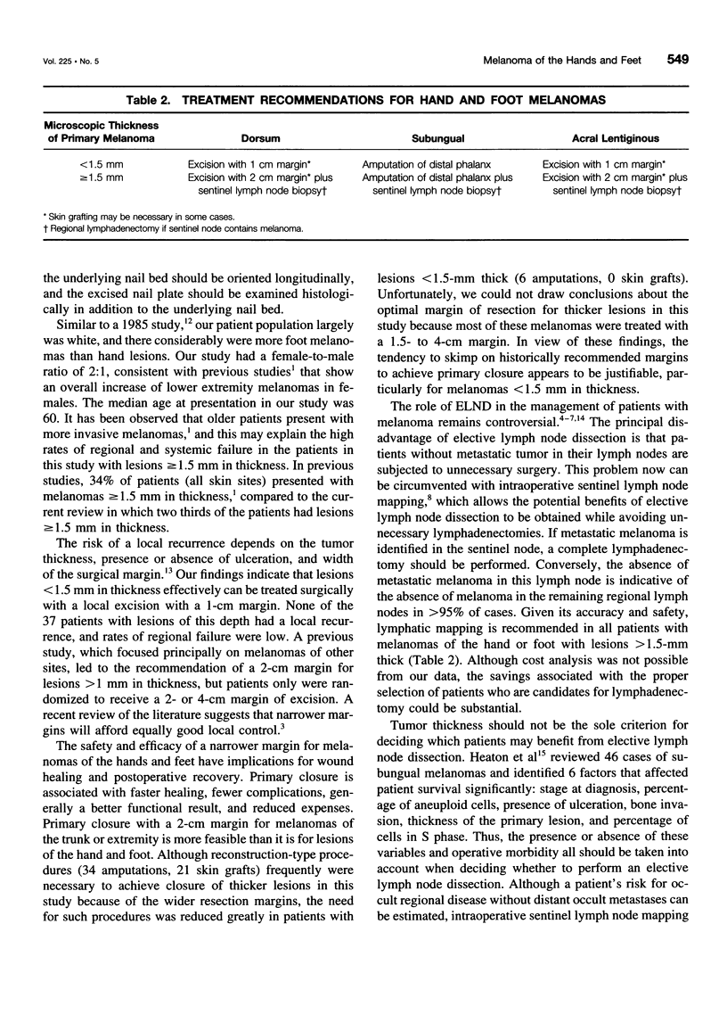
OBJECTIVE: The purpose of the study was to investigate the surgical management of cutaneous melanomas of the hands and feet. SUMMARY BACKGROUND DATA: Prior studies suggest that patients with melanomes > 1-mm thick should be treated with excision with a 2-cm margin and undergo elective lymphadenectomy in selected circumstances. These recommendations are based primarily on data from melanomas of the trunk and extremities. Melanomas of the hands and feet are less common and less well studied. They pose a surgical challenge because primary wound closure often is difficult, and the incidence and management of regional node metastases are unclear. METHODS: Charts of patients with melanomas of the hands or feet treated at the Massachusetts General Hospital between 1980 and 1994 were reviewed retrospectively. Local recurrence rates and the incidence of regional node metastases were analyzed as a function of histology, margin of excision, and microscopic thickness of the melanoma. RESULTS: Data from 116 patients (39 men, 77 women) with melanomas of the hands (n = 26) and feet (n = 90) were evaluated. Pathologic diagnoses were: acral lentiginous melanoma (48 patients); subungual melanoma (13 patients), and skin of dorsum of the hand or foot (n = 55). Digital amputation was required in all 13 patients with subungual melanoma to maintain local control; still, nodal metastases developed in 46% of patients within 1 year. Seventy-one percent of patients with acral lentiginous melanoma presented with lesions > or = 1.5 mm, and nodes or systemic disease or both developed in 56% of patients. Acral lentiginous melanoma lesions < 1.5-mm thick were treated principally by excision with a 1-cm margin; a local recurrence or metastases did not develop in any of the patients. None of the patients with melanomas on the dorsum of the hand or foot < 1.5-mm thick had a local recurrence, but regional or systemic disease developed in > 50%. Local control in patients with lesions > 1.5-mm thick frequently required skin grafting or amputation. The majority of patients with melanomas > or = 1.5 mm in thickness undergoing elective lymph node dissection had histologically positive nodes for melanoma. CONCLUSIONS: Melanomas of the hands and feet < 1.5-mm thick have a low incidence of nodal metastases and are treated effectively with wide excision of the primary with a 1-cm margin. Thicker melanomas are associated with a > 50% rate of regional or systemic failure. In the absence of metastatic disease, these individuals should undergo local excision with a 2-cm margin and intraoperative lymphatic mapping followed by lymphadenectomy if the sentinel node is positive.
Full text
PDF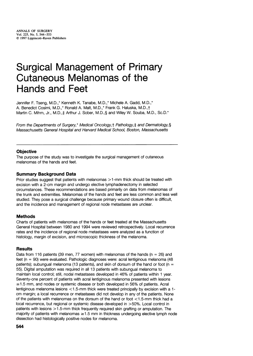
Selected References
These references are in PubMed. This may not be the complete list of references from this article.
- Balch C. M., Soong S. J., Bartolucci A. A., Urist M. M., Karakousis C. P., Smith T. J., Temple W. J., Ross M. I., Jewell W. R., Mihm M. C. Efficacy of an elective regional lymph node dissection of 1 to 4 mm thick melanomas for patients 60 years of age and younger. Ann Surg. 1996 Sep;224(3):255–266. doi: 10.1097/00000658-199609000-00002. [DOI] [PMC free article] [PubMed] [Google Scholar]
- Balch C. M. The role of elective lymph node dissection in melanoma: rationale, results, and controversies. J Clin Oncol. 1988 Jan;6(1):163–172. doi: 10.1200/JCO.1988.6.1.163. [DOI] [PubMed] [Google Scholar]
- Balch C. M., Urist M. M., Karakousis C. P., Smith T. J., Temple W. J., Drzewiecki K., Jewell W. R., Bartolucci A. A., Mihm M. C., Jr, Barnhill R. Efficacy of 2-cm surgical margins for intermediate-thickness melanomas (1 to 4 mm). Results of a multi-institutional randomized surgical trial. Ann Surg. 1993 Sep;218(3):262–269. doi: 10.1097/00000658-199309000-00005. [DOI] [PMC free article] [PubMed] [Google Scholar]
- Day C. L., Jr, Sober A. J., Kopf A. W., Lew R. A., Mihm M. C., Jr, Golomb F. M., Hennessey P., Harris M. N., Gumport S. L., Raker J. W. A prognostic model for clinical stage I melanoma of the lower extremity. Location on foot as independent risk factor for recurrent disease. Surgery. 1981 May;89(5):599–603. [PubMed] [Google Scholar]
- Heaton K. M., el-Naggar A., Ensign L. G., Ross M. I., Balch C. M. Surgical management and prognostic factors in patients with subungual melanoma. Ann Surg. 1994 Feb;219(2):197–204. doi: 10.1097/00000658-199402000-00012. [DOI] [PMC free article] [PubMed] [Google Scholar]
- Hughes L. E., Horgan K., Taylor B. A., Laidler P. Malignant melanoma of the hand and foot: diagnosis and management. Br J Surg. 1985 Oct;72(10):811–815. doi: 10.1002/bjs.1800721013. [DOI] [PubMed] [Google Scholar]
- McNeer G., Cantin J. Local failure in the treatment of melanoma. The Janeway lecture, 1966. Am J Roentgenol Radium Ther Nucl Med. 1967 Apr;99(4):791–808. doi: 10.2214/ajr.99.4.791. [DOI] [PubMed] [Google Scholar]
- Morton D. L., Wen D. R., Wong J. H., Economou J. S., Cagle L. A., Storm F. K., Foshag L. J., Cochran A. J. Technical details of intraoperative lymphatic mapping for early stage melanoma. Arch Surg. 1992 Apr;127(4):392–399. doi: 10.1001/archsurg.1992.01420040034005. [DOI] [PubMed] [Google Scholar]
- Piepkorn M., Barnhill R. L. A factual, not arbitrary, basis for choice of resection margins in melanoma. Arch Dermatol. 1996 Jul;132(7):811–814. [PubMed] [Google Scholar]
- Slingluff C. L., Jr, Stidham K. R., Ricci W. M., Stanley W. E., Seigler H. F. Surgical management of regional lymph nodes in patients with melanoma. Experience with 4682 patients. Ann Surg. 1994 Feb;219(2):120–130. doi: 10.1097/00000658-199402000-00003. [DOI] [PMC free article] [PubMed] [Google Scholar]
- Veronesi U., Cascinelli N., Adamus J., Balch C., Bandiera D., Barchuk A., Bufalino R., Craig P., De Marsillac J., Durand J. C. Thin stage I primary cutaneous malignant melanoma. Comparison of excision with margins of 1 or 3 cm. N Engl J Med. 1988 May 5;318(18):1159–1162. doi: 10.1056/NEJM198805053181804. [DOI] [PubMed] [Google Scholar]


