Abstract
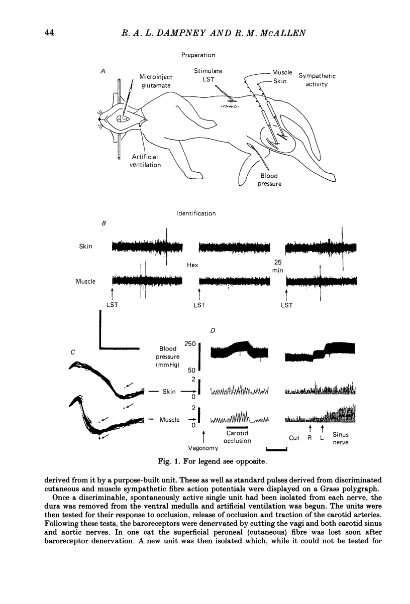
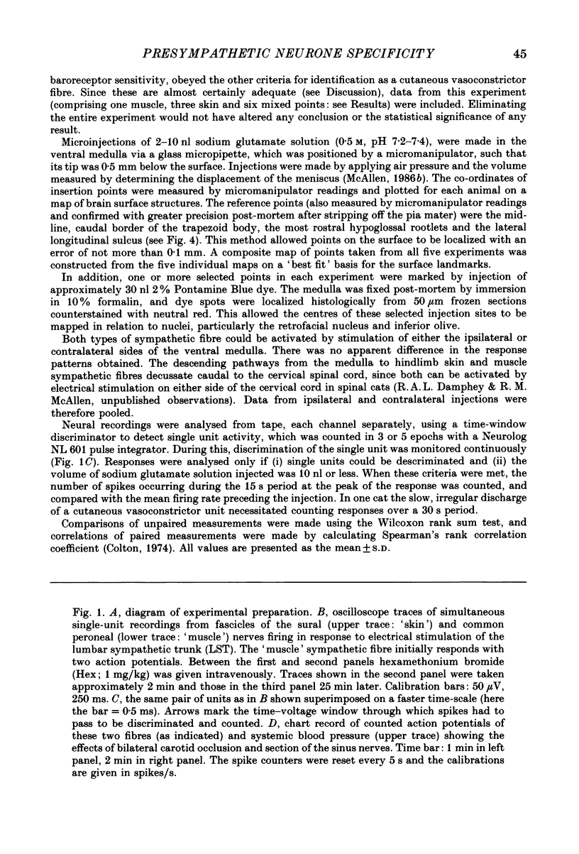
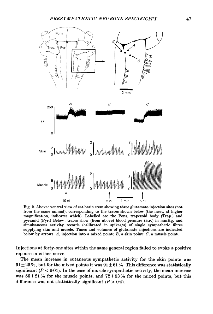
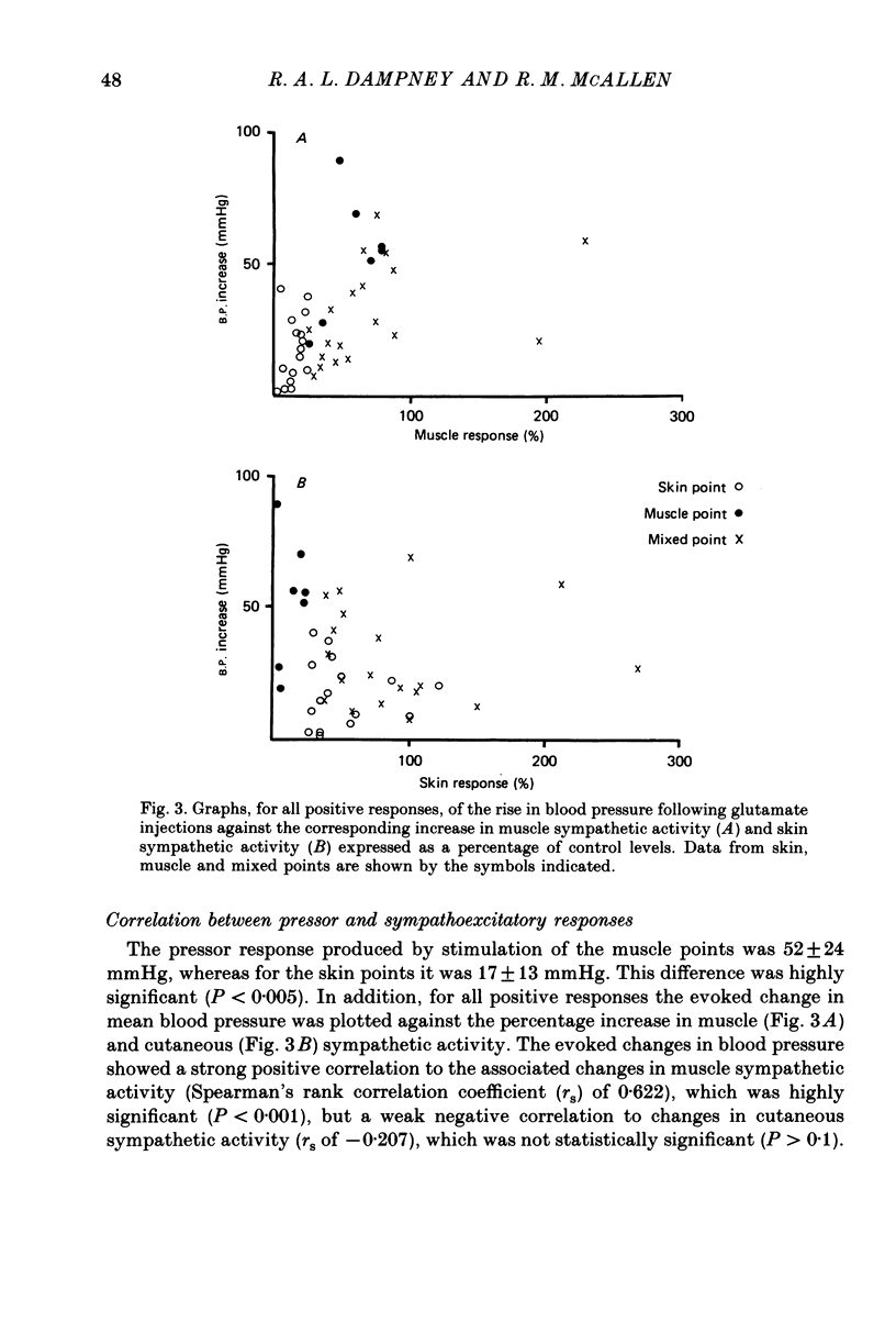
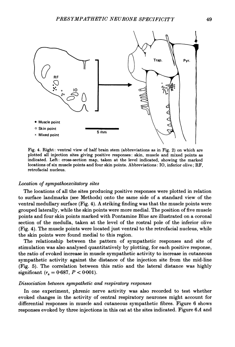
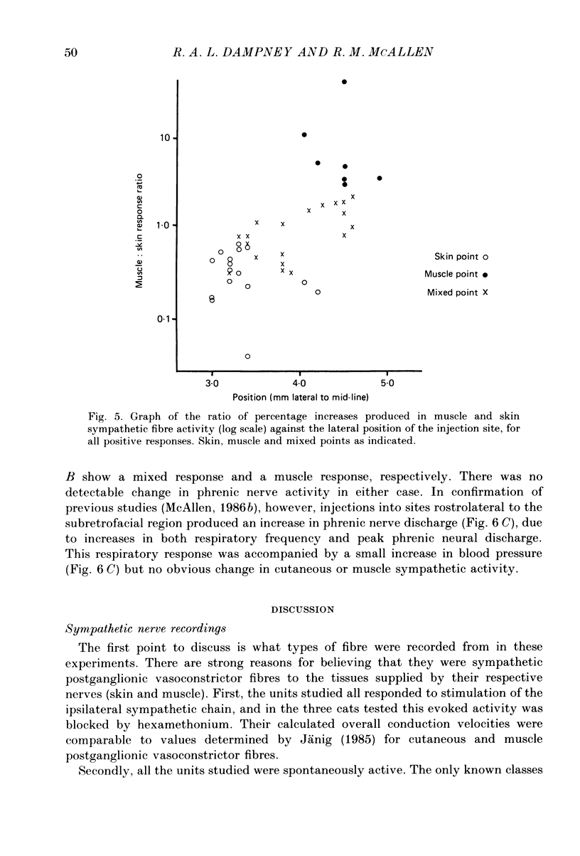
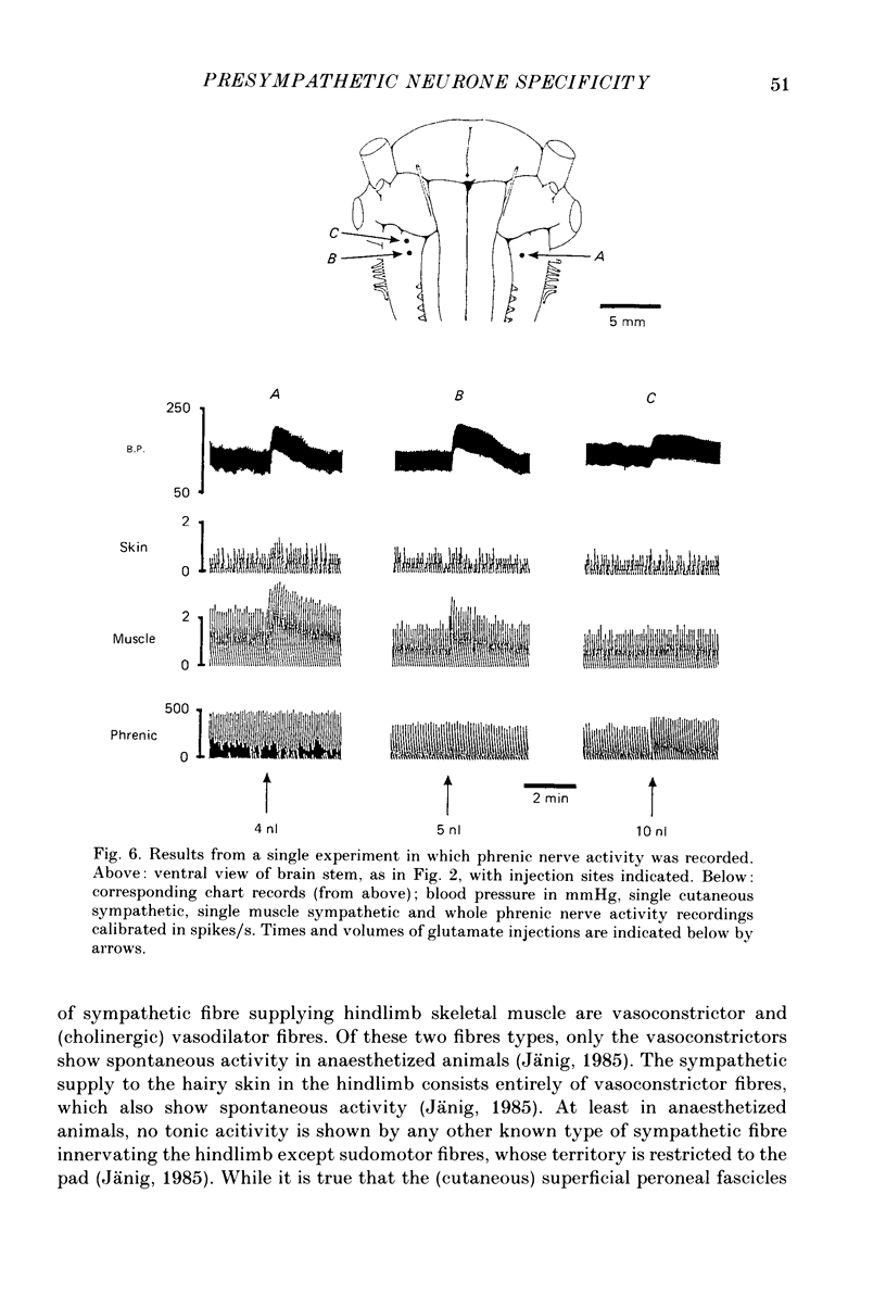
1. Simultaneous recordings were made from postganglionic sympathetic fibres supplying hindlimb skin and skeletal muscle in chloralose-anaesthetized, artificially ventilated cats. Single-fibre activity was either isolated by dissection or discriminated from few-fibre preparations of fascicles in the left superficial peroneal or sural nerve (innervating hairy skin) and common peroneal nerve (innervating muscle). Vasoconstrictor fibres were identified by their spontaneous activity as well as their responses to stimulation of the lumbar sympathetic chain and to changes in baroreceptor activity. The baroreceptors were then denervated by bilateral section of the vagi, carotid sinus and aortic nerves. 2. In five cats, neurones in the region of the subretrofacial nucleus were activated chemically by microinjections of 2-10 nl 0.5 M-sodium glutamate from a micropipette inserted into the ventral surface of the medulla. Both skin and muscle vasoconstrictor fibres were activated by glutamate injections into this region on either side of the medulla. Arterial pressure also rose. 3. Glutamate injections at forty-two sites evoked a positive response, defined as an increase in cutaneous and/or muscle vasoconstrictor fibre activity of at least 25%. This response was evoked only in the cutaneous fibre at sixteen of these sites ('skin points'), only in the muscle fibre at seven sites ('muscle points'), and in both fibres in the remainder ('mixed points'). The largest percentage increases in activity of either type of fibre were obtained from mixed points. 4. The blood pressure rises following glutamate stimulation of muscle points were significantly greater than those produced by stimulation of skin points. Analysis of all positive responses showed that the evoked rise in blood pressure was significantly correlated with muscle sympathetic activity but not with cutaneous sympathetic activity. 5. Glutamate stimulation at different sites could evoke differential responses in skin and muscle vasoconstrictor fibres without any detectable change in the pattern of phrenic nerve discharge. 6. Skin points were grouped in the medial part of the subretrofacial region, and muscle points in the lateral part. In addition, for all positive responses there was a highly significant correlation between the ratio of muscle to cutaneous sympathetic activity evoked, and the distance from the mid-line of the corresponding injection site. 7. These results demonstrate a functional differentiation among subretrofacial neurones in their relative control of the sympathetic vasoconstrictor supply to skin and skeletal muscle.(ABSTRACT TRUNCATED AT 400 WORDS)
Full text
PDF
Selected References
These references are in PubMed. This may not be the complete list of references from this article.
- Amendt K., Czachurski J., Dembowsky K., Seller H. Neurones within the "chemosensitive area" on the ventral surface of the brainstem which project to the intermediolateral column. Pflugers Arch. 1978 Aug;375(3):289–292. doi: 10.1007/BF00582443. [DOI] [PubMed] [Google Scholar]
- Barman S. M., Gebber G. L., Calaresu F. R. Differential control of sympathetic nerve discharge by the brain stem. Am J Physiol. 1984 Sep;247(3 Pt 2):R513–R519. doi: 10.1152/ajpregu.1984.247.3.R513. [DOI] [PubMed] [Google Scholar]
- Blessing W. W., Goodchild A. K., Dampney R. A., Chalmers J. P. Cell groups in the lower brain stem of the rabbit projecting to the spinal cord, with special reference to catecholamine-containing neurons. Brain Res. 1981 Sep 21;221(1):35–55. doi: 10.1016/0006-8993(81)91062-3. [DOI] [PubMed] [Google Scholar]
- CRAWFORD J. M., CURTIS D. R. THE EXCITATION AND DEPRESSION OF MAMMALIAN CORTICAL NEURONES BY AMINO ACIDS. Br J Pharmacol Chemother. 1964 Oct;23:313–329. doi: 10.1111/j.1476-5381.1964.tb01589.x. [DOI] [PMC free article] [PubMed] [Google Scholar]
- Curtis D. R., Ryall R. W. Pharmacological studies upon spinal presynaptic fibres. Exp Brain Res. 1966;1(2):195–204. doi: 10.1007/BF00236871. [DOI] [PubMed] [Google Scholar]
- Dampney R. A., Czachurski J., Dembowsky K., Goodchild A. K., Seller H. Afferent connections and spinal projections of the pressor region in the rostral ventrolateral medulla of the cat. J Auton Nerv Syst. 1987 Jul;20(1):73–86. doi: 10.1016/0165-1838(87)90083-x. [DOI] [PubMed] [Google Scholar]
- Dampney R. A., Goodchild A. K., Robertson L. G., Montgomery W. Role of ventrolateral medulla in vasomotor regulation: a correlative anatomical and physiological study. Brain Res. 1982 Oct 14;249(2):223–235. doi: 10.1016/0006-8993(82)90056-7. [DOI] [PubMed] [Google Scholar]
- Dampney R. A., Goodchild A. K., Tan E. Vasopressor neurons in the rostral ventrolateral medulla of the rabbit. J Auton Nerv Syst. 1985 Nov;14(3):239–254. doi: 10.1016/0165-1838(85)90113-4. [DOI] [PubMed] [Google Scholar]
- Engberg I., Flatman J. A., Lambert J. D. The actions of excitatory amino acids on motoneurones in the feline spinal cord. J Physiol. 1979 Mar;288:227–261. [PMC free article] [PubMed] [Google Scholar]
- Feldberg W. The ventral surface of the brain stem: a scarcely explored region of pharmacological sensitivity. Neuroscience. 1976 Dec;1(6):427–441. doi: 10.1016/0306-4522(76)90093-2. [DOI] [PubMed] [Google Scholar]
- Fries W., Zieglgänsberger W. A method to discriminate axonal from cellbody activity and to analyse "silent cells". Exp Brain Res. 1974;21(4):441–445. doi: 10.1007/BF00237906. [DOI] [PubMed] [Google Scholar]
- Goodchild A. K., Dampney R. A., Bandler R. A method for evoking physiological responses by stimulation of cell bodies, but not axons of passage, within localized regions of the central nervous system. J Neurosci Methods. 1982 Nov;6(4):351–363. doi: 10.1016/0165-0270(82)90036-x. [DOI] [PubMed] [Google Scholar]
- Jänig W. Organization of the lumbar sympathetic outflow to skeletal muscle and skin of the cat hindlimb and tail. Rev Physiol Biochem Pharmacol. 1985;102:119–213. doi: 10.1007/BFb0034086. [DOI] [PubMed] [Google Scholar]
- Jänig W., Sundlöf G., Wallin B. G. Discharge patterns of sympathetic neurons supplying skeletal muscle and skin in man and cat. J Auton Nerv Syst. 1983 Mar-Apr;7(3-4):239–256. doi: 10.1016/0165-1838(83)90077-2. [DOI] [PubMed] [Google Scholar]
- Lovick T. A., Hilton S. M. Vasodilator and vasoconstrictor neurones of the ventrolateral medulla in the cat. Brain Res. 1985 Apr 8;331(2):353–357. doi: 10.1016/0006-8993(85)91562-8. [DOI] [PubMed] [Google Scholar]
- McAllen R. M. Action and specificity of ventral medullary vasopressor neurones in the cat. Neuroscience. 1986 May;18(1):51–59. doi: 10.1016/0306-4522(86)90178-8. [DOI] [PubMed] [Google Scholar]
- McAllen R. M. Identification and properties of sub-retrofacial bulbospinal neurones: a descending cardiovascular pathway in the cat. J Auton Nerv Syst. 1986 Oct;17(2):151–164. doi: 10.1016/0165-1838(86)90090-1. [DOI] [PubMed] [Google Scholar]
- McAllen R. M. Location of neurones with cardiovascular and respiratory function, at the ventral surface of the cat's medulla. Neuroscience. 1986 May;18(1):43–49. doi: 10.1016/0306-4522(86)90177-6. [DOI] [PubMed] [Google Scholar]
- McAllen R. M., Neil J. J., Loewy A. D. Effects of kainic acid applied to the ventral surface of the medulla oblongata on vasomotor tone, the baroreceptor reflex and hypothalamic autonomic responses. Brain Res. 1982 Apr 22;238(1):65–76. doi: 10.1016/0006-8993(82)90771-5. [DOI] [PubMed] [Google Scholar]
- McAllen R. M., Spyer K. M. Two types of vagal preganglionic motoneurones projecting to the heart and lungs. J Physiol. 1978 Sep;282:353–364. doi: 10.1113/jphysiol.1978.sp012468. [DOI] [PMC free article] [PubMed] [Google Scholar]
- Ninomiya I., Nisimaru N., Irisawa H. Sympathetic nerve activity to the spleen, kidney, and heart in response to baroceptor input. Am J Physiol. 1971 Nov;221(5):1346–1351. doi: 10.1152/ajplegacy.1971.221.5.1346. [DOI] [PubMed] [Google Scholar]
- Preiss G., Polosa C. The relation between end-tidal CO2 and discharge patterns of sympathetic preganglionic neurons. Brain Res. 1977 Feb 18;122(2):255–267. doi: 10.1016/0006-8993(77)90293-1. [DOI] [PubMed] [Google Scholar]
- Reis D. J., Ross C. A., Ruggiero D. A., Granata A. R., Joh T. H. Role of adrenaline neurons of ventrolateral medulla (the C1 group) in the tonic and phasic control of arterial pressure. Clin Exp Hypertens A. 1984;6(1-2):221–241. doi: 10.3109/10641968409062562. [DOI] [PubMed] [Google Scholar]
- Ross C. A., Ruggiero D. A., Joh T. H., Park D. H., Reis D. J. Rostral ventrolateral medulla: selective projections to the thoracic autonomic cell column from the region containing C1 adrenaline neurons. J Comp Neurol. 1984 Sep 10;228(2):168–185. doi: 10.1002/cne.902280204. [DOI] [PubMed] [Google Scholar]
- Simon E., Riedel W. Diversity of regional sympathetic outflow in integrative cardiovascular control: patterns and mechanisms. Brain Res. 1975 Apr 11;87(2-3):323–333. doi: 10.1016/0006-8993(75)90429-1. [DOI] [PubMed] [Google Scholar]
- Willette R. N., Barcas P. P., Krieger A. J., Sapru H. N. Vasopressor and depressor areas in the rat medulla. Identification by microinjection of L-glutamate. Neuropharmacology. 1983 Sep;22(9):1071–1079. doi: 10.1016/0028-3908(83)90027-8. [DOI] [PubMed] [Google Scholar]


