

Abstract
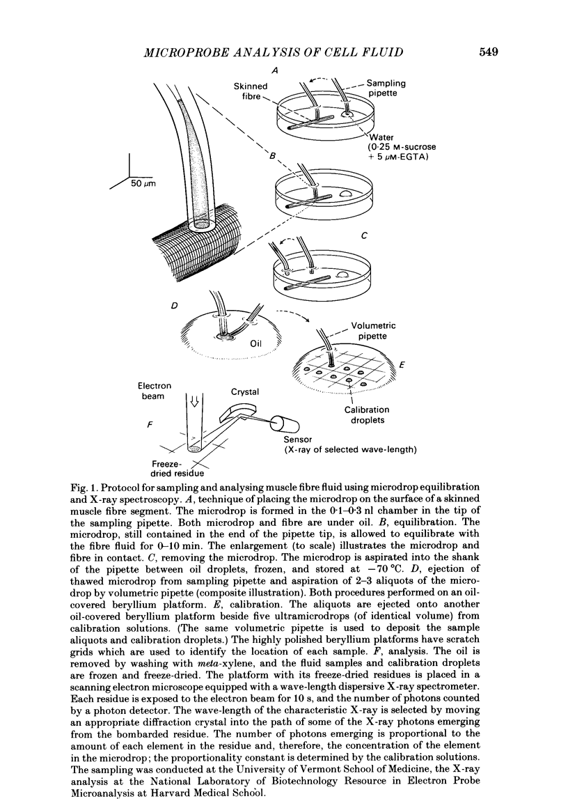
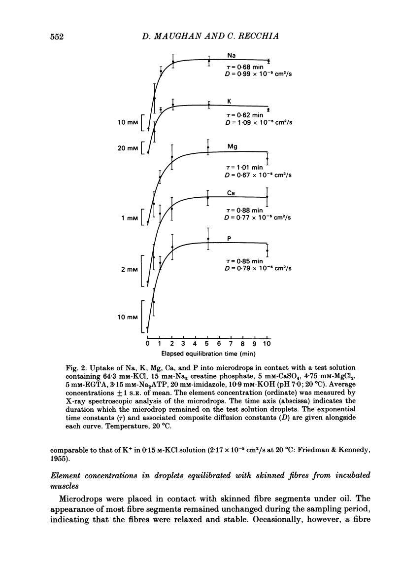
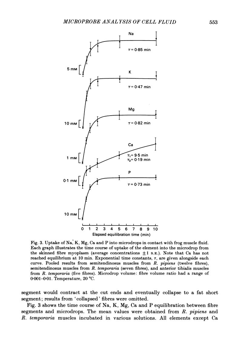
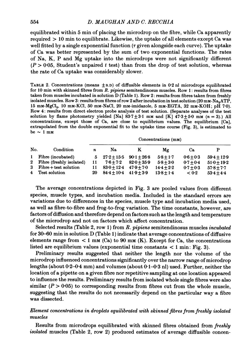
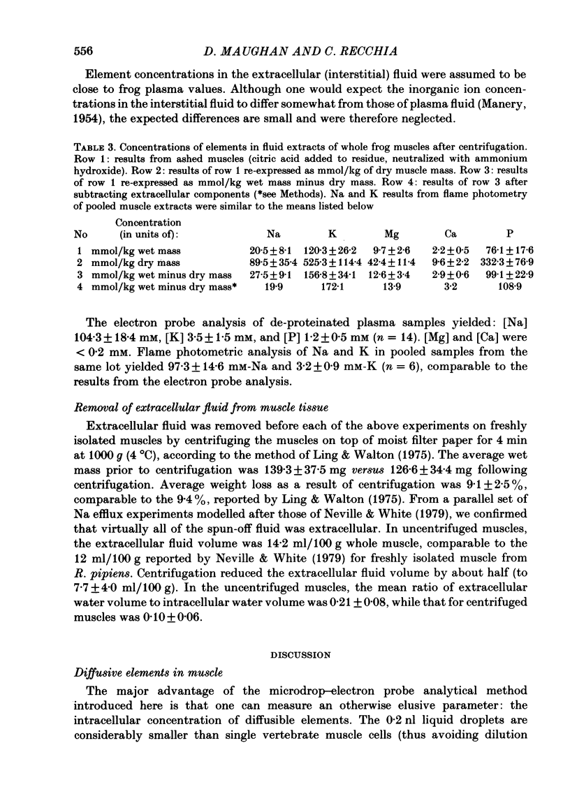
A microvolumetric analytical method has been developed to measure the endogenous concentrations of diffusible elements in muscle cells. Single twitch fibres from frog muscle were skinned under oil and 0.2 nl drops of isosmotic sucrose solution, held in the tips of specially constructed pipettes, were placed in contact with the skinned fibres. After 0-10 min, the microdrops were removed and analysed with a wave-length dispersive X-ray spectrometer. The uptake of Na, K, Mg, and P into the microdrops was well fitted by a single exponential function, while the uptake of Ca was better represented by the sum of two exponential functions. All elements analysed except Ca reached diffusional equilibrium within 5 min of placing the microdrop on the fibre, while Ca was still not equilibrated at 10 min. For freshly isolated muscle fibres, diffusible element concentrations in the microdrops at equilibrium were (in mM, mean +/- S.D.): Na, 7.6 +/- 7.2; K, 82 +/- 36; Mg, 5.8 +/- 3.0; P, 51 +/- 19. Diffusible Ca concentration (at 10 min elapsed sampling time) was 0.7 +/- 0.4 mM. Results from experiments in which microdrops were equilibrated with skinned fibres pre-soaked in an artificial (Ca-free) solution support the notion that the exogenous solutes can replace the endogenous diffusible contents of a skinned fibre by soaking the skinned fibre in a relatively large volume of the artificial solution. Total Na, K, Mg, Ca, and P content of whole muscle was measured by electron probe analysis of muscle extracts. In freshly isolated muscle, whole muscle element content was (in mmol/kg wet weight, mean +/- S.D.): Na, 21 +/- 8; K, 120 +/- 26; Mg, 9.7 +/- 2.6; Ca, 2.2 +/- 0.5; P, 76 +/- 18. Extracellular fluid volumes of freshly isolated whole muscles were estimated by compartmental analysis of Na efflux. Extracellular element concentrations were measured by electron probe analysis of frog plasma. Using the extracellular fluid volume and concentration estimates, extracellular contributions were subtracted from measurements of the element content of whole muscle to yield estimates of total intracellular element concentration (in mmol/l myoplasmic water). Based on the values for the intracellular total and diffusible element concentrations, the diffusible/total content fraction in freshly isolated muscle is estimated to be: Na, 0.38; K, 0.48; Mg, 0.42; Ca, 0.22; and P, 0.47.
Full text
PDF
Selected References
These references are in PubMed. This may not be the complete list of references from this article.
- Baylor S. M., Chandler W. K., Marshall M. W. Optical measurements of intracellular pH and magnesium in frog skeletal muscle fibres. J Physiol. 1982 Oct;331:105–137. doi: 10.1113/jphysiol.1982.sp014367. [DOI] [PMC free article] [PubMed] [Google Scholar]
- Baylor S. M., Chandler W. K., Marshall M. W. Sarcoplasmic reticulum calcium release in frog skeletal muscle fibres estimated from Arsenazo III calcium transients. J Physiol. 1983 Nov;344:625–666. doi: 10.1113/jphysiol.1983.sp014959. [DOI] [PMC free article] [PubMed] [Google Scholar]
- Borland R. M., Biggers J. D., Lechene C. P. Fluid transport by rabbit preimplantation blastocysts in vitro. J Reprod Fertil. 1977 Sep;51(1):131–135. doi: 10.1530/jrf.0.0510131. [DOI] [PubMed] [Google Scholar]
- CONWAY E. J. Nature and significance of concentration relations of potassium and sodium ions in skeletal muscle. Physiol Rev. 1957 Jan;37(1):84–132. doi: 10.1152/physrev.1957.37.1.84. [DOI] [PubMed] [Google Scholar]
- Close R. I., Lännergren J. I. Arsenazo III calcium transients and latency relaxation in frog skeletal muscle fibres at different sarcomere lengths. J Physiol. 1984 Oct;355:323–344. doi: 10.1113/jphysiol.1984.sp015422. [DOI] [PMC free article] [PubMed] [Google Scholar]
- Cohen S. M., Burt C. T. 31P nuclear magnetic relaxation studies of phosphocreatine in intact muscle: determination of intracellular free magnesium. Proc Natl Acad Sci U S A. 1977 Oct;74(10):4271–4275. doi: 10.1073/pnas.74.10.4271. [DOI] [PMC free article] [PubMed] [Google Scholar]
- Dawson M. J., Gadian D. G., Wilkie D. R. Mechanical relaxation rate and metabolism studied in fatiguing muscle by phosphorus nuclear magnetic resonance. J Physiol. 1980 Feb;299:465–484. doi: 10.1113/jphysiol.1980.sp013137. [DOI] [PMC free article] [PubMed] [Google Scholar]
- Dick D. A., McLaughlin S. G. The activities and concentrations of sodium and potassium in toad oocytes. J Physiol. 1969 Nov;205(1):61–78. doi: 10.1113/jphysiol.1969.sp008951. [DOI] [PMC free article] [PubMed] [Google Scholar]
- Elliott G. F. Donnan and osmotic effects in muscle fibres without membranes. J Mechanochem Cell Motil. 1973 May;2(1):83–89. [PubMed] [Google Scholar]
- Garfinkel L., Garfinkel D. Calculation of free-Mg2+ concentration in adenosine 5'-triphosphate containing solutions in vitro and in vivo. Biochemistry. 1984 Jul 17;23(15):3547–3552. doi: 10.1021/bi00310a025. [DOI] [PubMed] [Google Scholar]
- Gillis J. M., Piront A., Gosselin-Rey C. Parvalbumins. Distribution and physical state inside the muscle cell. Biochim Biophys Acta. 1979 Jul 4;585(3):444–450. doi: 10.1016/0304-4165(79)90089-8. [DOI] [PubMed] [Google Scholar]
- Godt R. E. A simple electrostatic model can explain the effect of pH upon the force-pCa relation of skinned frog skeletal muscle fibers. Biophys J. 1981 Aug;35(2):385–392. doi: 10.1016/S0006-3495(81)84797-2. [DOI] [PMC free article] [PubMed] [Google Scholar]
- Godt R. E., Baumgarten C. M. Potential and K+ activity in skinned muscle fibers. Evidence against a simple Donnan equilibrium. Biophys J. 1984 Feb;45(2):375–382. doi: 10.1016/S0006-3495(84)84161-2. [DOI] [PMC free article] [PubMed] [Google Scholar]
- Gordon A. M., Godt R. E., Donaldson S. K., Harris C. E. Tension in skinned frog muscle fibers in solutions of varying ionic strength and neutral salt composition. J Gen Physiol. 1973 Nov;62(5):550–574. doi: 10.1085/jgp.62.5.550. [DOI] [PMC free article] [PubMed] [Google Scholar]
- Gosselin-rey C., Gerday C. Parvalbumins from frog skeletal muscle (Rana temporaria L.). Isolation and characterization. Structural modifications associated with calcium binding. Biochim Biophys Acta. 1977 May 27;492(1):53–63. doi: 10.1016/0005-2795(77)90213-6. [DOI] [PubMed] [Google Scholar]
- Gupta R. K., Gupta P., Yushok W. D., Rose Z. B. On the noninvasive measurement of intracellular free magnesium by 31P NMR spectroscopy. Physiol Chem Phys Med NMR. 1983;15(3):265–280. [PubMed] [Google Scholar]
- Gupta R. K., Moore R. D. 31P NMR studies of intracellular free Mg2+ in intact frog skeletal muscle. J Biol Chem. 1980 May 10;255(9):3987–3993. [PubMed] [Google Scholar]
- Hess P., Metzger P., Weingart R. Free magnesium in sheep, ferret and frog striated muscle at rest measured with ion-selective micro-electrodes. J Physiol. 1982 Dec;333:173–188. doi: 10.1113/jphysiol.1982.sp014447. [DOI] [PMC free article] [PubMed] [Google Scholar]
- Horowitz S. B., Miller D. S. Solvent properties of ground substance studied by cryomicrodissection and intracellular reference-phase techniques. J Cell Biol. 1984 Jul;99(1 Pt 2):172s–179s. doi: 10.1083/jcb.99.1.172s. [DOI] [PMC free article] [PubMed] [Google Scholar]
- Horowitz S. B., Paine P. L., Tluczek L., Reynhout J. K. Reference phase analysis of free and bound intracellular solutes. I. Sodium and potassium in amphibian oocytes. Biophys J. 1979 Jan;25(1):33–44. doi: 10.1016/S0006-3495(79)85276-5. [DOI] [PMC free article] [PubMed] [Google Scholar]
- Ingram F. D., Ingram M. J., Hogben C. A. Quantitative electron probe analysis of soft biologic tissue for electrolytes. J Histochem Cytochem. 1972 Sep;20(9):716–722. doi: 10.1177/20.9.716. [DOI] [PubMed] [Google Scholar]
- Johnson J. D., Charlton S. C., Potter J. D. A fluorescence stopped flow analysis of Ca2+ exchange with troponin C. J Biol Chem. 1979 May 10;254(9):3497–3502. [PubMed] [Google Scholar]
- Kushmerick M. J., Podolsky R. J. Ionic mobility in muscle cells. Science. 1969 Dec 5;166(3910):1297–1298. doi: 10.1126/science.166.3910.1297. [DOI] [PubMed] [Google Scholar]
- LEV A. A. DETERMINATION OF ACTIVITY AND ACTIVITY COEFFICIENTS OF POTASSIUM AND SODIUM IONS IN FROG MUSCLE FIBRES. Nature. 1964 Mar 14;201:1132–1134. doi: 10.1038/2011132a0. [DOI] [PubMed] [Google Scholar]
- Lee C. O., Armstrong W. M. State and distribution of potassium and sodium ions in frog skeletal muscle. J Membr Biol. 1974;15(4):331–362. doi: 10.1007/BF01870094. [DOI] [PubMed] [Google Scholar]
- Ling G. N., Walton C. L. A simple rapid method for the quantitative separation of the extracellular fluid in frog muslces. Physiol Chem Phys. 1975;7(3):215–218. [PubMed] [Google Scholar]
- MANERY J. F. Water and electrolyte metabolism. Physiol Rev. 1954 Apr;34(2):334–417. doi: 10.1152/physrev.1954.34.2.334. [DOI] [PubMed] [Google Scholar]
- Macchia D. D., Baumgarten C. M. Is chloride passively distributed in skeletal muscle in vivo? Pflugers Arch. 1979 Nov;382(2):193–195. doi: 10.1007/BF00584222. [DOI] [PubMed] [Google Scholar]
- Maughan D. W., Godt R. E. A quantitative analysis of elastic, entropic, electrostatic, and osmotic forces within relaxed skinned muscle fibers. Biophys Struct Mech. 1980;7(1):17–40. doi: 10.1007/BF00538156. [DOI] [PubMed] [Google Scholar]
- Maughan D. W., Godt R. E. Stretch and radial compression studies on relaxed skinned muscle fibers of the frog. Biophys J. 1979 Dec;28(3):391–402. doi: 10.1016/S0006-3495(79)85188-7. [DOI] [PMC free article] [PubMed] [Google Scholar]
- Maughan D. Diffusible magnesium in frog skeletal muscle cells. Biophys J. 1983 Jul;43(1):75–80. doi: 10.1016/S0006-3495(83)84325-2. [DOI] [PMC free article] [PubMed] [Google Scholar]
- Neville M. C., White S. Extracellular space of frog skeletal muscle in vivo and in vitro: relation to proton magnetic resonance relaxation times. J Physiol. 1979 Mar;288:71–83. [PMC free article] [PubMed] [Google Scholar]
- Peachey L. D. The sarcoplasmic reticulum and transverse tubules of the frog's sartorius. J Cell Biol. 1965 Jun;25(3 Suppl):209–231. doi: 10.1083/jcb.25.3.209. [DOI] [PubMed] [Google Scholar]
- Somlyo A. V., Gonzalez-Serratos H. G., Shuman H., McClellan G., Somlyo A. P. Calcium release and ionic changes in the sarcoplasmic reticulum of tetanized muscle: an electron-probe study. J Cell Biol. 1981 Sep;90(3):577–594. doi: 10.1083/jcb.90.3.577. [DOI] [PMC free article] [PubMed] [Google Scholar]
- Somlyo A. V., Shuman H., Somlyo A. P. Elemental distribution in striated muscle and the effects of hypertonicity. Electron probe analysis of cryo sections. J Cell Biol. 1977 Sep;74(3):828–857. doi: 10.1083/jcb.74.3.828. [DOI] [PMC free article] [PubMed] [Google Scholar]
- Stephenson D. G., Wendt I. R., Forrest Q. G. Non-uniform ion distributions and electrical potentials in sarcoplasmic regions of skeletal muscle fibres. Nature. 1981 Feb 19;289(5799):690–692. doi: 10.1038/289690a0. [DOI] [PubMed] [Google Scholar]
- Stephenson E. W. Activation of fast skeletal muscle: contributions of studies on skinned fibers. Am J Physiol. 1981 Jan;240(1):C1–19. doi: 10.1152/ajpcell.1981.240.1.C1. [DOI] [PubMed] [Google Scholar]
- Weingart R., Hess P. Free calcium in sheep cardiac tissue and frog skeletal muscle measured with Ca2+-selective microelectrodes. Pflugers Arch. 1984 Sep;402(1):1–9. doi: 10.1007/BF00584824. [DOI] [PubMed] [Google Scholar]
- Yates L. D., Greaser M. L. Quantitative determination of myosin and actin in rabbit skeletal muscle. J Mol Biol. 1983 Jul 25;168(1):123–141. doi: 10.1016/s0022-2836(83)80326-x. [DOI] [PubMed] [Google Scholar]