



Abstract
Senescent cells accumulate throughout the body with advanced age, diseases and chronic conditions. They negatively impact health and function of multiple systems, including the central nervous system (CNS). Therapies that target senescent cells, broadly referred to as senotherapeutics, recently emerged as potentially important treatment strategies for the CNS. Promising therapeutic approaches involve clearing senescent cells by disarming their pro-survival pathways with ‘senolytics’; or dampening their toxic senescence-associated secretory phenotype (SASP) using ‘senomorphics’. Following the pioneering discovery of first-generation senolytics dasatinib and quercetin, dozens of additional therapies have been identified, and several promising targets are under investigation. Although potentially transformative, senotherapies are still in early stages and require thorough testing to ensure reliable target engagement, specificity, safety and efficacy. The limited brain penetrance and potential toxic side effects of CNS-acting senotherapeutics pose challenges for drug development and translation to the clinic. This Review assesses the potential impact of senotherapeutics for neurological conditions by summarizing preclinical evidence, innovative methods for target and biomarker identification, academic and industry drug development pipelines and progress in clinical trials.
Introduction
Cellular senescence was first described in 1961 as a phenomenon whereby primary cells in culture undergo a finite number of divisions1. This limited replicative potential was believed to be an artefact of cells in culture driven by telomere erosion2,3. The eventual discovery of molecular biomarkers enabled the identification of senescent cells in vivo4,5. However, the inclusion of cell cycle arrest in the definition of senescence cast doubt on the relevance of senescence in tissues with mostly non-dividing cells, such as the brain. The first study to report senescent neurons in mouse brain tissue was transformative to the field6 but was met with scepticism. Later discoveries of senescent-like neurons7–10 and astrocytes11,12 in post-mortem tissue from patients with Alzheimer disease (AD) and Parkinson disease supported the initial findings and highlighted the potential translational importance of senescent cells in the brain. These studies, along with the identification of postmitotic senescent-like cells in other tissues13, inspired the field to reconsider the senescence-defining criteria. Cellular senescence now refers to an end-stage change in cell fate, orchestrated through simultaneous and sustained activation of opposing pro-apoptotic and pro-survival molecular pathways14. For the context of this Review, we focus only on pathological senescence that arises through chronic stress activation, distinguishing it from physiological senescence, which occurs during development, tissue regeneration or wound healing15,16.
Similar to other changes in cell fate, senescent cells acquire distinct morphologies and remain metabolically active17. They often develop enlarged somas both in vitro18 and in vivo9,19, exhibit increased lysosomal senescence-associated β-galactosidase (SA-β-gal) activity20,21, elevated glycolysis22 and lipid metabolism23, and acquire a secretory phenotype16,24. The molecules they release, known as the senescence-messaging secretome (SMS)24, or more commonly as the senescence-associated secretory phenotype (SASP)16, communicate with their neighbouring environment and the immune system either to prepare the tissue for disease or to facilitate their own clearance, respectively. The presence of senescent cells perpetuates tissue inflammation and destruction through multiple mechanisms25. If senescent cells are not actively removed by the immune system, their sustained SASP signalling induces cytotoxicity or transforms healthy cells into senescent cells. Phagocytic cells can also become senescent after clearing senescent cells, which perpetuates inflammation and pathogenic processes (Fig. 1). Throughout the lifespan, tissues and organs accumulate senescent cells, and their contribution to disease and dysfunction earned them recognition as a “biological hallmark of aging”26.
Fig. 1 |. Senescence initiation and spread across brain cells.
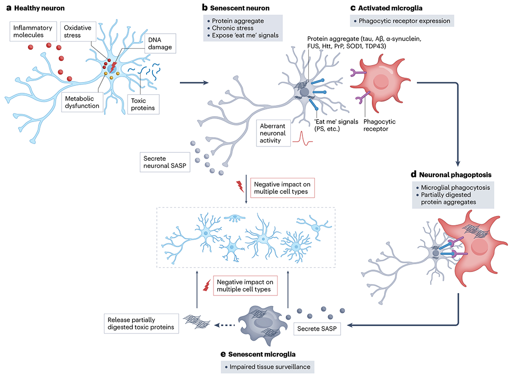
a, Age and disease-associated stressors that drive inflammation, reactive oxygen species, toxic protein accumulation, metabolic dysregulation or DNA damage may cause senescence in many cell types, including neurons. b, Senescent neurons exhibit altered excitability and/or activity, display ‘eat me’ signals and secrete deleterious molecules (the senescence-associated secretory phenotype (SASP)) that negatively impact neuronal, vascular and glial cells. They may also contain aggregate-prone, neurotoxic proteins that they transmit to other cells as neuronal SASP9,19. c, Activated microglia expressing phagocytic receptors recognize neuronal phagoptosis ‘eat me’ signals. d, Activated microglia engulf senescent neurons166 and their content, including difficult-to-digest protein aggregates, which may cause microglial senescence. e, Senescent microglia exhibit reduced phagocytic and surveillance function44. They release partially digested, neurotoxic fragments of protein aggregates and SASP factors that cause astrocytes42, microglia44,57, vasculature43 and oligodendrocyte precursor cells45 to become senescent. Aβ, amyloid-β; Htt, huntingtin; PrP, prion protein; PS, phosphatidylserine; SOD1, superoxide dismutase 1; TDP43, TAR DNA-binding protein 43.
During the transition to senescence, cells undergo significant changes in chromatin structure, alter their metabolism and morphology, and become resistant to apoptosis14. The features of senescent cells depend on the original cell type and the specific stressor involved27. Various markers have been used to identify senescent cells, but a confident identification requires evidence of multiple overlapping phenotypes within the same cell, such as changes in morphology, macromolecular damage, cell cycle arrest and an inflammatory secretome14,28. In addition to applying traditional senescence detection methods to tissues of interest, the advancement in single-cell and single-nucleus RNA sequencing, high-resolution multiplex spatial profiling techniques and machine learning (ML) and artificial intelligence (AI) enable the identification of rare senescent cells in complex tissues19,29–32. These strategies have propelled the discovery of novel biomarkers of senescence. Experimental evidence with new and established biomarkers indicates that several brain cell types, including neurons, glia, stem cells and cerebral vasculature cells, may undergo senescence owing to a range of stressors, such as physical, psychological, viral, oxidative and proteotoxic insults, DNA damage and protein accumulation (Table 1).
Table 1 |.
Evidence of cellular senescence in ageing and CNS conditions
| Condition | Astrocytes | Microglia | Neurons | Other CNS cells |
|---|---|---|---|---|
| Alzheimer disease | p16 (refs. 11,251), p53 (ref. 251) | p16 (ref. 202) morphology, DNA damage, HMGB1 (ref. 252); protein panel (ref. 251) | Transcriptome (refs. 9,19,208,253) p19+ NFTs, karyomegaly, lipofuscin (ref. 19) |
OPCs (ref. 45) Endothelial cells (ref. 200) |
| Ageing | p16 (refs. 11,254); p21, SA-β-gal254; loss of HMGB1, SASP, p16 (ref. 42) | p16 (ref. 255); protein panel (ref. 251) | p21 (ref. 6); Gdf11 knockout (ref. 104) | OPCs (ref. 255) VSMCs (ref. 31) Neuroblasts (ref. 256) |
| Amyotrophic lateral sclerosis | SA-β-gal (ref. 257), DNA damage (ref. 257) p16 (refs. 251,258–260), p53 (ref. 251) p21 (ref. 258); γH2AX (ref. 257) |
p16 (refs. 259,260), loss of lamin B1 (ref. 259) | p16 (ref. 259); p21 (ref. 258) | – |
| Chronic pain | Telomere erosion, p53 (ref. 53) | Telomere erosion, p53 (ref. 53) SA-β-gala (ref. 261) |
Telomere erosion, p53 (ref. 53) | – |
| Parkinson disease | Loss of lamin B1 (ref. 12) | – | p21 and loss of SATB1 (ref. 10) | – |
| Stroke | SA-β-gal (ref. 262), p16 (refs. 262,263); SASP (ref. 263) | p16, p21, SASP (ref. 264) | p16, p21, SASP (ref. 264) | Endothelial cells (ref. 263) |
| Tauopathy | FTLD: HMGB1, SASP, p16 (ref. 42) | SA-β-gal, SASP, MMP3 (ref. 44); p16 (ref. 57) | PSP: Cdkn2a, DNA damage, karyomegaly (ref. 9) | – |
| Traumatic brain injury or blast exposure | Cyclin D1, PCNA, p16, p21, SA-β-gal (ref. 265); γH2AX, loss of lamin B1, SASP (ref. 266) | Cyclin D1 (ref. 265); PCNA (ref. 265); p16 (refs. 265,267); p21 (refs. 265,267); SA-β-gal (ref. 265); Bcl-2 (ref. 267), lipofuscin (ref. 267) and γH2AX (ref. 267) | DNA damage (ref. 51); p16 (refs. 51,265); p21 (refs. 51;
265) Cyclin D1, PCNA, SA-β-gal (ref. 265) |
Ependymal cells (ref. 266) Oligodendrocytes (ref. 266) |
| Other conditions | Obesity (ref. 49) | – | Alcohol use disorder (ref. 48); Glaucoma (ref. 268) Insulin resistance (ref. 50) Spinal cord injury (ref. 52) SARS-CoV-2 (ref. 269) |
Glioblastoma (refs. 203,270,271) MS: NPCs (ref. 272) |
CNS, central nervous system; FTLD, frontotemporal lobar degeneration; MS, multiple sclerosis; NFT, neurofibrillary tangle; NPC, neural precursor cells; OPC, oligodendrocyte precursor cell; p16, p16INK4a; p19, p19INK4d; p21, p21CIP1; PSP, progressive supranuclear palsy; S-β-gal, senescence-associated β-galactosidase; SARS-CoV-2, severe acute respiratory syndrome coronavirus 2; SASP, senescence-associated secretory phenotype; VSMC, vascular smooth muscle cell. Evidence of senescence using bulk tissues without cell specificity not included.
Cell type inferred from in vitro model, but not confirmed in vivo.
In this Review, we describe mechanisms involved in central nervous system (CNS) cell senescence, and consider how these processes might be therapeutically targeted. We balance optimistic promise for their potential, including results from first clinical trials33, with an objective perspective on the challenges faced and strategies to overcome them.
Mechanisms leading to CNS cell senescence
Cell cycle dysfunction34,35, ‘loss of cell identity’36,37 or lysosomal stress38 may all represent key steps in the conversion from a healthy brain cell type into a final senescent state. Neurons, the cellular building blocks of the nervous system, communicate through electrical signals. They are necessary for the processing, storage and retrieval of information. Neurons are vulnerable to somatic mutations39,40 and insoluble protein accumulation41, which are known drivers of senescence through DNA damage pathway activation10 and protein aggregation9,19.
Non-neuronal brain cell types, such as glia, immune cells, endothelial cells and vascular smooth muscle cells, have crucial roles in maintaining the health and function of the nervous system by modulating neuronal activity, preventing and removing toxins, and nourishing the tissue. These cells may be especially vulnerable to cell non-autonomous stressors such as external protein aggregates and cellular by-products. For example, in AD and other tauopathies, intraneuronal tau aggregates drive neuronal senescence9,19, and tau oligomers released from neurons trigger astrocyte42 and vascular senescence43. Microglia can enter senescence after phagocytosing protein aggregates, including clearing senescent neurons that contain neurofibrillary tangles (NFTs) composed of aberrantly phosphorylated tau aggregates44. Similarly, amyloid-β, another hallmark of AD, can induce senescence in oligodendrocyte precursor cells45 and endothelial cells46. TNF, a cytokine secreted by many cell types, including brain microglia and senescent cells, can induce neuronal senescence, increasing the release of α-synuclein47, a synaptic protein that aberrantly accumulates in Parkinson disease and other neurodegenerative diseases. A model depicting senescent cell initiation and propagation across brain cell types is presented in Fig. 1, and the features of senescent cells, including postmitotic senescent cells, are depicted in Fig. 2.
Fig. 2 |. Cellular phenotypes of postmitotic senescent neurons.
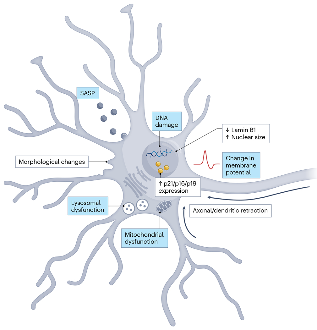
Cellular phenotypes of postmitotic senescent neurons include morphological changes, lysosomal and mitochondrial dysfunction, DNA damage, increased nuclear size, decreased expression of lamin B1, increased expression of p21, p16 and p19, changes in membrane potential and a senescence-associated secretory phenotype (SASP). The black arrows indicate axonal and dendritic retraction, which is a feature of postmitotic senescent neurons that does not occur in non-neuronal cell types. Processes highlighted in blue are being explored as senotherapeutic targets across senescent cell types. Increased β-galactosidase, lipofuscin accumulation, proliferation arrest and telomere attrition are senescence phenotypes that may occur in replicative or stress-induced senescence but are not specific markers for postmitotic senescent neurons and are not shown. p16: p16INK4a; p19: p19INK4d; p21: p21CIP1.
Senescent cells that accumulate within the brain or in peripheral tissues negatively impact brain health and function (Table 1). Senescent cell accumulation in the brain may be driven by comorbidities that increase the risk of developing cognitive impairment or brain dysfunction (for example, insulin resistance, obesity, alcohol use disorder48–50), or by physical stress to the nervous system such as traumatic brain injury51 or spinal cord injury52. Emerging evidence suggests that senescence is a contributing and causal factor for chronic pain and psychological stress-induced mental disorders, primarily through the release of inflammatory SASP factors49,50,53,54. Individuals with a high blood SASP index tend to experience more severe depressive episodes than individuals with low SASP indices55. Furthermore, elevated SASP expression levels have been linked to poorer treatment response and reduced remission rates in late-life depression56. These findings in humans are supported by mechanistic studies in mice, demonstrating a causal link between senescence and depressive behaviours49.
In 2018, studies began to report improvements in brain structure and function by elimination of senescent cells in transgenic mouse models of neurodegeneration 9,12,57,58. Mechanistic data indicated that clearing senescent cells improved brain structure, function and memory and reduced neuroinflammation and AD-associated neuropathology (Table 2). The clear link between senescent cell accumulation and organ dysfunction and healthy lifespan59–62, along with the potential for therapeutic interventions, led to a surge of funding to support research on cellular senescence in ageing and disease, including in the CNS63,64.
Table 2 |.
Selected senotherapeutic molecular targets under preclinical investigation for CNS conditions
| Drug target | Compounds (examples) | BBB penetrance | Condition/indication (CNS-specific only) | Mechanism |
|---|---|---|---|---|
| SRC kinase plus PI3K | Dasatinib–quercetin (ref. 65) | Dasatinib: yes (ref. 273) Quercetin: yes/weak (refs. 274,275) |
Parkinson disease mice (ref. 12) Overtraining-induced deficits in Learning and memory (ref. 276) Age-related cognitive decline (ref. 277) Tauopathy mice (ref. 9) AD amyLoid mice (ref. 45) |
Senolysis: activation of apoptosis in senescent cells |
|
| ||||
| mTOR | Rapamycin (ref. 148) | Yes/weak (ref. 278) | Brain health/ageing/memory (refs. 279,280) | Blocks NRF2-mediated senescence induction and reduces NF-κB-mediated SASP gene expression |
|
| ||||
| HSP90 | Geldanamycin (ref. 101) | No (ref. 281) | ND | Autophagy-mediated senolysis |
| Tanespimycin (ref. 101) | Yes (ref. 282) | ND | ||
| Alvespimycin (ref. 101) | Yes (ref. 282) | ND | ||
| Ansamycin (ref. 101) | ND | ND | ||
| Resorcinol (ref. 101) | ND | ND | ||
| Purine and pyrimidine-like N-terminal HSP90 inhibitors (ref. 101) | ND | ND | ||
|
| ||||
| Seno-antigens: Surface markers of senescent cells | uPAR targeted by CAR-T cells (ref. 169) | Yes (ref. 283) | ND | Phagocytosis-mediated clearance of senescent cells |
| Antibody-based approach for DPP4 (ref. 170) | No (ref. 284) | ND | ||
| GPNMB as immunization antigen (ref. 171) | No (ref. 284) | ND | ||
|
| ||||
| Na+K+-ATPase | Proscillaridin A (ref. 103) | ND | ND | Senolysis by modulating plasma membrane potential |
| Ouabain (refs. 103,105) | Yes (ref. 285) | TBI (ref. 289) | ||
| Bufalin (refs. 103,105) | Yes (ref. 286) | ND | ||
| Cinobufagin (ref. 103) | ND | ND | ||
| Peruvoside (ref. 103) | ND | ND | ||
| Digitoxin (refs. 103,105) | Weak (ref. 287) | ND | ||
| Convallatoxin (ref. 103) | Yes (ref. 288) | ND | ||
| Digoxin (refs. 103,105) | Weak (ref. 287) | Dementia (ref. 290) | ||
|
| ||||
| BCL-2 protein Family | ABT-737 (ref. 107) | No/weak (ref. 292) | COVID-19 neuropathology (ref. 294) | Senolysis: deactivation of anti-apoptotic pathway/activation of apoptosis in senescent cells |
| ABT-263 (navitoclax) (refs. 61,90,109) | No/weak (ref. 292) | BBB disruption (ref. 295) AD (ref. 57) COVID-19 neuropathoLogy (ref. 294) Learning and memory in age (ref. 296) |
||
| A-1331852 (ref. 90) | Yes (ref. 293) | ND | ||
| A-1155463 (ref. 90) | ND | ND | ||
| UBX1325 (ref. 291) | ND | ND | ||
|
| ||||
| PUMA/NOXA | PCC1 upregulates PUMA/NOXA (ref. 139) | ND | ND | Mitochondria-mediated senolysis |
|
| ||||
| USP7 | P5091 (ref. 121) | Modified (ref. 297) | Tau phosphoryLation (AD modeL)
(ref. 299) ND |
p53-mediated senolysis (elevated p53 expression) |
| P22077 (ref. 121) | Yes (ref. 298) | |||
|
| ||||
| MDM2 | Nutlin3a (ref. 300) and UBX1325 (ref. 223) attenuate SASP UBX0101 (ref. 301) RG-7112 (ref. 302) |
No/weak (ref. 303), unclear | ND | p53-mediated senoLysis (eLevated p53 expression) |
| ND | ||||
| ND | ND | |||
| Yes (ref. 304) | ND | |||
|
| ||||
| p53–FOXO4 Interaction | FOXO4-DRI (cell-penetrating peptide that disrupts interaction of p53 with FOXO4) (ref. 120) | ND | ND | p53-mediated senoLysis (p53 cytosolic translocation) |
|
| ||||
| HDAC | Panobinostat (ref. 129) | Yes (ref. 305) | ND | BCL-XL and H3-mediated senolysis |
|
| ||||
| NF-κB | BAY11-7082 (ref. 306) | ND | Aggregation of amyloid-β protein and memory deficits (ref. 307) | Reduced NF-κB activity to decrease SASP gene expression |
|
| ||||
| BRD4 | ARV825 (ref. 132) | Modified (ref. 308) | ND | PROTAC, senolysis mediated by elevated DNA damage |
|
| ||||
| IKK | EF24 (curcumin analogue) (ref. 309) | ND | ND | Reduced NF-κB activity to
decrease SASP gene expression |
|
| ||||
| ATM | KU-55933 suppressed senescence and SASP (ref. 143) | No (ref. 310) | Neuroprotection against hydrogen peroxide-induced cell damage (ref. 311) | Reduced NF-κB activity to
decrease SASP gene expression |
|
| ||||
| OXR1 | Piperlongumine (ref. 135) | Yes (ref. 312) | Age-related cognitive impairment (ref. 313) | Induces degradation of OXR1 to induce senolysis |
|
| ||||
| Lysosome | Cytotoxic drugs encapsulated with galacto-oligosaccharides (ref. 114) e.g., Nav-Gal (ref. 116), 5FURGal (ref. 314), SSK1 (ref. 315) | ND | ND | Cytotoxic drugs are released in the senescent lysosomes, causing senolysis |
|
| ||||
| SASP | SR12343 (ref. 154) | Yes (ref. 155) | Improved dystonia and motor function (ref. 154) | Blocks NF-κB-IKK; reduces
NF-κB-mediated SASP gene expression p38 inhibition; reduces NF-κB-mediated SASP gene expression Reduced C/EBPβ activity leading to lower expression of SASP genes |
| SB203580 (ref. 158) | ND | ND | ||
| UR-13756 (ref. 159) | ND | ND | ||
| BIRB 796 (ref. 159) | ND | ND | ||
| MW01-18-150SRM (ref. 316) | Yes (ref. 317) | Neuroinflammation, psychiatric and cognitive behaviour (AD) (refs. 316,319) | ||
| Ruxolitinib (suppressor of C/EBPβ activity) (ref. 161) | Yes (ref. 318) | Learning and memory (ref. 320) TBI (ref. 321) |
||
|
| ||||
| BCL-XL | PZ15227 (ref. 113) | ND | ND | ABT-263-based BCL-XL PROTAC; senolysis |
|
| ||||
| cGAS | Aspirin | Yes (ref. 322) | Reduced SASP and senescence in brain organoids (ref. 182) | Reduced SASP gene expression by inhibition of COX2 |
|
| ||||
| STING | H-151 | Yes (ref. 183) | Ameliorated SASP in aged mice (ref. 183) and reduced senescence in brain organoids (ref. 182) | Reduced SASP gene expression by reducing NF-κB activity |
AD, Alzheimer disease; ATM, ataxia–telangiectasia mutated; BBB, blood–brain barrier; CAR, chimeric antigen receptor; CNS, central nervous system; COVID-19, coronavirus disease 2019; COX2, cyclooxygenase 2; DPP4, dipeptidyl peptidase 4; FOXO4, forkhead box protein O4; GPNMB, glycoprotein nonmetastatic melanoma protein B; HDAC, histone deacetylase; HSP90, heat shock protein 90; IKK, IκB kinase; mTOR, mechanistic target of rapamycin; ND, not determined; OXR1, oxidation resistance gene 1; PCC1, procyanidin C1; PI3K, phosphatidylinositol 3-kinase; PROTAC, proteolysis-targeting chimera; SASP; senescence-associated secretory phenotype; TBI, traumatic brain injury; uPAR, urokinase-type plasminogen activator receptor; USP7, ubiquitin-specific peptidase 7.
The pioneering discovery of compounds that could eliminate senescent cells, including in the brain, stimulated great interest in drug development65. Therapeutic approaches under investigation include methods to clear senescent cells from tissue, to gently revert them back to their original cell type or to dampen their SASP. The type of senescent cell and its associated mechanism of toxicity influence the choice of therapeutic strategy. For instance, drugs designed to clear senescent cells may be most suitable for replaceable cell types, such as microglia; whereas non-clearing strategies may be more appropriate to target cells that are difficult to replace, such as neurons. Research into lifestyle interventions and other non-pharmacological strategies to reduce senescent cell burden, including approaches such as mindfulness to treat insomnia in AD caregivers (NCT03538574), is also active and promising, and may be effective for some individuals66–68. However, this Review focuses on the various pharmacological approaches under investigation (Box 1).
Box 1 |. Senescent cell-targeting strategies.
Senotherapeutic drug strategies broadly fall into two categories, senolytics and senomorphics. Senolytics clear senescent cells from the tissue, whereas senomorphics allow senescent cells to remain but mitigate their pathogenic properties. Non-pharmacological strategies are also gaining attention, but are beyond the scope of this Review.
Senolytics
- Senescence-associated pathway inhibition
- SRC kinase and phosphatidylinositol 3-kinase inhibitors
- Heat shock protein 90 inhibitors
- Na+K+-ATPase inhibitors
- BCL-2 protein family inhibitors
- Ubiquitin-specific peptidase 7 (USP7), MDM2 and p53–forkhead box protein O4 (FOXO4) interaction inhibitors
- Histone deacetylase inhibitors
- BRD4 inhibitors
- Harnessing the immune system
- Glycoprotein nonmetastatic melanoma protein B vaccine
- Chimeric antigen receptor-T therapy (urokinase-type plasminogen activator receptor (uPAR))
- Prodrugs
- Galactose-modified duocarmycin prodrugs
- Proteolysis-targeting chimeras
- PZ15227
- ARV825
Senomorphics
- Senescence-associated secretory phenotype modulation
- Rapamycin
- Metformin
- NF-κB inhibitors
- p38 inhibitors
- JAK–STAT inhibitors
- Ataxia–telangiectasia mutated (ATM) inhibitors
- Cell reprogramming
- CDGSH iron–sulfur domain 2
- G-quadruplex structures
- DNA methylation
- Mitochondrial decay
Non-pharmacological senotherapeutics
Caloric restriction
Intermittent fasting
Exercise
CNS senotherapeutics in development
To develop effective senotherapeutics for the CNS, strategies need to incorporate various drug modalities against diverse targets and senescent cell types. This section describes therapeutic strategies that range from inhibition of specific senescence-associated signalling pathways, such as those that control SASP, to activation of apoptosis-inducing pathways. Several molecules have been identified as potential senotherapeutic targets (Table 2 and Fig. 3). Small-molecule approaches are the most straightforward option for targeting senescent cells in the CNS owing to their potential for oral bioavailability and relatively favourable profile regarding CNS penetration across the blood–brain barrier (BBB). Candidate molecular processes can be targeted with senolytics, which destroy senescent cells, or with senomorphics, which modulate senescent cells (Box 1). We also explore innovative approaches such as targeting proteins specific to senescent cells for degradation. Although not all the targets and approaches discussed in the following paragraphs have been directly tested in the CNS, they describe basic principles for tackling senescent cells and lay the groundwork for the future development of CNS-targeted applications.
Fig. 3 |. Potential molecular drug targets for CNS senotherapeutics.
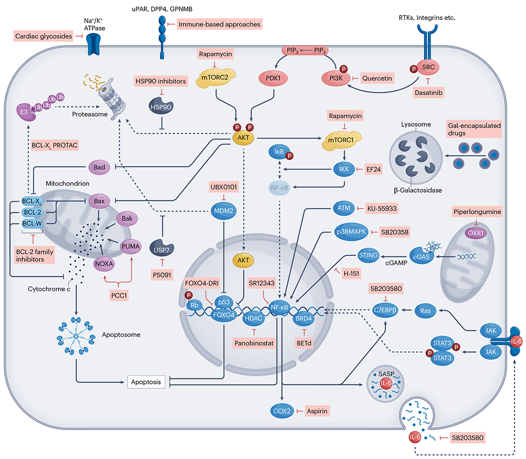
Senescent cells feature upregulated pro-survival pathways and a senescence-associated secretory phenotype (SASP). Several molecular players in these pathways are under investigation as potential targets for senotherapeutic strategies. Drug-targeting approaches to activate (red arrows), inhibit (red inhibitory lines) or promote (dashed arrows) protein transport within the cell can result in cell clearance (senolytics) or modulate the SASP (senomorphics). A combination of dasatinib and quercetin, which target Src tyrosine kinase signalling, were the first generation of potential senolytics to be discovered. Other promising approaches include targeted inhibition of the pro-survival BCL-2-related pathways, or NF-κB signalling to ameliorate the expression of SASP genes. Selected compounds are shown as examples (red boxes); see Table 2 for further details. Note that galacto-oligosaccharide (gal)-encapsulated drugs, which target raised β-galactosidase levels in the lysosomes of senescent cells, might not be suitable for central nervous system (CNS) applications owing to high β-galactosidase expression in non-senescent neurons. COX2, cyclooxygenase 2; DPP4, dipeptidyl peptidase 4; GPNMB, glycoprotein nonmetastatic melanoma protein B; HDAC, histone deacetylase; HSP90, heat shock protein 90; OXR1, oxidation resistance gene 1; RTK, receptor tyrosine kinase; Ub, ubiquitin; uPAR, urokinase-type plasminogen activator receptor; USP7, ubiquitin-specific peptidase 7.
Senolytics
Senolytics are a class of small-molecule therapies designed to selectively eliminate senescent cells. The term ‘senolytics’ originates from combining ‘senescence’ and ‘lytic,’ indicating the literal destruction of senescent cells. These cells are inherently damaged with upregulated apoptotic pathways. Their long-term survival relies on concurrent upregulation of pro-survival pathways inhibiting full engagement of apoptosis. Many senolytics function by deactivating these survival pathways, referred to as senescent cell anti-apoptotic pathways (SCAPs), allowing senescent cells to undergo apoptosis (Table 2 and Fig. 3). The therapeutic druggability of senescence survival pathways is explored further below.
PI3K and Src kinase inhibition.
Phosphatidylinositol 3-kinases (PI3Ks) are lipid kinases that have been shown to take crucial roles in the regulation of cell cycle, apoptosis, DNA repair, angiogenesis, cellular metabolism and cellular senescence69. Dysregulation of the PI3K pathway is observed in one-third of human tumours, which has led to the development of PI3K inhibitors as cancer therapeutics70. Similarly, the Src family kinases are involved in regulation of important cellular functions such as cell proliferation, differentiation, apoptosis, migration and metabolism71. Early activation of Src has been implicated in shifting the cell fate from apoptosis to senescence72.
A significant advancement in the field of senolytic agents was the discovery of a combinatorial therapeutic approach involving dasatinib and quercetin. Dasatinib, a Src tyrosine kinase inhibitor with FDA approval for leukaemia treatment, was combined with quercetin, a naturally occurring flavonoid that inhibits PI3K65. Although dasatinib had already demonstrated its ability to induce apoptosis in cancer cells, it initially showed no significant effects on senescent cell clearance when used alone. Similarly, quercetin, a relatively nonspecific PI3K inhibitor known for its anti-inflammatory properties and broad kinase inhibitory profile, demonstrated only modest senolytic potential as a monotherapy. However, the combination of dasatinib with quercetin synergistically influenced SCAPs and demonstrated significant senolytic potential65.
This combination of dasatinib and quercetin has since proved effective in eradicating senescent cells and improving health outcomes in mouse models of tau-associated neurodegeneration9, AD-associated amyloid-β accumulation45, frailty65, osteoporosis65, vasomotor dysregulation65,73, hepatic steatosis74, insulin resistance75, pulmonary fibrosis76, adipose tissue inflammation77, intervertebral disc degeneration78 and skeletal muscle debility79. The success of dasatinib–quercetin has stimulated considerable interest in combatting age-related CNS conditions in human trials (Table 3). Although the side effects of long-term use of dasatinib–quercetin remain unknown, to date all the clinical trials have reported favourable safety outcomes. Chronic complications of dasatinib treatment in cancer therapy include recurrent pleural effusions in up to 37% of patients, as well as possible side effects affecting platelet function, leading to a bleeding tendency80. To minimize potential severe side effects such as these, dasatinib–quercetin senolysis treatment is administered intermittently. Despite dasatinib’s short elimination half-life of less than 11 h, and quercetin’s poor bioavailbility81, early data from clinical trials indicate that dasatinib–quercetin is potent enough to significantly reduce the senescent cell burden in humans using this ‘hit-and-run’ regimen82, which simultaneously improves the safety profile.
Table 3 |.
Selected senotherapeutics under clinical investigation for central nervous system conditions
| Compounds | Targets | Indication | Sponsor | Study phase/type | NCT identifier |
|---|---|---|---|---|---|
| Dasatinib–quercetin combination therapy | Primary target Dasatinib: SRC kinase (ref. 65) Quercetin: PI3K (ref. 65) |
MCI with slow gait speed | Hebrew Senior Life | Phase I/II open label | NCT05422885 |
| Other drug
targets Dasatinib: ABL kinases (ref. 323) (ABL1, BCR–ABL); receptor tyrosine kinases (ref. 324) (EGFR, EPHA2, DDR1); non-receptor tyrosine kinases (ref. 324) (FRK, BRK, ACK), p90RSK (ref. 324) Quercetin: suppression of COX2, NF-κB and AP-1 pathways (ref. 325), nonspecific, multiple targets (ref. 326) |
MCI/early AD | University of Texas Health Science Center at San Antonio | Phase I/II POC open label | NCT04063124 | |
| MCI/early AD | Wake Forest University Health Sciences | Phase II RCT | NCT04685590 | ||
| MCI/early AD | Mayo Clinic | Phase I/II open label | NCT04785300 | ||
| Older adults with depression or schizophrenia | Washington University School of Medicine | Phase II RCT | NCT05838560 | ||
| UBX1325 | BCL-XL | DME or neovascular AMD | Unity Biotechnology | Phase I open label | NCT04537884 |
| Wet AMD | Unity Biotechnology | Phase II RCT | NCT05275205 | ||
| DME | Unity Biotechnology | Phase IIb | NCT06011798 |
Non-pharmacological trials or those using compounds with senotherapeutic potential (e.g., polyphenols, rapamycin, etc.) but not in a senescence indication are not included. AD, Alzheimer disease; AMD, age-related macular degeneration; DME, diabetic macular oedema; MCI, mild cognitive impairment; PI3K, phosphatidylinositol 3-kinase; POC, proof-of-concept; RCT, randomized controlled trial.
The first-in-human trial of dasatinib–quercetin demonstrated safety in a clinical population of idiopathic pulmonary fibrosis83, as did the first phase I trial in older adults with mild cognitive impairment (MCI) or early AD33. The MCI/early AD open-label study enrolled five participants to assess the safety of an intermittent dasatinib–quercetin treatment protocol and determine BBB penetrance of the compounds. Dasatinib and quercetin were administered orally for two consecutive days followed by a 2-week break, repeated for six cycles. Drug levels were measured in plasma and cerebrospinal fluid (CSF) under fasting conditions at baseline, before drug administration and 80–150 min after the final dose of study drug. After treatment, both dasatinib and quercetin were detectable in the plasma of all participants at higher levels than baseline. Dasatinib was also detected in the CSF in 80% of the participants after treatment; however, quercetin was not. The lack of quercetin detection could be attributed to its low bioavailability, levels being below detection sensitivity, the timing of CSF collection not capturing quercetin owing to its short half-life, or quercetin not penetrating the BBB. Two additional open-label phase I trials testing dasatinib–quercetin in AD (NCT04785300 and NCT05422885) will be important for providing confirmatory safety data, and the ongoing phase II SToMP-AD study (NCT04685590 (ref. 84)) will help to establish efficacy and timing of senescent cell re-emergence (Table 3). An interventional trial has also been initiated using dasatinib–quercetin in treatment-resistant depression, given the observed association between SASP and psychiatric symptoms55,56. Collectively, the data generated in these early trials are crucial for informing safety, dosing strategies and outcome measures that will guide future trials85.
Other nonspecific PI3K-inhibiting flavonoids have been investigated for their senolytic properties. Luteolin and curcumin have shown weak senolytic activity at a dose at which quercetin was ineffective86. Fisetin, a natural compound structurally related to quercetin, is found in various fruits, vegetables and teas, has gained considerable attention in the area of senolytic therapies87,88. Fisetin exhibits a diverse pharmacological profile including antioxidant, anti-inflammatory, antimicrobial as well as neuroprotective properties87–89. In addition to PI3K, fisetin acts on multiple signalling pathways that have pivotal roles in the senescence process, including BCL-2, PI3K–AKT, p53 and NF-κB. This multifaceted action has led to the recognition of fisetin as a senolytic compound, a discovery that emerged from systematic screening studies involving flavonoid polyphenols and senescent human and murine fibroblasts86,90. Notably, fisetin has demonstrated the ability to restore tissue homeostasis, mitigate age-related pathologies and extend lifespan in mice86. In relevance to neurodegeneration, fisetin has been shown to modulate inflammatory pathways and preserve cognitive function in AD transgenic mouse models91. It also enhances memory and induces hippocampal long-term potentiation in rats, suggesting its positive action in the brain and ability to ameliorate AD-related symptoms in vivo92. Fisetin is currently undergoing clinical testing in approximately 20 clinical trials targeting a range of age-related diseases, including MCI (NCT02741804)93.
Of note, like quercetin, fisetin has low oral bioavailability owing to factors such as low aqueous solubility, high lipophilicity and high first-pass metabolism94. To improve the bioavailability of these drugs, strategies may include careful modification of their chemical structures to enhance their pharmacokinetic profiles while maintaining their efficacy95, or using novel drug delivery systems96,97.
HSP90 inhibition.
The essential family of heat shock protein 90 (HSP90) is involved in multiple cellular processes and regulatory pathways such as apoptosis, cell cycle control, cell viability, protein folding and degradation, and various signalling cascades98. Dysregulation of HSP90 is implicated in diverse pathologies, including cancer and neurodegenerative diseases such as Parkinson disease99 and AD100. In a senescence assay-based screen of compounds that regulate autophagy, inhibitors of the HSP90 chaperone were identified to have senolytic activity in mouse and human cells, including fibroblasts, mesenchymal stem cells and umbilical vein endothelial cells101. Various HSP90 inhibitors showed dose-dependent senolytic potential, and inhibition of HSP90 delayed the onset of age-related symptoms, extended health span and reduced p16INK4a levels in a mouse model of a human progeroid (premature ageing) syndrome101.
Na+K+-ATPase inhibitors.
The Na+K+-ATPase pump is a crucial transmembrane protein that actively transports sodium and potassium ions against their respective concentration gradients to maintain the cellular osmotic balance and membrane potential102. Recent studies have reported that senescent cells possess a marginally depolarized plasma membrane and elevated concentrations of H+ compared with control cells103. A similar aberrant membrane potential phenotype has been described in senescent neurons10,37,104.
A high-throughput approach was used to determine the in vitro and in vivo senolytic potential of cardiac glycosides, which inhibit the Na+K+-ATPase103. In vivo treatment with cardiac glycosides induced the clearance of senescent tumour cells and of senescent fibroblasts in a mouse model of lung fibrosis103. The authors acknowledge that the therapeutic range for cardiac glycosides such as digoxin is relatively narrow. However, they also emphasize that digoxin has been successfully used for many years, and that monoclonal antibodies capable of reversing potential drug overdoses have been developed103. Another cardiac glycoside, ouabain, was shown to selectively eliminate cells undergoing oncogene-induced senescence in a mouse model of liver cancer105. Both studies suggest that the senolytic effects of cardiac glycosides are due to on-target inhibition of the Na+K+-ATPase, but further research is required to explore the molecular senolytic properties of these compounds, including for application to the CNS.
BCL-2 protein family inhibitors.
The BCL-2 family has a key role in regulation of apoptosis and has broader functions. These include neuronal activity, autophagy, calcium regulation, mitochondrial dynamics and energy production, as well as various other processes essential for maintenance of the health and functionality of cells106. The BCL-2 family includes both inhibitors and promoters of cell death, collectively orchestrating the intrinsic apoptosis pathway, with mitochondria having a pivotal role. Senescent cells use an anti-apoptotic programme driven by members of the BCL-2 family, including BCL-2, BCL-XL and BCL-W, to persist within tissues and contribute to chronic inflammation107.
Inhibition of the BCL-2 pathway can induce apoptosis in some, but not all, senescent cell types61,107. This discovery led to the identification of BCL-2 family inhibitors as senolytic compounds, including ABT-737, ABT-263 (navitoclax), A-1331852 and A-1155463. ABT-737, an inhibitor of BCL-W, BCL-XL and BCL-2, was the first in this class shown to specifically induce apoptosis in senescent cells through a BCL family-dependent mechanism107. In mice, ABT-737 effectively removed senescent cells in the lungs and epidermis, resulting in increased proliferation of hair-follicle stem cells107. However, ABT-737 has limitations such as poor oral bioavailability and low aqueous solubility, leading to the development of an orally bioavailable derivative, ABT-263, as a pan-BCL inhibitor108. In addition to its in vitro senolytic activity, ABT-263 has been shown to rejuvenate aged haematopoietic stem cells and effectively clear senescent cells in irradiated or aged mice109. Nonetheless, notable side effects, including thrombocytopenia and transient thrombocytopathy, have been reported for ABT-263 (ref. 110).
Inhibiting BCL-2 family members can pose challenges in diseases of the CNS. Treatment with ABT-737 has been found to be toxic to stem cell-derived dopaminergic neurons in vitro10, likely due to the reliance of neuronal development and survival on BCL-XL activity111. ABT-263 does not easily penetrate the BBB, hindering its ability to reach the brain parenchyma112. Nevertheless, studies in tau transgenic mice indicate that ABT-263 reduces senescent cells in the brain and improves behaviour57. Despite their demonstrated senolytic effects, the clinical translation of BCL-2 family inhibitors to CNS disorders is collectively constrained by potential side effects, off-target effects on platelets and limited CNS penetrance. Although systemic removal of senescent cells might positively impact the brain regardless of BBB penetrance (discussed below), future research efforts are still needed to develop more specific, safe and brain-penetrant BCL-2 family inhibitors for use as CNS senolytic therapies.
More recently, an ABT-263-based BCL-XL proteolysis-targeting chimera (PROTAC) senolytic has been developed to target BCL-XL to the cereblon E3 ligase for degradation113. Additionally, to selectively target senescent cells, researchers leveraged the elevated activity of the lysosomal β-galactosidase that is common in senescent cells, developing a drug delivery system based on encapsulating drugs with galacto-oligosaccharides114,115. In a mouse model involving chemotherapy-induced senescence, gal-encapsulated cytotoxic drugs effectively targeted palbociclib-induced senescent tumour cells, leading to improved regression of tumour xenografts. In another mouse model of pulmonary fibrosis, gal-encapsulated cytotoxic drugs or ABT-263 specifically targeted senescent cells, resulting in reduced collagen deposition and restored pulmonary function. Notably, the gal-encapsulation approach mitigated the toxic side effects associated with senolytic drugs114. Furthermore, Nav-Gal, engineered by linking the BCL-2 family inhibitor ABT-263 to an acetylated galactose group, selectively eliminated senescent cells both in vitro and in vivo, demonstrating reduced platelet toxicity and ameliorating the common issue of thrombocytopenia associated with ABT-263 treatment116. Although this approach is promising, targeting β-galactosidase for CNS conditions may not be appropriate as many non-senescent neuronal cell populations express high levels of β-galactosidase activity9,117 (Fig. 2), as discussed further below.
USP7, MDM2 and p53–FOXO4 interaction inhibitors.
Modulators of the p53 pathway have gained attention as potential senolytic agents owing to the role of p53 in controlling senescence and apoptosis, particularly through its regulation of BCL-2 family proteins118,119. One note-worthy example is forkhead box protein O4 (FOXO4), an anti-apoptotic transcription factor that is upregulated in some senescent cell types. FOXO4 prevents cell death by binding to and sequestering p53 within the nucleus120. To target this interaction, a senolytic peptide named FOXO4-d-Retro-Inverso (FOXO4-DRI) was developed. FOXO4-DRI disrupts the binding between FOXO4 and p53, which allows p53 to translocate to the cytosol and induce apoptosis in senescent cells. FOXO4-DRI has demonstrated selective removal of senescent cells and has shown promise in alleviating age-related symptoms across organ systems in animal models120.
p53 protein levels are tightly regulated through ubiquitylation by the E3 ubiquitin ligase murine double minute 2 (MDM2). Inhibitors that disrupt the MDM2–p53 interaction, such as UBX0101 and RG7112 (RO5045337), as well as inhibitors of the de-ubiquitinating enzyme ubiquitin-specific peptidase 7 (USP7), serve to increase and stabilize p53 levels. RG7112, UBX0101 and USP7 inhibitors, such as P5091 and P22077, cleared various peripheral senescent cells by upregulating p53 (ref. 121).
Moving into clinical applications, however, has faced challenges. Although UBX0101 has been shown to induce apoptosis of senescent synoviocytes, and a phase I study (NCT03513016) suggested that it has clinically meaningful effects on pain and function in patients with knee osteoarthritis, preliminary reports from the follow-up phase II trial of UBX0101 for osteoarthritis suggest that it has not demonstrated significant efficacy compared with placebo122. The authors speculated that possible reasons for this could include the dosing route, patient and/or investigator expectedness, and gender dimorphism in pain reporting. Manipulating p53 levels in senescence regulation requires careful consideration, as p53 can have both promoting and inhibitory effects depending on cell type and stress levels. Adding complexity to the therapeutic approach, p53 activity can induce p21-dependent senescence. Additionally, some p53-targeting agents are peptide based, which may limit their applicability to address brain senescence. As mentioned above, senescent cells have been reported in Parkinson disease post-mortem human brain tissue and model organisms10,12,47,123. Elevated p53 levels and activity were observed in the brains of patients with Parkinson disease, as well as in Parkinson disease animal and cellular models, mostly correlating with DNA damage124. p53-dependent brain senescence has also been reported in Huntington disease and AD124, suggesting that p53 may be an especially appealing target for CNS senescence. Recently, it was reported that a ketogenic diet induced p53-dependent cellular senescence in multiple tissues, including the brain125. These findings may have serious implications for humans on the ketogenic diet; however, we note that immunoblotting with an antibody to β-galactosidase was the primary readout, which may reflect changes in lysosomal function rather than senescence. Further research is needed to unravel the complicated molecular mechanisms by which p53 regulates senescence, with potential implications for clinical applications.
HDAC inhibitors.
Histone deacetylases (HDACs) modulate gene expression by removing acetyl groups at the lysine residues of histones and non-histone proteins. They impact many cellular functions including differentiation, proliferation and survival126. Senescent cells show changes in H3 acetylation and BCL-XL expression107,127,128. In vitro, the FDA-approved HDAC inhibitor panobinostat cleared cancer cells with chemotherapy-induced senescence. This activity was associated with decreased BCL-XL expression and increased H3 acetylation129.
BRD4 inhibitors.
Bromodomain and extra-terminal domain (BET) family proteins act as transcriptional regulators130. The family member BRD4 is well-characterized for its role in transcriptional elongation131. In a high-throughput screen of compounds alongside bio-functional analysis, the BET family protein degrader (BETd) was identified as senolytic132. The authors showed that inhibition of the BET family protein BRD4, either with chemical inhibitors or gene-specific RNA interference, induced senolysis. The hetero-bifunctional PROTAC ARV825 proved particularly effective, as it recruits BET family proteins to the E3 ubiquitin ligase cereblon, leading to their degradation133. The senolytic effects of ARV825 were mediated by attenuating non-homologous end joining (NHEJ) and enhancing autophagic gene expression. Specifically, the authors noted that ARV825 induces senolysis by exacerbating DNA double-strand breaks through targeting at least two independent mechanisms of the NHEJ machinery in senescent cells: first, inhibition of gene expression of XRCC4, which encodes a protein required for NHEJ repair; and secondly, blockade of the recruitment of 53BP1, an adaptor protein necessary for the assembly and activation of the DNA repair machinery, to double-strand break sites132. Treatment with ARV825 was sufficient to eliminate senescent cells by targeting BRD4, as confirmed by small interfering RNA (siRNA)-based gene-specific experiments, which showed that ARV825 reduced levels of BRD3 and BRD4, but not BRD2. Furthermore, the compound showed senolytic activity in the livers of obese mice and cleared senescent hepatic stellate cells, as well as chemotherapy-induced senescent cells in mice132.
OXR1 inhibitors.
Oxidation resistance gene 1 (OXR1) acts as a sensor and regulator of cellular oxidative stress. It is involved in the transcriptional networks needed to detoxify reactive oxygen species and modulate cell cycle and apoptosis134 and is upregulated in senescent human fibroblasts135. Interestingly, OXR1 is the target of the previously identified senolytic piperlongumine, a natural compound found in long pepper135,136. Piperlongumine was identified by screening a library of rationally selected compounds for their senolytic activity in human senescent fibroblasts136. The initial study reported that piperlongumine kills senescent fibroblasts without the induction of reactive oxygen species; however, it was later demonstrated that piperlongumine binds to OXR1, leading to its degradation via the ubiquitin–proteasome system specifically within senescent cells. The reduction of OXR1 induces apoptosis exclusively in senescent cells, likely by increasing their vulnerability to oxidative stress135. In a recent computational study investigating natural senotherapeutics to find candidates that could replace dasatinib, based on their similarity in gene expression effects, piperlongumine was identified as the highest-potential substitute, suggesting that further development of the compound is warranted137.
PUMA/NOXA activators.
PUMA and NOXA are pro-apoptotic genes that are functionally repressed by BCL-2 family molecules in senescent cells138. Through screening natural products, procyanidin C1 (PCC1), a polyphenolic component of grape seed extract, was identified as being capable of ameliorating the SASP at low concentrations and exhibiting senolytic effects at higher concentrations139. It was hypothesized that PCC1-induced apoptosis in senescent cells is partially mediated by NOXA and PUMA, accompanied by increased reactive oxygen species production and mitochondrial dysfunction139. Additionally, PCC1 ameliorated physical dysfunction and prolonged survival in mice.
Senomorphics
Senomorphics, also known as senostatics, mitigate the toxicity of senescent cells without removing them from the tissue, with the ultimate goal of retaining the cells’ initial physiological functions (Table 2 and Fig. 3). This section focuses on the molecular pathways targeted in senomorphic drug development.
ATM inhibitors.
The ataxia–telangiectasia mutated (ATM) kinase is crucial for DNA damage response signalling and is known to activate NF-κB in response to stress. NF-κB is a transcription factor involved in the regulation of the immune response, inflammation, cell survival, cellular senescence and organismal ageing140. ATM kinase mediates the equilibrium between senescence and apoptosis. Active ATM promotes autophagy, with a specific emphasis on sustaining the lysosome–mitochondrion connection, boosting senescence while restraining apoptosis141. Disruptions in autophagy mechanisms, however, have been demonstrated to elevate DNA damage levels, thereby promoting the development of cancer and neurodegenerative diseases142. Activated ATM has been reported in senescent cells, and genetic and pharmacological (using KU-55933) inhibition of ATM resulted in a reduction in NF-κB activity, markers of senescence and the SASP in cells as well as in mouse models of ageing143. This treatment also extended the health span of progeroid mice, indicating that ameliorating ATM-mediated NF-κB activity can slow the progression of ageing143.
mTOR inhibitors.
The PI3K-related kinase mechanistic target of rapamycin (mTOR) regulates numerous cellular pathways including apoptosis, growth and autophagy144,145. mTOR inhibition has emerged as an avenue to improve longevity and ameliorate the impact of cellular senescence. The mTOR inhibitor rapamycin, an FDA-approved immunosuppressive drug, has repeatedly proved its ability to extend lifespan in various organisms, ranging from yeast to mice146,147. Rapamycin promotes autophagy, a cellular process that has a crucial role in recycling damaged components and maintaining cellular health. Although not a senolytic, rapamycin has gained attention for its potential to suppress the SASP148. Although rapamycin holds promise for extending lifespan and curbing the SASP, clinical applications have encountered challenges, including metabolic dysregulation; impaired wound healing, a process in which senescent cells appear to have a beneficial role149; and the development of hyperlipidaemia150. Despite these challenges, the positive outcomes observed in various animal models of ageing and neurodegeneration have propelled rapamycin into clinical trials to assess its potential in treatment of AD (NCT04200911, NCT04629495), although it is not known whether senescence-associated outcomes will be assessed.
NF-κB inhibition and SASP modulation.
The transcription factor NF-κB has a dual role, acting as both a guardian against and a potential contributor to neuroinflammation and the accumulation of senescent cells in the brain151. It mediates neuroinflammation by enhancing the expression of pro-inflammatory molecules, many of which are part of the SASP, such as inducible NO synthase (iNOS), cyclooxygenase 2, TNF and interleukins (for example, IL-1 and IL-6). Under normal conditions, the activity of NF-κB is restrained by inhibitors known as IκB proteins. However, in response to stress or inflammation, IκB inhibitors are phosphorylated by the IκB kinase (IKK) complex, unleashing NF-κB and triggering a cascade of immune responses152. Studies have shown that enhanced NF-κB activity can lead to neuroinflammation and an increased burden of senescent cells in the brain151. Conversely, inhibition of the binding between NF-κB and IKK, using a peptide inhibitor, ameliorated senescence in progeroid mice153. Building on this, researchers developed a small molecule, SR12343, which disrupts the interaction between NF-κB and IKK. This molecule significantly reduced the release of SASP factors, alleviated senescence and improved markers of senescence in mice154. Importantly, SR12343 can traverse the BBB, making it a compelling candidate for evaluation in the context of neurodegenerative conditions155. In a recent study using SR12343, it was found to lower markers of cellular senescence and inflammation in the liver, skeletal muscle and blood, which were elevated in response to chemotherapy. Additionally, it was reported that SR12343 effectively counteracted chemotherapy-induced skeletal muscle wasting and dysfunction156.
The p38 pathways regulates the transcriptional activity of NF-κB157. Consequently, inhibiting the p38 pathway has emerged as a promising avenue to address the detrimental effects of the SASP and reduce age-related inflammation, particularly in the context of neurodegenerative conditions. The p38 inhibitor SB203580 has been shown to reduce the expression of SASP factors in human senescent cells158. Similarly, next-generation p38 inhibitors, such as UR-13756 and BIRB 796, have demonstrated promising results in limiting the expression of IL-6. By reducing the p38-dependent transcriptional activity of NF-κB on pro-inflammatory genes, these inhibitors mitigate SASP-related effects, marking them as potential therapeutic candidates for age-related inflammatory conditions159.
The regulation of the SASP is complex. Recent findings indicate that the immune gene transcriptional effector C/EBPβ cooperates with NF-κB to activate SASP genes160. JAK–STAT signalling is involved in the regulation of C/EBPβ and vice versa. Inhibition of the JAK–STAT signalling pathway by ruxolitinib suppresses the transcriptional activity of C/EBPβ. This suppression led to a reduction in systemic inflammation, attributed to SASP repression, and an improvement in the overall fitness of aged (24-month-old) mice161. These findings underscore the potential of modulating stress kinase p38 as a senomorphic approach to combat neuroinflammation, a common feature of neurodegenerative disorders. To tackle neuroinflammation in neurodegenerative diseases, a brain-permeable and orally available p38 inhibitor, MW150 (also known as MW01-18-150SRM), is currently in phase II trials for AD (NCT05194163).
Although strategies to reduce NF-κB activity hold promise in the realm of senomorphic therapies, inhibition may lead to side effects owing to NF-κB involvement in diverse biological processes. An alternative approach to reducing inflammation, perhaps with a better safety profile, involves the use of the nonsteroidal anti-inflammatory drug, ibuprofen. This strategy has been shown to mitigate senescent cell accumulation, neuroinflammation and cognitive dysfunction in mice151. However, clinical trials using ibuprofen as monotherapy for AD have not significantly impacted disease progression or outcomes162,163, but perhaps could be a useful combination therapy adjunct.
Harnessing the immune system
A healthy immune system actively clears senescent cells and prevents their accumulation. Therapeutic approaches to boost senescent cell clearance by the immune system are under investigation. These include enhancing the recognition of senescent cells by the immune system, promoting immune cell activity or modulating immune responses to improve the clearance of senescent cells from tissues. Generally, modulating the immune system for senolysis represents a promising approach.
The body’s natural defence against senescent cells involves immune surveillance mechanisms, with immune cells such as macrophages, T cells, natural killer cells (NK cells)164,165 and microglia in the brain44,166 tasked with removing these cells. However, age-related immunosenescence and the ability of senescent cells to evade immune detection – for example, by expressing immunosuppressive molecules such as PDL1 and PDL2, by producing tolerogenic major histocompatibility complex class I variants and by secreting factors that attract immunosuppressive cells167 – can lead to their accumulation in tissues168.
Notably, some senescent cells upregulate specific surface markers that are being explored as therapeutic targets. Urokinase-type plasminogen activator receptor (uPAR) has been shown to be upregulated in several senescent cells, including mouse lung adenocarcinoma cells induced to senesce by MEK and CDK4/6 inhibition, oncogene-induced senescence in mouse hepatocytes and culture-induced senescence in mouse hepatic stellate cells169. Mass spectrometry analyses comparing proliferating human fibroblasts with those undergoing replicative senescence revealed an increase in the surface molecule dipeptidyl peptidase 4 (DPP4; also known as CD26) in senescent cells170. Transcriptome data from senescent vascular endothelial cells indicated an enrichment of the transmembrane glycoprotein nonmetastatic melanoma protein B (GPNMB)171. Collectively, uPAR, DPP4 and GPNMB were identified as ‘seno-antigens’ and targeted for immune-based interventions as described below.
To bolster immune clearance of senescent cells, chimeric antigen receptor-T (CAR-T) cells have been used. In this approach, T cells from patients are genetically engineered to target and eliminate cells expressing specific antigens. Utilizing uPAR as a target antigen, CAR-T cells have been shown to effectively clear senescent cells, mitigating various pathologies in mouse models of senescent lung adenocarcinoma and liver fibrosis169. Similarly, antibody-based therapies targeting the senescent cell surface protein DPP4 have demonstrated efficacy in eliminating senescent cells in vitro170.
A vaccine-like protocol aimed at senolysis has emerged as another possible approach to mitigate senescent cell burden. Immunizing mice against GPNMB, which is elevated in senescent vascular endothelial cells and in samples from patients with atherosclerosis, reduced GPNMB-positive cells, improved metabolic parameters in mice fed a high-fat diet, reduced atherosclerotic plaque burden in Apoe-knockout mice, improved age-related phenotypes and ultimately extended the lifespan of progeroid mice171. Developing immune- and vaccine-based approaches to eliminate senescent cells is intriguing, but identifying senescence-specific antigens is crucial to avoid potential autoimmunity against non-senescent cells. To date, none of the senescence-associated surface markers seems truly exclusive for senescent cells, as GPNMB is expressed across tissue and cell types and has diverse roles in inflammation and disease172,173; DPP4 is known to be ubiquitously expressed and involved in immunoregulatory actions174; and uPAR is present on many immune cells and is detected in various bodily fluids including plasma and seminal fluid, and in extracellular matrix175.
In addition to the challenge of identifying markers exclusively expressed on senescent cells, targeting CNS senescence through immune modulation faces other challenges. Immune cell trafficking into the CNS is tightly regulated by the BBB, permitting entry only for specific immune cell subsets176. Additionally, immune-based approaches trigger an inflammatory response, which could be problematic in the context of neurodegenerative diseases, where neuroinflammation is present. Therefore, while harnessing the immune system holds great potential for senescent cell clearance, the impact of on-target, off-tissue effects and unintended immune responses must be carefully evaluated to minimize potential harm.
Future perspective
Many ‘first-generation’ senolytic drugs such as quercetin, fisetin and piperlongumine, lack a single precise target and instead affect multiple pathways that are implicated in cellular ageing. These broad-spectrum senolytics have the capacity to impact multiple senescence pathways simultaneously177; however, they present a challenge in predicting efficacy owing to the considerable heterogeneity between senescent cell populations. Drug discovery programmes based on identifying molecules that are exclusively upregulated in senescent cells offer opportunities for more precisely targeted senolytic therapeutics. Other exciting senotherapeutic approaches, currently in preclinical stages, include cellular reprogramming178; targeting mitochondrial function179–181; targeting the cGAS–STING pathway182,183; and various genome-targeting approaches including G-quadruplex structures184, DNA methylation185 and retrotransposon activity186. These diverse and innovative approaches reflect a growing enthusiasm for senotherapeutics and underscore the need for continued research to fully realize their potential for CNS conditions.
Challenges in developing CNS senotherapies
Senotherapeutic drug development faces several challenges, some of which are common to CNS drug development more broadly and contribute to the long development timelines of such therapeutics187. Here, we consider challenges that are particularly important in the development of CNS senotherapeutic drugs and discuss strategies to address them.
Senescence-specific targeting
The need to achieve selectivity for therapeutic targets and specific cells, while sparing healthy cells, to achieve a favourable safety profile is a common challenge for drug discovery. Senotherapeutics that impact cellular mechanisms that are vital for neuronal viability and function, such as Na+K+-ATPase inhibitors188, BCL-2 inhibitors189 and NF-κB inhibitors190, pose a particularly substantial risk. Understanding cellular senescence in various disease states and identifying relevant cell types are crucial to identify and select optimal senotherapeutic targets191. Drug development, and most senescent cell identification, occurs in laboratory models. Discerning the differences between human disease and experimental models is a crucial step in safely and effectively translating senotherapies to the clinic. We anticipate that technological advances in single-cell and spatial multi-omics, along with advanced computational and AI approaches capable of analysing the vast datasets, will enable the identification of tractable and specific targets for senotherapeutic interventions28.
New drug modalities in development could help to increase specificity for therapeutic targeting. For example, PROTACs, as mentioned earlier, are an innovative mechanism that capitalizes on the cell’s ubiquitin–proteasome system192. They hold promise for specificity, reduced side effects and the ability to hit targets previously deemed ‘undruggable’. However, their size presents a significant hurdle to optimization of CNS delivery, necessitating research efforts to improve their design for such applications. Beyond modalities that directly engage the target proteins, others such as gene therapy and RNA-based therapies offer versatile approaches for gene silencing or functional restoration and are in early stages of development193.
BBB penetrance
Favourable properties of molecules that target the CNS include small size, hydrophobicity optimal for penetrating the BBB without becoming trapped in the lipid bilayer or losing blood solubility, and low affinity for the P-glycoprotein transporter194. Peptides and antibodies offer higher specificity compared with small molecules, but they have low BBB penetrance and face challenges related to stability, solubility and incompatibility with oral administration. Advances in engineering antibodies and designing peptides that are more resistant to degradation, along with new delivery systems and BBB-crossing strategies, such as tagging with molecules that allow uptake via receptor-mediated transcytosis (for example, TfR195 and CD98hc196) are making them more viable for CNS applications197, although this is a complex and ongoing challenge. Even when drugs penetrate the brain, determining the correct dosage is challenging because drug metabolism in the brain can differ from that in other organs198,199.
Notably, the cells that comprise the BBB are susceptible to becoming senescent. Vascular smooth muscle cells were identified as the most prominent senescent cell type across 50 healthy human tissues31. A separate study identified a significant increase in senescence gene expression in vascular cells isolated from post-mortem human brains with advanced Braak stages of AD compared with control cases200. These findings suggest that senotherapies may benefit the brain by targeting vascular cells, potentially without the need for BBB penetration or direct brain exposure. Although BBB penetrance is a key consideration for CNS drug development, there is some evidence that clearing senescent cells in peripheral tissues may benefit brain function. For example, ABT-263, which does not easily penetrate the BBB, has shown benefits to the CNS following whole-body irradiation201 in mouse models that develop AD neuropathology57,202 and in glioblastoma models203,204. Although the senescence-inducing insults (for example, radiation, toxic tau and amyloid-β, or cancer, respectively) may have allowed ABT-263 to penetrate the BBB, it is also possible that the benefits were an indirect result of clearance of senescent peripheral cells, which have been shown to impact senescent cell accumulation in the brain205. Thus, senolytic therapies may improve brain function by clearing senescent immune cells and cells of brain vasculature without a need to fully penetrate the parenchyma.
Although the ‘hit-and-run’ strategy of intermittent dosing has been used to minimize potential adverse events owing to prolonged systemic levels of senolytics, an alternative approach is local delivery. This strategy was used in a recent open-label phase I trial for UBX1325 (foselutoclax), a senolytic small-molecule inhibitor of BCL-XL, in patients with advanced diabetic macular oedema (DME)206. In the study, UBX1325 was administered via intravitreal injection, keeping plasma levels of UBX1325 and its active parent molecule below the lower limit of quantification and minimizing adverse events linked to systemic BCL-XL inhibition. Visual acuity improvements were observed in six of eight patients at 12 weeks, and in five of eight patients at 24 weeks. The favourable safety profile supports the ongoing phase IIb study efforts (Table 3).
Risks of neuronal senolysis
The question of whether postmitotic neurons that have entered a senescence-like state should be eliminated requires careful consideration. The therapeutic elimination of neurons is not unprecedented, with the best-known example of this being the surgical removal of small volumes of brain tissue to treat drug-resistant epilepsy207. Evidence to support the clearance of senescent neurons includes preclinical studies in tauopathy mice that demonstrate benefits from the clearance of NFT-bearing neurons9. Neurons with NFTs maintain the ability to respond to inputs and remain active in vivo208 but contribute to pathogenic tau spread in an activity-dependent manner209. Similarly, senescent neurons maintain electrical activity, but with altered function. For example, inducing a senescence phenotype in excitatory neurons by deleting Gdf11 causes neuronal hyperexcitability with reduced synaptic inputs in vivo104. A similar hyperexcitable phenotype with decreased synaptic protein expression was observed in an in vitro model of neuronal senescence generated by transfecting iNeurons with CDKN2A208. Removing SATB1 in human dopaminergic neurons drives a senescence-like phenotype in vitro, whereby the cells maintain spontaneous pacemaking activity but show significant differences in maintenance of response to positive current injections10. Regulated neuronal activity is crucial for proper brain function, warranting a better understanding of the consequences of altered excitability in senescent neurons.
Senescent cells induce paracrine senescence in neighbouring cells through their SASP. Neuronal SASP may contain toxic aggregate-prone proteins47, which could facilitate senescence spread between distant regions particularly via well-connected, anatomically linked neurons210. A link between intraneuronal tau19,98 and α-synuclein deposition123 and senescence has been established. Given that neurons increase the release of α-synuclein after becoming senescent47, it is tempting to speculate that neuronal senescence is a key process mediating the transfer of aggregate-prone proteins between neurons and across circuits. Senescent neurons also spread senescence to non-neuronal cells including to astrocytes through traditional SASP mechanisms208 and to microglia through phagoptosis44,166. Specifically, neurons that contain NFTs expose phosphatidylserine on their cell membranes, which signals microglial phagocytosis166. Upon engulfing the NFT-bearing neuron, the microglia become senescent, resulting in their decreased tissue surveillance and their release of partially digested NFT toxic fragments into their environment, which amplifies pathogenesis44 (Fig. 1). In summary, the connectivity of senescent neurons within complex neuronal circuits and their close interaction with glia cells may allow them to propagate neuropathology to multiple brain cell types, which provides rationale in favour of their clearance. However, in some neurodegenerative models associated with aberrant microglial activity, positive outcomes have been seen from retaining damaged neurons by interrupting phagoptosis211–214. These studies highlight the nuanced, and important, choice of senotherapy approach (senolytic versus senomorphic) depending on context and condition, especially when targeting cells and tissues with limited regenerative capacity.
Measuring CNS target engagement
Target engagement, which measures the interaction of a compound with its intended molecular target within a living system, provides insights into the drug’s pharmacological profile, mechanism of action and potential efficacy. Evaluation of precise target engagement in preclinical senolytic studies often involves histological analysis, as well as protein and gene expression measurements on mouse brain tissue9,45,57. These methods offer detailed insights into the cellular and tissue distribution of senolytic effects. However, assessment of target engagement in living humans requires in vivo imaging strategies or surrogate measures in accessible biofluids.
MRI provides information on the effects of a drug on brain morphology and physiology215 and has been used to assess the impact of senolytics in a tauopathy mouse model9. Although MRI cannot provide direct evidence for the action of senotherapeutics on pro-survival or SASP pathways within senescent cells, it allows for the longitudinal tracking of disease progression in the brain, providing a surrogate marker of drug effects in the target tissue. Positron emission tomography (PET) tracks the accumulation of radiolabelled drugs in the body216, with ongoing development of senescence-specific PET tracers focusing on labelling lipofuscin217 or β-galactosidase218, which appear when mitotically competent cells enter senescence. However, these tracers may have limited applicability to cerebral tissues as lipofuscin deposition and elevated β-galactosidase activity can also be detected in healthy CNS tissue throughout the lifespan9,117,219 (Fig. 2).
Other strategies for identifying surrogate markers of target engagement include characterization of the surface proteome of senescent cells to discover seno-antigen-specific proteins unique to these cell populations220,221, which can then be visualized using clinical imaging techniques, such as immuno-PET222. A recent report suggests that senescent cells produce unique oxylipins that are released upon their death, raising the possibility of developing senolysis-specific biomarkers223. Although the identification of PET tracers specific to brain senescent cells could represent a significant advance, it remains unclear whether the small percentage (2%) of sporadically dispersed senescent cells19 would produce a strong enough signal to be detectable in PET imaging.
The limitations with imaging senescent cells in the brain have increased the reliance on biofluid analytes as surrogate markers of target engagement. Mouse studies suggest that senescent T cell levels in blood could serve as a potential surrogate for senescent burden in solid tissues, including the brain205. In this regard, p16INK4a (p16) mRNA expression in peripheral blood T lymphocytes has emerged as a quantifiable measure of total senescent cell load224 and is being used in clinical trials62,225,226, including an ongoing phase II trial for AD84. Other commonly used surrogate markers for senescent cells include circulating SASP factors227 such as cytokines, chemokines, proteases and growth factors82,83. Similar to measuring a SASP index, as mentioned earlier, the first phase I clinical trial to test senolytics in older adults with MCI or early AD, used a SASP cytokine panel to evaluate senescence-associated markers of inflammation in the blood and CSF33. However, the challenge with using SASP factors is that they can be secreted for reasons other than cellular senescence, complicating the interpretation of outcomes. Ongoing efforts to understand how the secreted milieu of senescent cells varies by cell type or context will help to clarify the utility of these promising biomarkers228,229. Moreover, results from placebo-controlled trials, such as the phase II SToMP-AD study, are needed to interpret how SASP readouts relate to senolytic effects84.
Methodological advances with omics technologies are paving the way for a deeper understanding of molecular changes associated with CNS diseases230. These approaches may allow for the identification of rare and distinct senescent cell populations and omics changes in response to senolytic therapies. For example, whereas traditional SASP protein markers did not change significantly in the first phase I trial for AD33, lipidomics and transcriptomics analyses revealed analytes that may be more sensitive to senolytic treatment85. While we are cautious not to overinterpret the results from the open-label trial, especially given the small sample size, the approach underscores the ongoing efforts to identify biomarkers specific to senescent cells and their clearance.
Applying AI to senotherapy development
The complex and heterogeneous phenotypes of senescent cells requires innovative approaches in developing effective senotherapeutics. Advances in experimental platforms and omics technologies, coupled with AI and ML, enable the extraction of relevant information for chemical activity and functional genomics in drug discovery231. AI refers to the simulation of human intelligence in machines for performing tasks, making decisions and adapting by learning from their experiences. ML, a specific AI technique, involves training computer algorithms to learn from data and take actions based on the learned knowledge231. Predictive modelling using ML algorithms expedites decision-making232, identifies potential drug candidates with a higher likelihood of success, and reduces costs and development time233,234.
In this section, we briefly review AI and ML applications in key steps of senotherapeutic development, including identification of senescent cells, identification of senescence targets, assessment of druggability and virtual screening compounds, and highlight major challenges in these areas. Overall, the application of AI and ML in senescence research is still in its early stages but holds potential to accelerate drug development in this area.
Identifying senescent cells
Senotherapeutic drug development requires accurate identification of senescent cells. ML approaches can be used to classify and characterize senescent cells using morphological and omics (transcriptomics, proteomics and methylomics) data235. For instance, ML methods such as multivariate regression analysis236,237 and k-means clustering238 have been used to identify senescent cells using bulk transcriptomic data. In multivariate regression analysis, the ‘known’ senescent genes were treated as variates. For single-cell RNA-sequencing data, Teo et al.32 used pseudotime trajectory analysis to identify senescent cell subpopulations. This is an especially powerful approach as it captures cells and cell functions at various stages of senescence progression. Traditional methodologies focus primarily on the end point senescent state at which lysosomal function and cytoskeletal abnormalities are enriched. However, trajectory analyses suggest that cellular respiration and active transportation are transiently elevated in the middle trajectories, which could be interesting targets to explore as their modulation could potentially prevent cells from entering the terminal senescence state.
Others are investigating morphology-based approaches to identify and classify senescent cells. A convolutional neural network (CNN) system, Deep Learning-Based Senescence Scoring System by Morphology (Deep-SeSMo), was trained on phase-contrast images of human umbilical vein endothelial cells treated with senescence-inducing agents239. Thousands of single cells were given a senescence probability score, predicted as senescent or control, and then compared with predetermined answers. Weighted, automatic and iterative optimization was used to improve the accuracy of the system, resulting in an accuracy of 0.93 and area under the curve of the receiver operating characteristic of 0.98.
Although these approaches hold great potential, limitations include a lack of rigorous wet-lab experiments to confirm the informatic predictions. For example, studies with cell culture validation data commonly rely on SA-β-gal as a gold-standard marker for senescence, which may be misleading, especially for brain cells.
Predicting novel senescence-associated genes
Intersecting cancer driver genes and ageing-associated genes identified dual-purpose targets for the treatment of cancer and ageing and revealed that senescence pathways were upregulated in one subgroup240. A comprehensive transcriptomic network analysis of human tissue-specific RNA-sequencing data from the Genotype-Tissue Expression Project (GTEx) identified co-expressed gene modules enriched for senescence-associated genes31. This approach revealed 51 senescence-associated genes conserved across all human tissues and identified regulators centred around CDKN1A (p21)31. Although p21 is not a novel senescence-associated molecule, the study represents a first effort in systematically identifying senescence pathways and regulators across multiple human tissues31.
Predicting novel senotherapeutic compounds
Research teams are also using ML approaches to discover novel senolytics239,241. A dataset with 58 known senolytics and 2,465 non-senolytic drugs, each characterized by 200 molecular and structural properties241, was used to train SVM and Random Forest-based classifiers to predict new senolytics. The trained classifiers were then used to screen diverse chemical libraries and identified several potential new senolytic compounds. Three compounds, all plant-derived molecules (oleandrin, periplocin and ginkgetin), showed evidence of senolysis using in vitro models. Mechanistically, oleandrin is a cardiac glycoside that inhibits Na+K+-ATPase; periplocin, also a glycoside, induces apoptosis through AMPK–mTOR or ERK signalling; while ginkgetin, a naturally occurring biflavonoid, acts on multiple pathways crucial to senescence, including apoptosis induction, cell cycle arrest and JAK–STAT and MAPK signalling.
A separate approach used a morphology-based CNN model, Deep-SeSMo, to calculate senescence scores in a drug screen of 80 compounds tested for their senolytic ability. The readout was percentage of SA-β-gal-positive cells, without presenting data on total cell counts, making it unclear whether senolysis occurred. One possibility is that these experiments may have identified senomorphics that impact lysosomal function and SA-β-gal activity, which is consistent with the senotherapeutic agents chosen to train the system, metformin and nicotinamide mononucleotide, both of which act as senomorphics, not senolytics (Box 1).
These early studies showcase the power of AI and/or ML to rapidly identify multiple potential senotherapeutics, increasing the need for validation in appropriate brain cell culture systems and in vivo models to understand the true predictive potential of these innovative models.
Predicting druggability
Although identifying and selecting a target is a crucial initial step in combatting senescence, the target’s druggability remains a decisive factor in determining the viability of potential therapeutic interventions. Druggability refers to the likelihood that a specific biological target can be effectively modulated by a drug-like molecule242,243. This aspect is particularly important when exploring potential therapeutic interventions for senescence owing to the complex pathways involved and the diverse nature of cellular senescent responses. For a target to be considered druggable, it should be accessible to the intended drug, whether that is a small molecule or a larger biological agent. The probability of successfully drugging a given target is greatly improved if its structure can be elucidated using methods such as NMR, X-ray crystallography or cryo-electron microscopy179, as it allows for the use of structure-based drug design. This approach, which uses computer algorithms to identify potential binding pockets in the relevant domains of the target protein and virtually screen them for chemical matter that is likely to engage with these regions before commencing experimental work, greatly cuts down on the number of compounds needed in phenotypic screening approaches relative to the conventional ‘brute-force’ approach, in which large chemical libraries are screened244. Notably, recent advancements in AI systems, such as AlphaFold245 have the potential to revolutionize protein structure prediction by reducing reliance on laborious experimental methods, thereby significantly broadening the scope of available targets for drug design explorations.
Predicting off-target effects
Using systems biology approaches246,247, including network modelling and integration of multi-omics data, can provide comprehensive insights into the global effects of senescence-targeting drugs beyond the intended target. These tools can facilitate the identification of potential off-target effects, discover novel pathways impacted by the drug or drug combinations and optimize therapeutic efficacy while minimizing adverse effects.
Conclusions
Over the past 5 years, research on cellular senescence in the brain has evolved from phenomenology to clinical trials. Overcoming challenges in CNS drug delivery, deepening our understanding of senescence-associated mechanisms and appropriate targets, and discerning when to use senolytics versus senomorphics are essential to advance these strategies. Similarly, refinement of terminology in the field to reflect specific types of senescent cell (for example, replicative, physiological, stress-induced and postmitotic senescent), will enable more precise drug development as each may require different treatment strategies including choice of senotherapeutic, route of administration, dose and scheduling protocol.
Developing dementia has become the most feared health concern among adults worldwide248–250. As the global population continues to live longer, the prevalence of dementia is expected to rise significantly, creating an urgent need for effective therapeutic strategies. Interdisciplinary approaches, AI and ML advancements and a comprehensive view of CNS senotherapeutics as described in Box 1 can pave the way for innovative treatments in age-related neurodegenerative disorders. Currently, more than 20 senolytic trials are under way, with four focusing on CNS conditions. Simply stated, the true potential benefits of these interventions are yet to be determined. While acknowledging the challenges involved with advancing this new class of treatments to the CNS, we maintain optimism and enthusiasm for the future of senotherapy development and its application to nervous system health. Addressing these challenges not only holds promise for improving the quality of life for ageing individuals but also has the potential to alleviate the broader societal and economic impacts of an ageing population.
Acknowledgements
M.R. is supported by the Thomas Hartman Foundation and the National Institute of Neurological Disorders and Stroke (R01NS124735-01A1). M.E.O. is supported by the Alzheimer’s Drug Discovery Foundation (GC-201908-2019443), Cure Alzheimer’s Fund, Hevolution/American Federation for Aging Research, National Institute on Aging (R01AG068293, R01AG065839, U54AG079754, R24AG073199), National Institute of Neurological Disorders and Stroke (R21NS125171), Rainwater Charitable Foundation and US Department of Veterans Affairs (I01BX005717).
Glossary
- Cell cycle arrest
The interruption or halting of the cell cycle at specific checkpoints to prevent the completion of cell division
- Immunosenescence
The ageing-related decline in efficacy of the adaptive and innate immune systems, leading to increased susceptibility to infections, reduced vaccine efficacy, less efficient clearing of senescent cells and a higher incidence of age-related diseases
- Oxylipin
A large class of bioactive lipid metabolites derived from the oxidation of polyunsaturated fatty acids
- Phagoptosis
A form of cell death characterized by the removal of living cells by phagocytic cells, such as macrophages and microglia
- Progeroid mice
Genetically engineered or naturally occurring mouse models that exhibit accelerated ageing processes similar to those seen in human progeroid syndromes
- PROTAC senolytics
Proteolysis-targeting chimeras that enable the targeted degradation of specific pro-survival proteins within senescent cells
- SASP index
A quantitative composite score of cellular senescence that measures the expression of senescence-associated secretory phenotype (SASP) components using techniques such as quantitative PCR, enzyme-linked immunosorbent assay or mass spectrometry
- Senescence-associated secretory phenotype (SASP)
The complex molecular milieu secreted by senescent cells affects neighbouring cells through extracellular matrix remodelling, chronic inflammation, attracting immune cells and altering function. SASP factors include cytokines, chemokines, growth factors, proteases, bioactive lipids, exosomes and free non-coding nucleic acids
- Senescence-messaging secretome (SMS)
Secreted proteins that contribute to the induction and maintenance of senescence; later updated to reflect a greater variety of molecules and renamed the senescence-associated secretory phenotype
Footnotes
Competing interests
M.X. is an employee of Merck Sharp & Dohme LLC, a subsidiary of Merck & Co., Inc., Rahway, NJ, USA. A.A.J. is an employee of Merck Sharp & Dohme (UK) Limited. M.E.O. has a patent pending, ‘Detecting and treating conditions associated with neuronal senescence’.
Related links
References
- 1.Hayflick L & Moorhead PS The serial cultivation of human diploid cell strains. Exp. Cell Res 25, 585–621 (1961). [DOI] [PubMed] [Google Scholar]
- 2.Lundblad V & Szostak JW A mutant with a defect in telomere elongation leads to senescence in yeast. Cell 57, 633–643 (1989). [DOI] [PubMed] [Google Scholar]
- 3.Harley CB, Futcher AB & Greider CW Telomeres shorten during ageing of human fibroblasts. Nature 345, 458–460 (1990). [DOI] [PubMed] [Google Scholar]
- 4.Herbig U, Ferreira M, Condel L, Carey D & Sedivy JM Cellular senescence in aging primates. Science 311, 1257 (2006). [DOI] [PubMed] [Google Scholar]
- 5.Minamino T et al. Endothelial cell senescence in human atherosclerosis: role of telomere in endothelial dysfunction. Circulation 105, 1541–1544 (2002). [DOI] [PubMed] [Google Scholar]
- 6.Jurk D et al. Postmitotic neurons develop a p21-dependent senescence-like phenotype driven by a DNA damage response. Aging Cell 11, 996–1004 (2012). [DOI] [PMC free article] [PubMed] [Google Scholar]
- 7.Arendt T, Rodel L, Gartner U & Holzer M Expression of the cyclin-dependent kinase inhibitor p16 in Alzheimer’s disease. Neuroreport 7, 3047–3049 (1996). [DOI] [PubMed] [Google Scholar]
- 8.McShea A, Harris PL, Webster KR, Wahl AF & Smith MA Abnormal expression of the cell cycle regulators P16 and CDK4 in Alzheimer’s disease. Am. J. Pathol 150, 1933–1939 (1997). [PMC free article] [PubMed] [Google Scholar]
- 9.Musi N et al. Tau protein aggregation is associated with cellular senescence in the brain. Aging Cell 17, e12840 (2018). [DOI] [PMC free article] [PubMed] [Google Scholar]
- 10.Riessland M et al. Loss of SATB1 induces p21-dependent cellular senescence in post-mitotic dopaminergic neurons. Cell Stem Cell 25, 514–530.e518 (2019). [DOI] [PMC free article] [PubMed] [Google Scholar]
- 11.Bhat R et al. Astrocyte senescence as a component of Alzheimer’s disease. PLoS ONE 7, e45069 (2012). [DOI] [PMC free article] [PubMed] [Google Scholar]
- 12.Chinta SJ et al. Cellular senescence is induced by the environmental neurotoxin paraquat and contributes to neuropathology linked to Parkinson’s disease. Cell Rep. 22, 930–940 (2018). [DOI] [PMC free article] [PubMed] [Google Scholar]
- 13.von Zglinicki T, Wan T & Miwa S Senescence in post-mitotic cells: a driver of aging? Antioxid. Redox Signal 34, 308–323 (2021). [DOI] [PMC free article] [PubMed] [Google Scholar]
- 14.Gorgoulis V et al. Cellular senescence: defining a path forward. Cell 179, 813–827 (2019). [DOI] [PubMed] [Google Scholar]
- 15.Munoz-Espin D et al. Programmed cell senescence during mammalian embryonic development. Cell 155, 1104–1118 (2013). [DOI] [PubMed] [Google Scholar]
- 16.Coppe JP, Desprez PY, Krtolica A & Campisi J The senescence-associated secretory phenotype: the dark side of tumor suppression. Annu. Rev. Pathol 5, 99–118 (2010). [DOI] [PMC free article] [PubMed] [Google Scholar]
- 17.Wiley CD & Campisi J The metabolic roots of senescence: mechanisms and opportunities for intervention. Nat. Metab 3, 1290–1301 (2021). [DOI] [PMC free article] [PubMed] [Google Scholar]
- 18.Nishio K, Inoue A, Qiao S, Kondo H & Mimura A Senescence and cytoskeleton: overproduction of vimentin induces senescent-like morphology in human fibroblasts. Histochem. Cell Biol 116, 321–327 (2001). [DOI] [PubMed] [Google Scholar]
- 19.Dehkordi SK et al. Profiling senescent cells in human brains reveals neurons with CDKN2D/p19 and tau neuropathology. Nat. Aging 1, 1107–1116 (2021). [DOI] [PMC free article] [PubMed] [Google Scholar]
- 20.Lee BY et al. Senescence-associated beta-galactosidase is lysosomal beta-galactosidase. Aging Cell 5, 187–195 (2006). [DOI] [PubMed] [Google Scholar]
- 21.Dimri GP et al. A biomarker that identifies senescent human cells in culture and in aging skin in vivo. Proc. Natl Acad. Sci. USA 92, 9363–9367 (1995). [DOI] [PMC free article] [PubMed] [Google Scholar]
- 22.Bittles AH & Harper N Increased glycolysis in ageing cultured human diploid fibroblasts. Biosci. Rep 4, 751–756 (1984). [DOI] [PubMed] [Google Scholar]
- 23.Flor AC, Wolfgeher D, Wu D & Kron SJ A signature of enhanced lipid metabolism, lipid peroxidation and aldehyde stress in therapy-induced senescence. Cell Death Discov. 3, 17075 (2017). [DOI] [PMC free article] [PubMed] [Google Scholar]
- 24.Kuilman T & Peeper DS Senescence-messaging secretome: SMS-ing cellular stress. Nat. Rev. Cancer 9, 81–94 (2009). [DOI] [PubMed] [Google Scholar]
- 25.Acosta JC et al. A complex secretory program orchestrated by the inflammasome controls paracrine senescence. Nat. Cell Biol 15, 978–990 (2013). [DOI] [PMC free article] [PubMed] [Google Scholar]
- 26.Lopez-Otin C, Blasco MA, Partridge L, Serrano M & Kroemer G Hallmarks of aging: an expanding universe. Cell 186, 243–278 (2023). [DOI] [PubMed] [Google Scholar]
- 27.Wiley CD et al. Analysis of individual cells identifies cell-to-cell variability following induction of cellular senescence. Aging Cell 16, 1043–1050 (2017). [DOI] [PMC free article] [PubMed] [Google Scholar]
- 28.Suryadevara V et al. SenNet recommendations for detecting senescent cells in different tissues. Nat. Rev. Mol. Cell Biol 10.1038/s41580-024-00738-8(2024). [DOI] [PMC free article] [PubMed] [Google Scholar]
- 29.Song Q et al. Integrated multi-omics approach revealed cellular senescence landscape. Nucleic Acids Res. 50, 10947–10963 (2022). [DOI] [PMC free article] [PubMed] [Google Scholar]
- 30.Gureghian V et al. A multi-omics integrative approach unravels novel genes and pathways associated with senescence escape after targeted therapy in NRAS mutant melanoma. Cancer Gene Ther. 30, 1330–1345 (2023). [DOI] [PMC free article] [PubMed] [Google Scholar]
- 31.Xu P et al. The landscape of human tissue and cell type specific expression and co-regulation of senescence genes. Mol. Neurodegener 17, 5 (2022). [DOI] [PMC free article] [PubMed] [Google Scholar]
- 32.Tao W, Yu Z & Han JJ Single-cell senescence identification reveals senescence heterogeneity, trajectory, and modulators. Cell Metab. 36, 1126–1143.e1125 (2024). [DOI] [PubMed] [Google Scholar]
- 33.Gonzales MM et al. Senolytic therapy in mild Alzheimer’s disease: a phase 1 feasibility trial. Nat. Med 36, 2481–2488 (2023). [DOI] [PMC free article] [PubMed] [Google Scholar]
- 34.Moh C et al. Cell cycle deregulation in the neurons of Alzheimer’s disease. Results Probl. Cell Differ 53, 565–576 (2011). [DOI] [PMC free article] [PubMed] [Google Scholar]
- 35.Yang Y, Mufson EJ & Herrup K Neuronal cell death is preceded by cell cycle events at all stages of Alzheimer’s disease. J. Neurosci 23, 2557–2563 (2003). [DOI] [PMC free article] [PubMed] [Google Scholar]
- 36.Xiong X et al. Epigenomic dissection of Alzheimer’s disease pinpoints causal variants and reveals epigenome erosion. Cell 186, 4422–4437.e4421 (2023). [DOI] [PMC free article] [PubMed] [Google Scholar]
- 37.Mertens J et al. Age-dependent instability of mature neuronal fate in induced neurons from Alzheimer’s patients. Cell Stem Cell 28, 1533–1548.e1536 (2021). [DOI] [PMC free article] [PubMed] [Google Scholar]
- 38.Fujimaki K et al. Graded regulation of cellular quiescence depth between proliferation and senescence by a lysosomal dimmer switch. Proc. Natl Acad. Sci. USA 116, 22624–22634 (2019). [DOI] [PMC free article] [PubMed] [Google Scholar]
- 39.Lodato MA et al. Aging and neurodegeneration are associated with increased mutations in single human neurons. Science 359, 555–559 (2018). [DOI] [PMC free article] [PubMed] [Google Scholar]
- 40.Abascal F et al. Somatic mutation landscapes at single-molecule resolution. Nature 593, 405–410 (2021). [DOI] [PubMed] [Google Scholar]
- 41.Goedert M, Clavaguera F & Tolnay M The propagation of prion-like protein inclusions in neurodegenerative diseases. Trends Neurosci. 33, 317–325 (2010). [DOI] [PubMed] [Google Scholar]
- 42.Gaikwad S et al. Tau oligomer induced HMGB1 release contributes to cellular senescence and neuropathology linked to Alzheimer’s disease and frontotemporal dementia. Cell Rep. 36, 109419 (2021). [DOI] [PMC free article] [PubMed] [Google Scholar]
- 43.Hussong SA et al. Soluble pathogenic tau enters brain vascular endothelial cells and drives cellular senescence and brain microvascular dysfunction in a mouse model of tauopathy. Nat. Commun 14, 2367 (2023). [DOI] [PMC free article] [PubMed] [Google Scholar]
- 44.Brelstaff JH et al. Microglia become hypofunctional and release metalloproteases and tau seeds when phagocytosing live neurons with P301S tau aggregates. Sci. Adv 7, eabg4980 (2021). [DOI] [PMC free article] [PubMed] [Google Scholar]
- 45.Zhang P et al. Senolytic therapy alleviates Aβ-associated oligodendrocyte progenitor cell senescence and cognitive deficits in an Alzheimer’s disease model. Nat. Neurosci 22, 719–728 (2019). [DOI] [PMC free article] [PubMed] [Google Scholar]
- 46.Ting KK et al. Vascular senescence and leak are features of the early breakdown of the blood-brain barrier in Alzheimer’s disease models. Geroscience 45, 3307–3331 (2023). [DOI] [PMC free article] [PubMed] [Google Scholar]
- 47.Bae EJ et al. TNF-alpha promotes alpha-synuclein propagation through stimulation of senescence-associated lysosomal exocytosis. Exp. Mol. Med 54, 788–800 (2022). [DOI] [PMC free article] [PubMed] [Google Scholar]
- 48.Sun JK et al. Chronic alcohol metabolism results in DNA repair infidelity and cell cycle-induced senescence in neurons. Aging Cell 22, e13772 (2023). [DOI] [PMC free article] [PubMed] [Google Scholar]
- 49.Ogrodnik M et al. Obesity-induced cellular senescence drives anxiety and impairs neurogenesis. Cell Metab. 29, 1061–1077.e1068 (2019). [DOI] [PMC free article] [PubMed] [Google Scholar]
- 50.Chow HM et al. Age-related hyperinsulinemia leads to insulin resistance in neurons and cell-cycle-induced senescence. Nat. Neurosci 22, 1806–1819 (2019). [DOI] [PubMed] [Google Scholar]
- 51.Schwab N et al. Neurons and glial cells acquire a senescent signature after repeated mild traumatic brain injury in a sex-dependent manner. Front. Neurosci 16, 1027116 (2022). [DOI] [PMC free article] [PubMed] [Google Scholar]
- 52.Paramos-de-Carvalho D et al. Targeting senescent cells improves functional recovery after spinal cord injury. Cell Rep. 36, 109334 (2021). [DOI] [PubMed] [Google Scholar]
- 53.Muralidharan A et al. Long-term male-specific chronic pain via telomere- and p53-mediated spinal cord cellular senescence. J. Clin. Invest 132, e151817 (2022). [DOI] [PMC free article] [PubMed] [Google Scholar]
- 54.Razzoli M et al. Social stress shortens lifespan in mice. Aging Cell 17, e12778 (2018). [DOI] [PMC free article] [PubMed] [Google Scholar]
- 55.Diniz BS, Reynolds Iii CF, Sibille E, Bot M & Penninx B Major depression and enhanced molecular senescence abnormalities in young and middle-aged adults. Transl. Psychiatry 9, 198 (2019). [DOI] [PMC free article] [PubMed] [Google Scholar]
- 56.Diniz BS et al. Association of molecular senescence markers in late-life depression with clinical characteristics and treatment outcome. JAMA Netw. Open 5, e2219678 (2022). [DOI] [PMC free article] [PubMed] [Google Scholar]
- 57.Bussian TJ et al. Clearance of senescent glial cells prevents tau-dependent pathology and cognitive decline. Nature 562, 578–582 (2018). [DOI] [PMC free article] [PubMed] [Google Scholar]
- 58.Zhang P et al. Senolytic therapy alleviates Abeta-associated oligodendrocyte progenitor cell senescence and cognitive deficits in an Alzheimer’s disease model. Nat. Neurosci 22, 719–728 (2019). [DOI] [PMC free article] [PubMed] [Google Scholar]
- 59.Baker DJ et al. Naturally occurring p16(Ink4a)-positive cells shorten healthy lifespan. Nature 530, 184–189 (2016). [DOI] [PMC free article] [PubMed] [Google Scholar]
- 60.Baker DJ et al. Clearance of p16Ink4a-positive senescent cells delays ageing-associated disorders. Nature 479, 232–236 (2011). [DOI] [PMC free article] [PubMed] [Google Scholar]
- 61.Zhu Y et al. Identification of a novel senolytic agent, navitoclax, targeting the Bcl-2 family of anti-apoptotic factors. Aging Cell 15, 428–435 (2016). [DOI] [PMC free article] [PubMed] [Google Scholar]
- 62.Demaria M et al. Cellular senescence promotes adverse effects of chemotherapy and cancer relapse. Cancer Discov. 7, 165–176 (2017). [DOI] [PMC free article] [PubMed] [Google Scholar]
- 63.DiBattista AM, Sierra F & Masliah E NIA workshop on senescence in brain aging and Alzheimer’s disease and its related dementias. Geroscience 42, 389–396 (2020). [DOI] [PMC free article] [PubMed] [Google Scholar]
- 64.Roy AL et al. A blueprint for characterizing senescence. Cell 183, 1143–1146 (2020). [DOI] [PMC free article] [PubMed] [Google Scholar]
- 65.Zhu Y et al. The Achilles’ heel of senescent cells: from transcriptome to senolytic drugs. Aging Cell 14, 644–658 (2015). [DOI] [PMC free article] [PubMed] [Google Scholar]
- 66.Erlangga Z et al. The effect of prolonged intermittent fasting on autophagy, inflammasome and senescence genes expressions: an exploratory study in healthy young males. Hum. Nutr. Metab 32, 200189 (2023). [Google Scholar]
- 67.Fontana L, Nehme J & Demaria M Caloric restriction and cellular senescence. Mech. Ageing Dev 176, 19–23 (2018). [DOI] [PubMed] [Google Scholar]
- 68.Jang Y et al. Endurance exercise prevents metabolic distress-induced senescence in the hippocampus. Med. Sci. Sports Exerc 51, 2012–2024 (2019). [DOI] [PubMed] [Google Scholar]
- 69.Cantley LC The phosphoinositide 3-kinase pathway. Science 296, 1655–1657 (2002). [DOI] [PubMed] [Google Scholar]
- 70.Akinleye A, Avvaru P, Furqan M, Song Y & Liu D Phosphatidylinositol 3-kinase (PI3K) inhibitors as cancer therapeutics. J. Hematol. Oncol 6, 88 (2013). [DOI] [PMC free article] [PubMed] [Google Scholar]
- 71.Ortiz MA et al. Src family kinases, adaptor proteins and the actin cytoskeleton in epithelial-to-mesenchymal transition. Cell Commun. Signal 19, 67 (2021). [DOI] [PMC free article] [PubMed] [Google Scholar]
- 72.Anerillas C et al. Early SRC activation skews cell fate from apoptosis to senescence. Sci. Adv 8, eabm0756 (2022). [DOI] [PMC free article] [PubMed] [Google Scholar]
- 73.Roos CM et al. Chronic senolytic treatment alleviates established vasomotor dysfunction in aged or atherosclerotic mice. Aging Cell 15, 973–977 (2016). [DOI] [PMC free article] [PubMed] [Google Scholar]
- 74.Ogrodnik M et al. Cellular senescence drives age-dependent hepatic steatosis. Nat. Commun 8, 15691 (2017). [DOI] [PMC free article] [PubMed] [Google Scholar]
- 75.Palmer AK et al. Targeting senescent cells alleviates obesity-induced metabolic dysfunction. Aging Cell 18, e12950 (2019). [DOI] [PMC free article] [PubMed] [Google Scholar]
- 76.Schafer MJ et al. Cellular senescence mediates fibrotic pulmonary disease. Nat. Commun 8, 14532 (2017). [DOI] [PMC free article] [PubMed] [Google Scholar]
- 77.Islam MT et al. Senolytic drugs, dasatinib and quercetin, attenuate adipose tissue inflammation, and ameliorate metabolic function in old age. Aging Cell 22, e13767 (2023). [DOI] [PMC free article] [PubMed] [Google Scholar]
- 78.Novais EJ et al. Long-term treatment with senolytic drugs dasatinib and quercetin ameliorates age-dependent intervertebral disc degeneration in mice. Nat. Commun 12, 5213 (2021). [DOI] [PMC free article] [PubMed] [Google Scholar]
- 79.Zhang X et al. Characterization of cellular senescence in aging skeletal muscle. Nat. Aging 2, 601–615 (2022). [DOI] [PMC free article] [PubMed] [Google Scholar]
- 80.Hochhaus A et al. European leukemiaNet 2020 recommendations for treating chronic myeloid leukemia. Leukemia 34, 966–984 (2020). [DOI] [PMC free article] [PubMed] [Google Scholar]
- 81.Wróbel-Biedrawa D, Grabowska K, Galanty A, Sobolewska D & Podolak I A flavonoid on the brain: quercetin as a potential therapeutic agent in central nervous system disorders. Life 12, 591 (2022). [DOI] [PMC free article] [PubMed] [Google Scholar]
- 82.Hickson LTJ et al. Senolytics decrease senescent cells in humans: preliminary report from a clinical trial of dasatinib plus quercetin in individuals with diabetic kidney disease. EBioMedicine 47, 446–456 (2019). [DOI] [PMC free article] [PubMed] [Google Scholar]
- 83.Justice JN et al. Senolytics in idiopathic pulmonary fibrosis: results from a first-in-human, open-label, pilot study. EBioMedicine 40, 554–563 (2019). [DOI] [PMC free article] [PubMed] [Google Scholar]
- 84.Riessland M & Orr ME Translating the biology of aging into new therapeutics for Alzheimer’s disease: senolytics. J. Prev. Alzheimers Dis 10, 633–646 (2023). [DOI] [PMC free article] [PubMed] [Google Scholar]
- 85.Garbarino VR et al. Evaluation of exploratory fluid biomarker results from a phase 1 senolytic trial in mild Alzheimer’s disease. Preprint at Res. Sq 10.21203/rs.3.rs-3994894/v1(2024). [DOI] [Google Scholar]
- 86.Yousefzadeh MJ et al. Fisetin is a senotherapeutic that extends health and lifespan. EBioMedicine 36, 18–28 (2018). [DOI] [PMC free article] [PubMed] [Google Scholar]
- 87.Kumar RM et al. Fisetin in cancer: attributes, developmental aspects, and nanotherapeutics. Pharmaceuticals 16, 196 (2023). [DOI] [PMC free article] [PubMed] [Google Scholar]
- 88.Viñas P, Martínez-Castillo N, Campillo N & Hernández-Córdoba M Directly suspended droplet microextraction with in injection-port derivatization coupled to gas chromatography–mass spectrometry for the analysis of polyphenols in herbal infusions, fruits and functional foods. J. Chromatogr. A 1218, 639–646 (2011). [DOI] [PubMed] [Google Scholar]
- 89.Sundarraj K, Raghunath A & Perumal E A review on the chemotherapeutic potential of fisetin: in vitro evidences. Biomed. Pharmacother 97, 928–940 (2018). [DOI] [PubMed] [Google Scholar]
- 90.Zhu Y et al. New agents that target senescent cells: the flavone, fisetin, and the BCL-X(L) inhibitors, A1331852 and A1155463. Aging 9, 955–963 (2017). [DOI] [PMC free article] [PubMed] [Google Scholar]
- 91.Currais A et al. Modulation of p25 and inflammatory pathways by fisetin maintains cognitive function in Alzheimer’s disease transgenic mice. Aging Cell 13, 379–390 (2014). [DOI] [PMC free article] [PubMed] [Google Scholar]
- 92.He WB, Abe K & Akaishi T Oral administration of fisetin promotes the induction of hippocampal long-term potentiation in vivo. J. Pharmacol. Sci 136, 42–45 (2018). [DOI] [PubMed] [Google Scholar]
- 93.Zhang L, Pitcher LE, Prahalad V, Niedernhofer LJ & Robbins PD Targeting cellular senescence with senotherapeutics: senolytics and senomorphics. FEBS J. 290, 1362–1383 (2023). [DOI] [PubMed] [Google Scholar]
- 94.Mehta P, Pawar A, Mahadik K & Bothiraja C Emerging novel drug delivery strategies for bioactive flavonol fisetin in biomedicine. Biomed. Pharmacother 106, 1282–1291 (2018). [DOI] [PubMed] [Google Scholar]
- 95.Jornada DH et al. The prodrug approach: a successful tool for improving drug solubility. Molecules 21, 42 (2015). [DOI] [PMC free article] [PubMed] [Google Scholar]
- 96.Herrmann IK, Wood MJA & Fuhrmann G Extracellular vesicles as a next-generation drug delivery platform. Nat. Nanotechnol 16, 748–759 (2021). [DOI] [PubMed] [Google Scholar]
- 97.Torchilin VP Multifunctional, stimuli-sensitive nanoparticulate systems for drug delivery. Nat. Rev. Drug Discov 13, 813–827 (2014). [DOI] [PMC free article] [PubMed] [Google Scholar]
- 98.Hoter A, El-Sabban ME & Naim HY The HSP90 family: structure, regulation, function, and implications in health and disease. Int. J. Mol. Sci 19, 2560 (2018). [DOI] [PMC free article] [PubMed] [Google Scholar]
- 99.Uryu K et al. Convergence of heat shock protein 90 with ubiquitin in filamentous alpha-synuclein inclusions of alpha-synucleinopathies. Am. J. Pathol 168, 947–961 (2006). [DOI] [PMC free article] [PubMed] [Google Scholar]
- 100.Tortosa E et al. Binding of Hsp90 to tau promotes a conformational change and aggregation of tau protein. J. Alzheimers Dis 17, 319–325 (2009). [DOI] [PubMed] [Google Scholar]
- 101.Fuhrmann-Stroissnigg H et al. Identification of HSP90 inhibitors as a novel class of senolytics. Nat. Commun 8, 422 (2017). [DOI] [PMC free article] [PubMed] [Google Scholar]
- 102.Pirahanchi Y, Jessu R & Aeddula NR Physiology, sodium potassium pump. StatPearls (2024). [PubMed] [Google Scholar]
- 103.Triana-Martínez F et al. Identification and characterization of cardiac glycosides as senolytic compounds. Nat. Commun 10, 4731 (2019). [DOI] [PMC free article] [PubMed] [Google Scholar]
- 104.Wang DX et al. GDF11 slows excitatory neuronal senescence and brain ageing by repressing p21. Nat. Commun 14, 7476 (2023). [DOI] [PMC free article] [PubMed] [Google Scholar]
- 105.Guerrero A et al. Cardiac glycosides are broad-spectrum senolytics. Nat. Metab 1, 1074–1088 (2019). [DOI] [PMC free article] [PubMed] [Google Scholar]
- 106.Hardwick JM & Soane L Multiple functions of BCL-2 family proteins. Cold Spring Harb. Perspect. Biol 5, a008722 (2013). [DOI] [PMC free article] [PubMed] [Google Scholar]
- 107.Yosef R et al. Directed elimination of senescent cells by inhibition of BCL-W and BCL-XL. Nat. Commun 7, 11190 (2016). [DOI] [PMC free article] [PubMed] [Google Scholar]
- 108.Tse C et al. ABT-263: a potent and orally bioavailable Bcl-2 family inhibitor. Cancer Res. 68, 3421–3428 (2008). [DOI] [PubMed] [Google Scholar]
- 109.Chang J et al. Clearance of senescent cells by ABT263 rejuvenates aged hematopoietic stem cells in mice. Nat. Med 22, 78–83 (2016). [DOI] [PMC free article] [PubMed] [Google Scholar]
- 110.Schoenwaelder SM et al. Bcl-xL-inhibitory BH3 mimetics can induce a transient thrombocytopathy that undermines the hemostatic function of platelets. Blood 118, 1663–1674 (2011). [DOI] [PubMed] [Google Scholar]
- 111.Nakamura A et al. Bcl-xL is essential for the survival and function of differentiated neurons in the cortex that control complex behaviors. J. Neurosci 36, 5448–5461 (2016). [DOI] [PMC free article] [PubMed] [Google Scholar]
- 112.Koessinger AL et al. Increased apoptotic sensitivity of glioblastoma enables therapeutic targeting by BH3-mimetics. Cell Death Differ. 29, 2089–2104 (2022). [DOI] [PMC free article] [PubMed] [Google Scholar]
- 113.He Y et al. Using proteolysis-targeting chimera technology to reduce navitoclax platelet toxicity and improve its senolytic activity. Nat. Commun 11, 1996 (2020). [DOI] [PMC free article] [PubMed] [Google Scholar]
- 114.Muñoz-Espín D et al. A versatile drug delivery system targeting senescent cells. EMBO Mol. Med 10, e9355 (2018). [DOI] [PMC free article] [PubMed] [Google Scholar]
- 115.Agostini A et al. Targeted cargo delivery in senescent cells using capped mesoporous silica nanoparticles. Angew. Chem. Int. Ed. Engl 51, 10556–10560 (2012). [DOI] [PubMed] [Google Scholar]
- 116.González-Gualda E et al. Galacto-conjugation of navitoclax as an efficient strategy to increase senolytic specificity and reduce platelet toxicity. Aging Cell 19, e13142 (2020). [DOI] [PMC free article] [PubMed] [Google Scholar]
- 117.Piechota M et al. Is senescence-associated beta-galactosidase a marker of neuronal senescence? Oncotarget 7, 81099–81109 (2016). [DOI] [PMC free article] [PubMed] [Google Scholar]
- 118.Hemann MT & Lowe SW The p53-Bcl-2 connection. Cell Death Differ. 13, 1256–1259 (2006). [DOI] [PMC free article] [PubMed] [Google Scholar]
- 119.Kruiswijk F, Labuschagne CF & Vousden KH p53 in survival, death and metabolic health: a lifeguard with a licence to kill. Nat. Rev. Mol. Cell Biol 16, 393–405 (2015). [DOI] [PubMed] [Google Scholar]
- 120.Baar MP et al. Targeted apoptosis of senescent cells restores tissue homeostasis in response to chemotoxicity and aging. Cell 169, 132–147.e116 (2017). [DOI] [PMC free article] [PubMed] [Google Scholar]
- 121.He Y et al. Inhibition of USP7 activity selectively eliminates senescent cells in part via restoration of p53 activity. Aging Cell 19, e13117 (2020). [DOI] [PMC free article] [PubMed] [Google Scholar]
- 122.Lane N et al. A phase 2, randomized, double-blind, placebo-controlled study of senolytic molecule UBX0101 in the treatment of painful knee osteoarthritis. Osteoarthr. Cartil 29, S52–S53 (2021). [Google Scholar]
- 123.Yoon YS et al. Senescence and impaired DNA damage responses in alpha-synucleinopathy models. Exp. Mol. Med 54, 115–128 (2022). [DOI] [PMC free article] [PubMed] [Google Scholar]
- 124.Mijit M, Caracciolo V, Melillo A, Amicarelli F & Giordano A Role of p53 in the regulation of cellular senescence. Biomolecules 10, 420 (2020). [DOI] [PMC free article] [PubMed] [Google Scholar]
- 125.Wei SJ et al. Ketogenic diet induces p53-dependent cellular senescence in multiple organs. Sci. Adv 10, eado1463 (2024). [DOI] [PMC free article] [PubMed] [Google Scholar]
- 126.West AC & Johnstone RW New and emerging HDAC inhibitors for cancer treatment. J. Clin. Invest 124, 30–39 (2014). [DOI] [PMC free article] [PubMed] [Google Scholar]
- 127.Crochemore C, Fernandez-Molina C, Montagne B, Salles A & Ricchetti M CSB promoter downregulation via histone H3 hypoacetylation is an early determinant of replicative senescence. Nat. Commun 10, 5576 (2019). [DOI] [PMC free article] [PubMed] [Google Scholar]
- 128.Sen P et al. Histone acetyltransferase p300 induces de novo super-enhancers to drive cellular senescence. Mol. Cell 73, 684–698.e688 (2019). [DOI] [PMC free article] [PubMed] [Google Scholar]
- 129.Samaraweera L, Adomako A, Rodriguez-Gabin A & McDaid HM A novel indication for panobinostat as a senolytic drug in NSCLC and HNSCC. Sci. Rep 7, 1900 (2017). [DOI] [PMC free article] [PubMed] [Google Scholar]
- 130.Cheung KL, Kim C & Zhou MM The functions of BET proteins in gene transcription of biology and diseases. Front. Mol. Biosci 8, 728777 (2021). [DOI] [PMC free article] [PubMed] [Google Scholar]
- 131.Hargreaves DC, Horng T & Medzhitov R Control of inducible gene expression by signal-dependent transcriptional elongation. Cell 138, 129–145 (2009). [DOI] [PMC free article] [PubMed] [Google Scholar]
- 132.Wakita M et al. A BET family protein degrader provokes senolysis by targeting NHEJ and autophagy in senescent cells. Nat. Commun 11, 1935 (2020). [DOI] [PMC free article] [PubMed] [Google Scholar]
- 133.Lu J et al. Hijacking the E3 ubiquitin ligase cereblon to efficiently target BRD4. Chem. Biol 22, 755–763 (2015). [DOI] [PMC free article] [PubMed] [Google Scholar]
- 134.Yang M et al. Transcriptome analysis of human OXR1 depleted cells reveals its role in regulating the p53 signaling pathway. Sci. Rep 5, 17409 (2015). [DOI] [PMC free article] [PubMed] [Google Scholar]
- 135.Zhang X et al. Oxidation resistance 1 is a novel senolytic target. Aging Cell 17, e12780 (2018). [DOI] [PMC free article] [PubMed] [Google Scholar]
- 136.Wang Y et al. Discovery of piperlongumine as a potential novel lead for the development of senolytic agents. Aging 8, 2915–2926 (2016). [DOI] [PMC free article] [PubMed] [Google Scholar]
- 137.Meiners F et al. Computational identification of natural senotherapeutic compounds that mimic dasatinib based on gene expression data. Sci. Rep 14, 6286 (2024). [DOI] [PMC free article] [PubMed] [Google Scholar]
- 138.Childs BG, Baker DJ, Kirkland JL, Campisi J & van Deursen JM Senescence and apoptosis: dueling or complementary cell fates? EMBO Rep. 15, 1139–1153 (2014). [DOI] [PMC free article] [PubMed] [Google Scholar]
- 139.Xu Q et al. The flavonoid procyanidin C1 has senotherapeutic activity and increases lifespan in mice. Nat. Metab 3, 1706–1726 (2021). [DOI] [PMC free article] [PubMed] [Google Scholar]
- 140.Salminen A & Kaarniranta K Control of p53 and NF-κB signaling by WIP1 and MIF: role in cellular senescence and organismal aging. Cell Signal. 23, 747–752 (2011). [DOI] [PubMed] [Google Scholar]
- 141.Stagni V, Ferri A, Cirotti C & Barilá D ATM kinase-dependent regulation of autophagy: a key player in senescence? Front. Cell Dev. Biol 8, 599048 (2020). [DOI] [PMC free article] [PubMed] [Google Scholar]
- 142.Hewitt G & Korolchuk VI Repair, reuse, recycle: the expanding role of autophagy in genome maintenance. Trends Cell Biol. 27, 340–351 (2017). [DOI] [PubMed] [Google Scholar]
- 143.Zhao J et al. ATM is a key driver of NF-κB-dependent DNA-damage-induced senescence, stem cell dysfunction and aging. Aging 12, 4688–4710 (2020). [DOI] [PMC free article] [PubMed] [Google Scholar]
- 144.Zou Z, Tao T, Li H & Zhu X mTOR signaling pathway and mTOR inhibitors in cancer: progress and challenges. Cell Biosci. 10, 31 (2020). [DOI] [PMC free article] [PubMed] [Google Scholar]
- 145.Neufeld TP TOR-dependent control of autophagy: biting the hand that feeds. Curr. Opin. Cell Biol 22, 157–168 (2010). [DOI] [PMC free article] [PubMed] [Google Scholar]
- 146.Lamming DW, Ye L, Sabatini DM & Baur JA Rapalogs and mTOR inhibitors as anti-aging therapeutics. J. Clin. Invest 123, 980–989 (2013). [DOI] [PMC free article] [PubMed] [Google Scholar]
- 147.Harrison DE et al. Rapamycin fed late in life extends lifespan in genetically heterogeneous mice. Nature 460, 392–395 (2009). [DOI] [PMC free article] [PubMed] [Google Scholar]
- 148.Wang R et al. Rapamycin inhibits the secretory phenotype of senescent cells by a Nrf2-independent mechanism. Aging Cell 16, 564–574 (2017). [DOI] [PMC free article] [PubMed] [Google Scholar]
- 149.Demaria M et al. An essential role for senescent cells in optimal wound healing through secretion of PDGF-AA. Dev. Cell 31, 722–733 (2014). [DOI] [PMC free article] [PubMed] [Google Scholar]
- 150.Li J, Kim SG & Blenis J Rapamycin: one drug, many effects. Cell Metab. 19, 373–379 (2014). [DOI] [PMC free article] [PubMed] [Google Scholar]
- 151.Fielder E et al. Anti-inflammatory treatment rescues memory deficits during aging in Nfkb1−/− mice. Aging Cell 19, e13188 (2020). [DOI] [PMC free article] [PubMed] [Google Scholar]
- 152.Israël A The IKK complex, a central regulator of NF-kappaB activation. Cold Spring Harb. Perspect. Biol 2, a000158 (2010). [DOI] [PMC free article] [PubMed] [Google Scholar]
- 153.Tilstra JS et al. NF-κB inhibition delays DNA damage-induced senescence and aging in mice. J. Clin. Invest 122, 2601–2612 (2012). [DOI] [PMC free article] [PubMed] [Google Scholar]
- 154.Zhang L et al. Novel small molecule inhibition of IKK/NF-κB activation reduces markers of senescence and improves healthspan in mouse models of aging. Aging Cell 20, e13486 (2021). [DOI] [PMC free article] [PubMed] [Google Scholar]
- 155.Zhao J et al. Development of novel NEMO-binding domain mimetics for inhibiting IKK/NF-κB activation. PLoS Biol. 16, e2004663 (2018). [DOI] [PMC free article] [PubMed] [Google Scholar]
- 156.Englund DA et al. Senotherapeutic drug treatment ameliorates chemotherapy-induced cachexia. JCI Insight 9, e169512 (2024). [DOI] [PMC free article] [PubMed] [Google Scholar]
- 157.Saha RN, Jana M & Pahan K MAPK p38 regulates transcriptional activity of NF-kappaB in primary human astrocytes via acetylation of p65. J. Immunol 179, 7101–7109 (2007). [DOI] [PMC free article] [PubMed] [Google Scholar]
- 158.Khalil R, Diab-Assaf M & Lemaitre JM Emerging therapeutic approaches to target the dark side of senescent cells: new hopes to treat aging as a disease and to delay age-related pathologies. Cells 12, 915 (2023). [DOI] [PMC free article] [PubMed] [Google Scholar]
- 159.Alimbetov D et al. Suppression of the senescence-associated secretory phenotype (SASP) in human fibroblasts using small molecule inhibitors of p38 MAP kinase and MK2. Biogerontology 17, 305–315 (2016). [DOI] [PMC free article] [PubMed] [Google Scholar]
- 160.Salotti J & Johnson PF Regulation of senescence and the SASP by the transcription factor C/EBPbeta. Exp. Gerontol 128, 110752 (2019). [DOI] [PubMed] [Google Scholar]
- 161.Xu M et al. JAK inhibition alleviates the cellular senescence-associated secretory phenotype and frailty in old age. Proc. Natl Acad. Sci. USA 112, E6301–E6310 (2015). [DOI] [PMC free article] [PubMed] [Google Scholar]
- 162.Pasqualetti P et al. A randomized controlled study on effects of ibuprofen on cognitive progression of Alzheimer’s disease. Aging Clin. Exp. Res 21, 102–110 (2009). [DOI] [PubMed] [Google Scholar]
- 163.Tabet N & Feldmand H Ibuprofen for Alzheimer’s disease. Cochrane Database Syst. Rev 2, CD004031 (2003). [DOI] [PMC free article] [PubMed] [Google Scholar]
- 164.Marin I, Serrano M & Pietrocola F Recent insights into the crosstalk between senescent cells and CD8 T lymphocytes. NPJ Aging 9, 8 (2023). [DOI] [PMC free article] [PubMed] [Google Scholar]
- 165.Arora S et al. Invariant natural killer T cells coordinate removal of senescent cells. Med 2, 938–950 (2021). [DOI] [PMC free article] [PubMed] [Google Scholar]
- 166.Brelstaff J, Tolkovsky AM, Ghetti B, Goedert M & Spillantini MG Living neurons with tau filaments aberrantly expose phosphatidylserine and are phagocytosed by microglia. Cell Rep. 24, 1939–1948.e1934 (2018). [DOI] [PMC free article] [PubMed] [Google Scholar]
- 167.Giannoula Y, Kroemer G & Pietrocola F Cellular senescence and the host immune system in aging and age-related disorders. Biomed. J 46, 100581 (2023). [DOI] [PMC free article] [PubMed] [Google Scholar]
- 168.Lee S et al. A guide to senolytic intervention in neurodegenerative disease. Mech. Ageing Dev 200, 111585 (2021). [DOI] [PMC free article] [PubMed] [Google Scholar]
- 169.Amor C et al. Senolytic CAR T cells reverse senescence-associated pathologies. Nature 583, 127–132 (2020). [DOI] [PMC free article] [PubMed] [Google Scholar]
- 170.Kim KM et al. Identification of senescent cell surface targetable protein DPP4. Genes Dev. 31, 1529–1534 (2017). [DOI] [PMC free article] [PubMed] [Google Scholar]
- 171.Suda M et al. Senolytic vaccination improves normal and pathological age-related phenotypes and increases lifespan in progeroid mice. Nat. Aging 1, 1117–1126 (2021). [DOI] [PubMed] [Google Scholar]
- 172.Lear TB & Finkel T Senolytic vaccination: a new mandate for cardiovascular health? J. Cardiovasc. Aging 2, 17 (2022). [DOI] [PMC free article] [PubMed] [Google Scholar]
- 173.Saade M, Araujo de Souza G, Scavone C & Kinoshita PF The role of GPNMB in inflammation. Front. Immunol 12, 674739 (2021). [DOI] [PMC free article] [PubMed] [Google Scholar]
- 174.Huang J et al. Emerging role of dipeptidyl peptidase-4 in autoimmune disease. Front. Immunol 13, 830863 (2022). [DOI] [PMC free article] [PubMed] [Google Scholar]
- 175.Hamada M et al. Urokinase-type plasminogen activator receptor (uPAR) in inflammation and disease: a unique inflammatory pathway activator. Biomedicines 12, 1167 (2024). [DOI] [PMC free article] [PubMed] [Google Scholar]
- 176.Marchetti L & Engelhardt B Immune cell trafficking across the blood-brain barrier in the absence and presence of neuroinflammation. Vasc. Biol 2, H1–H18 (2020). [DOI] [PMC free article] [PubMed] [Google Scholar]
- 177.Chaib S, Tchkonia T & Kirkland JL Cellular senescence and senolytics: the path to the clinic. Nat. Med 28, 1556–1568 (2022). [DOI] [PMC free article] [PubMed] [Google Scholar]
- 178.Kaur P et al. Combining stem cell rejuvenation and senescence targeting to synergistically extend lifespan. Aging 14, 8270–8291 (2022). [DOI] [PMC free article] [PubMed] [Google Scholar]
- 179.Ziegler DV, Martin N & Bernard D Cellular senescence links mitochondria-ER contacts and aging. Commun. Biol 4, 1323 (2021). [DOI] [PMC free article] [PubMed] [Google Scholar]
- 180.Kim S et al. Supramolecular senolytics via intracellular oligomerization of peptides in response to elevated reactive oxygen species levels in aging cells. J. Am. Chem. Soc 145, 21991–22008 (2023). [DOI] [PubMed] [Google Scholar]
- 181.Liao KM et al. Senomorphic effect of diphenyleneiodonium through AMPK/MFF/DRP1 mediated mitochondrial fission. Biomed. Pharmacother 162, 114616 (2023). [DOI] [PubMed] [Google Scholar]
- 182.Aguado J et al. Inhibition of the cGAS-STING pathway ameliorates the premature senescence hallmarks of ataxia-telangiectasia brain organoids. Aging Cell 20, e13468 (2021). [DOI] [PMC free article] [PubMed] [Google Scholar]
- 183.Gulen MF et al. cGAS–STING drives ageing-related inflammation and neurodegeneration. Nature 620, 374–380 (2023). [DOI] [PMC free article] [PubMed] [Google Scholar]
- 184.Vijay Kumar MJ, Morales R & Tsvetkov AS G-quadruplexes and associated proteins in aging and Alzheimer’s disease. Front. Aging 4, 1164057 (2023). [DOI] [PMC free article] [PubMed] [Google Scholar]
- 185.Saunderson EA et al. Hit-and-run epigenetic editing prevents senescence entry in primary breast cells from healthy donors. Nat. Commun 8, 1450 (2017). [DOI] [PMC free article] [PubMed] [Google Scholar]
- 186.De Cecco M et al. L1 drives IFN in senescent cells and promotes age-associated inflammation. Nature 566, 73–78 (2019). [DOI] [PMC free article] [PubMed] [Google Scholar]
- 187.Mohs RC & Greig NH Drug discovery and development: role of basic biological research. Alzheimers Dement. 3, 651–657 (2017). [DOI] [PMC free article] [PubMed] [Google Scholar]
- 188.Kurauchi Y, Noma K, Hisatsune A, Seki T & Katsuki H Na+, K+-ATPase inhibition induces neuronal cell death in rat hippocampal slice cultures: association with GLAST and glial cell abnormalities. J. Pharmacol. Sci 138, 167–175 (2018). [DOI] [PubMed] [Google Scholar]
- 189.Robinson EJ et al. Survival of midbrain dopamine neurons depends on the Bcl2 factor Mcl1. Cell Death Discov. 4, 107 (2018). [DOI] [PMC free article] [PubMed] [Google Scholar]
- 190.Kassed CA, Willing AE, Garbuzova-Davis S, Sanberg PR & Pennypacker KR Lack of NF-kappaB p50 exacerbates degeneration of hippocampal neurons after chemical exposure and impairs learning. Exp. Neurol 176, 277–288 (2002). [DOI] [PubMed] [Google Scholar]
- 191.Childs BG et al. Senescent cells: an emerging target for diseases of ageing. Nat. Rev. Drug Discov 16, 718–735 (2017). [DOI] [PMC free article] [PubMed] [Google Scholar]
- 192.Sakamoto KM et al. Protacs: chimeric molecules that target proteins to the Skp1-Cullin-F box complex for ubiquitination and degradation. Proc. Natl Acad. Sci. USA 98, 8554–8559 (2001). [DOI] [PMC free article] [PubMed] [Google Scholar]
- 193.Lopez-Seijas J et al. Development of new non-viral systems for genetic modification of senescent cells. Mol. Ther. Nucleic Acids 32, 302–317 (2023). [DOI] [PMC free article] [PubMed] [Google Scholar]
- 194.Marchi N et al. Significance of MDR1 and multiple drug resistance in refractory human epileptic brain. BMC Med. 2, 37 (2004). [DOI] [PMC free article] [PubMed] [Google Scholar]
- 195.Friden PM et al. Anti-transferrin receptor antibody and antibody-drug conjugates cross the blood-brain barrier. Proc. Natl Acad. Sci. USA 88, 4771–4775 (1991). [DOI] [PMC free article] [PubMed] [Google Scholar]
- 196.Pornnoppadol G et al. Bispecific antibody shuttles targeting CD98hc mediate efficient and long-lived brain delivery of IgGs. Cell Chem. Biol 31, 361–372.e368 (2024). [DOI] [PMC free article] [PubMed] [Google Scholar]
- 197.Pardridge WM Drug transport across the blood–brain barrier. J. Cereb. Blood Flow Metab 32, 1959–1972 (2012). [DOI] [PMC free article] [PubMed] [Google Scholar]
- 198.Pardridge WM The blood-brain barrier: bottleneck in brain drug development. NeuroRx 2, 3–14 (2005). [DOI] [PMC free article] [PubMed] [Google Scholar]
- 199.Alavijeh MS, Chishty M, Qaiser MZ & Palmer AM Drug metabolism and pharmacokinetics, the blood–brain barrier, and central nervous system drug discovery. NeuroRx 2, 554–571 (2005). [DOI] [PMC free article] [PubMed] [Google Scholar]
- 200.Bryant AG et al. Cerebrovascular senescence is associated with tau pathology in Alzheimer’s disease. Front. Neurol 11, 575953 (2020). [DOI] [PMC free article] [PubMed] [Google Scholar]
- 201.Yabluchanskiy A et al. Pharmacological or genetic depletion of senescent astrocytes prevents whole brain irradiation-induced impairment of neurovascular coupling responses protecting cognitive function in mice. Geroscience 42, 409–428 (2020). [DOI] [PMC free article] [PubMed] [Google Scholar]
- 202.Ng PY, Zhang C, Li H & Baker DJ Senescence targeting methods impact Alzheimer’s disease features in 3xTg mice. J. Alzheimers Dis 97, 1751–1763 (2024). [DOI] [PMC free article] [PubMed] [Google Scholar]
- 203.Fletcher-Sananikone E et al. Elimination of radiation-induced senescence in the brain tumor microenvironment attenuates glioblastoma recurrence. Cancer Res. 81, 5935–5947 (2021). [DOI] [PMC free article] [PubMed] [Google Scholar]
- 204.Karpel-Massler G et al. Induction of synthetic lethality in IDH1-mutated gliomas through inhibition of Bcl-xL. Nat. Commun 8, 1067 (2017). [DOI] [PMC free article] [PubMed] [Google Scholar]
- 205.Yousefzadeh MJ et al. An aged immune system drives senescence and ageing of solid organs. Nature 594, 100–105 (2021). [DOI] [PMC free article] [PubMed] [Google Scholar]
- 206.Crespo-Garcia S et al. Therapeutic targeting of cellular senescence in diabetic macular edema: preclinical and phase 1 trial results. Nat. Med 30, 443–454 (2024). [DOI] [PubMed] [Google Scholar]
- 207.West S, Nolan SJ & Newton R Surgery for epilepsy: a systematic review of current evidence. Epileptic Disord. 18, 113–121 (2016). [DOI] [PubMed] [Google Scholar]
- 208.Herdy JR et al. Increased post-mitotic senescence in aged human neurons is a pathological feature of Alzheimer’s disease. Cell Stem Cell 29, 1637–1652.e1636 (2022). [DOI] [PMC free article] [PubMed] [Google Scholar]
- 209.Wu JW et al. Neuronal activity enhances tau propagation and tau pathology in vivo. Nat. Neurosci 19, 1085–1092 (2016). [DOI] [PMC free article] [PubMed] [Google Scholar]
- 210.Franzmeier N et al. Tau deposition patterns are associated with functional connectivity in primary tauopathies. Nat. Commun 13, 1362 (2022). [DOI] [PMC free article] [PubMed] [Google Scholar]
- 211.Neher JJ et al. Inhibition of microglial phagocytosis is sufficient to prevent inflammatory neuronal death. J. Immunol 186, 4973–4983 (2011). [DOI] [PubMed] [Google Scholar]
- 212.Puigdellivol M et al. The microglial P2Y6 receptor mediates neuronal loss and memory deficits in neurodegeneration. Cell Rep. 37, 110148 (2021). [DOI] [PMC free article] [PubMed] [Google Scholar]
- 213.Milde S et al. Inflammatory neuronal loss in the substantia nigra induced by systemic lipopolysaccharide is prevented by knockout of the P2Y6 receptor in mice. J. Neuroinflamm 18, 225 (2021). [DOI] [PMC free article] [PubMed] [Google Scholar]
- 214.Dundee JM, Puigdellivol M, Butler R, Cockram TOJ & Brown GC P2Y6 receptor-dependent microglial phagocytosis of synapses mediates synaptic and memory loss in aging. Aging Cell 22, e13761 (2023). [DOI] [PMC free article] [PubMed] [Google Scholar]
- 215.Maynard J & Hart P The opportunities and use of imaging to measure target engagement. SLAS Discov. 25, 127–136 (2020). [DOI] [PubMed] [Google Scholar]
- 216.Matthews PM, Rabiner EA, Passchier J & Gunn RN Positron emission tomography molecular imaging for drug development. Br. J. Clin. Pharmacol 73, 175–186 (2012). [DOI] [PMC free article] [PubMed] [Google Scholar]
- 217.Brickute D et al. Design, synthesis, and evaluation of a novel PET imaging agent targeting lipofuscin in senescent cells. RSC Adv. 12, 26372–26381 (2022). [DOI] [PMC free article] [PubMed] [Google Scholar]
- 218.Krueger MA et al. Abstract 1146: [18F]FPyGal: a novel ß-galactosidase specific PET tracer for in vivo imaging of tumor senescence. Cancer Res. 79, 1146 (2019). [Google Scholar]
- 219.Benavides SH, Monserrat AJ, Farina S & Porta EA Sequential histochemical studies of neuronal lipofuscin in human cerebral cortex from the first to the ninth decade of life. Arch. Gerontol. Geriatr 34, 219–231 (2002). [DOI] [PubMed] [Google Scholar]
- 220.Rossi M & Abdelmohsen K The emergence of senescent surface biomarkers as senotherapeutic targets. Cells 10, 1740 (2021). [DOI] [PMC free article] [PubMed] [Google Scholar]
- 221.Poblocka M et al. Targeted clearance of senescent cells using an antibody-drug conjugate against a specific membrane marker. Sci. Rep 11, 20358 (2021). [DOI] [PMC free article] [PubMed] [Google Scholar]
- 222.Fu R, Carroll L, Yahioglu G, Aboagye EO & Miller PW Antibody fragment and affibody immunopet imaging agents: radiolabelling strategies and applications. ChemMedChem 13, 2466–2478 (2018). [DOI] [PMC free article] [PubMed] [Google Scholar]
- 223.Wiley CD et al. Small-molecule MDM2 antagonists attenuate the senescence-associated secretory phenotype. Sci. Rep 8, 2410 (2018). [DOI] [PMC free article] [PubMed] [Google Scholar]
- 224.Liu Y et al. Expression of p16INK4a in peripheral blood T-cells is a biomarker of human aging. Aging Cell 8, 439–448 (2009). [DOI] [PMC free article] [PubMed] [Google Scholar]
- 225.Justice JN et al. Caloric restriction intervention alters specific circulating biomarkers of the senescence-associated secretome in middle-aged and older adults with obesity and prediabetes in an 18-week randomized controlled trial. J. Gerontol. A Biol. Sci. Med. Sci 79, glad214 (2024). [DOI] [PMC free article] [PubMed] [Google Scholar]
- 226.Mitin N et al. A biomarker of aging, p16, predicts peripheral neuropathy in women receiving adjuvant taxanes for breast cancer. NPJ Breast Cancer 8, 103 (2022). [DOI] [PMC free article] [PubMed] [Google Scholar]
- 227.Schafer MJ et al. The senescence-associated secretome as an indicator of age and medical risk. JCI insight 5, e133668 (2020). [DOI] [PMC free article] [PubMed] [Google Scholar]
- 228.Basisty N et al. A proteomic atlas of senescence-associated secretomes for aging biomarker development. PLoS Biol. 18, e3000599 (2020). [DOI] [PMC free article] [PubMed] [Google Scholar]
- 229.Basisty N, Kale A, Patel S, Campsi J & Schilling B The power of proteomics to monitor senescence-associated secretory phenotypes and beyond: toward clinical applications. Expert Rev. Proteomics 17, 297–308 (2020). [DOI] [PMC free article] [PubMed] [Google Scholar]
- 230.Gurkar AU et al. Spatial mapping of cellular senescence: emerging challenges and opportunities. Nat. Aging 3, 776–790 (2023). [DOI] [PMC free article] [PubMed] [Google Scholar]
- 231.Vatansever S et al. Artificial intelligence and machine learning-aided drug discovery in central nervous system diseases: state-of-the-arts and future directions. Med. Res. Rev 41, 1427–1473 (2021). [DOI] [PMC free article] [PubMed] [Google Scholar]
- 232.Vamathevan J et al. Applications of machine learning in drug discovery and development. Nat. Rev. Drug Discov 18, 463–477 (2019). [DOI] [PMC free article] [PubMed] [Google Scholar]
- 233.DiMasi JA, Grabowski HG & Hansen RW Innovation in the pharmaceutical industry: new estimates of R&D costs. J. Health Econ 47, 20–33 (2016). [DOI] [PubMed] [Google Scholar]
- 234.Waring MJ et al. An analysis of the attrition of drug candidates from four major pharmaceutical companies. Nat. Rev. Drug Discov 14, 475–486 (2015). [DOI] [PubMed] [Google Scholar]
- 235.Hughes BK, Wallis R & Bishop CL Yearning for machine learning: applications for the classification and characterisation of senescence. Cell Tissue Res. 394, 1–16 (2023). [DOI] [PMC free article] [PubMed] [Google Scholar]
- 236.Ito TK et al. A crucial role for CDC42 in senescence-associated inflammation and atherosclerosis. PLoS ONE 9, e102186 (2014). [DOI] [PMC free article] [PubMed] [Google Scholar]
- 237.Kerber RA, O’Brien E & Cawthon RM Gene expression profiles associated with aging and mortality in humans. Aging Cell 8, 239–250 (2009). [DOI] [PMC free article] [PubMed] [Google Scholar]
- 238.Khadirnaikar S, Chatterjee A & Shukla S Identification and characterization of senescence phenotype in lung adenocarcinoma with high drug sensitivity. Am. J. Pathol 191, 1966–1973 (2021). [DOI] [PubMed] [Google Scholar]
- 239.Kusumoto D et al. Anti-senescent drug screening by deep learning-based morphology senescence scoring. Nat. Commun 12, 257 (2021). [DOI] [PMC free article] [PubMed] [Google Scholar]
- 240.Pun FW et al. A comprehensive AI-driven analysis of large-scale omic datasets reveals novel dual-purpose targets for the treatment of cancer and aging. Aging Cell 22, e14017 (2023). [DOI] [PMC free article] [PubMed] [Google Scholar]
- 241.Smer-Barreto V et al. Discovery of senolytics using machine learning. Nat. Commun 14, 3445 (2023). [DOI] [PMC free article] [PubMed] [Google Scholar]
- 242.Hopkins AL & Groom CR The druggable genome. Nat. Rev. Drug Discov 1, 727–730 (2002). [DOI] [PubMed] [Google Scholar]
- 243.Hughes JP, Rees S, Kalindjian SB & Philpott KL Principles of early drug discovery. Br. J. Pharmacol 162, 1239–1249 (2011). [DOI] [PMC free article] [PubMed] [Google Scholar]
- 244.Batool M, Ahmad B & Choi S A structure-based drug discovery paradigm. Int. J. Mol. Sci 20, 2783 (2019). [DOI] [PMC free article] [PubMed] [Google Scholar]
- 245.Jumper J et al. Highly accurate protein structure prediction with AlphaFold. Nature 596, 583–589 (2021). [DOI] [PMC free article] [PubMed] [Google Scholar]
- 246.Avelar RA et al. A multidimensional systems biology analysis of cellular senescence in aging and disease. Genome Biol. 21, 91 (2020). [DOI] [PMC free article] [PubMed] [Google Scholar]
- 247.Hasin Y, Seldin M & Lusis A Multi-omics approaches to disease. Genome Biol. 18, 83 (2017). [DOI] [PMC free article] [PubMed] [Google Scholar]
- 248.No place like home The Live-in Care Hub https://www.liveincarehub.co.uk/no-place-like-home-report/ (2021).
- 249.Mehegan L & Rainville C 2021 AARP survey on the perceptions related to a dementia diagnosis: adults age 40+. aarp.org https://www.aarp.org/content/dam/aarp/research/surveys_statistics/health/2021/dementia-diagnosis-perceptions.doi.10.26419-2Fres.00471.001.pdf (AARP Research, 2021). [Google Scholar]
- 250.Watson R, Sanson-Fisher R, Bryant J & Mansfield E Dementia is the second most feared condition among Australian health service consumers: results of a cross-sectional survey. BMC Public Health 23, 876 (2023). [DOI] [PMC free article] [PubMed] [Google Scholar]
- 251.Turnquist C et al. p53 isoforms regulate astrocyte-mediated neuroprotection and neurodegeneration. Cell Death Differ. 23, 1515–1528 (2016). [DOI] [PMC free article] [PubMed] [Google Scholar]
- 252.Choi I et al. Autophagy enables microglia to engage amyloid plaques and prevents microglial senescence. Nat. Cell Biol 25, 963–974 (2023). [DOI] [PMC free article] [PubMed] [Google Scholar]
- 253.Wu D, Sun JK & Chow KH Neuronal cell cycle reentry events in the aging brain are more prevalent in neurodegeneration and lead to cellular senescence. PLoS Biol. 22, e3002559 (2024). [DOI] [PMC free article] [PubMed] [Google Scholar]
- 254.Bitto A et al. Stress-induced senescence in human and rodent astrocytes. Exp. Cell Res 316, 2961–2968 (2010). [DOI] [PubMed] [Google Scholar]
- 255.Ogrodnik M et al. Whole-body senescent cell clearance alleviates age-related brain inflammation and cognitive impairment in mice. Aging Cell 20, e13296 (2021). [DOI] [PMC free article] [PubMed] [Google Scholar]
- 256.Jin WN et al. Neuroblast senescence in the aged brain augments natural killer cell cytotoxicity leading to impaired neurogenesis and cognition. Nat. Neurosci 24, 61–73 (2021). [DOI] [PubMed] [Google Scholar]
- 257.Das MM & Svendsen CN Astrocytes show reduced support of motor neurons with aging that is accelerated in a rodent model of ALS. Neurobiol. Aging 36, 1130–1139 (2015). [DOI] [PubMed] [Google Scholar]
- 258.Vazquez-Villasenor I et al. Expression of p16 and p21 in the frontal association cortex of ALS/MND brains suggests neuronal cell cycle dysregulation and astrocyte senescence in early stages of the disease. Neuropathol. Appl. Neurobiol 46, 171–185 (2020). [DOI] [PMC free article] [PubMed] [Google Scholar]
- 259.Trias E et al. Emergence of microglia bearing senescence markers during paralysis progression in a rat model of inherited ALS. Front. Aging Neurosci 11, 42 (2019). [DOI] [PMC free article] [PubMed] [Google Scholar]
- 260.Torres P et al. A motor neuron disease mouse model reveals a non-canonical profile of senescence biomarkers. Dis. Model. Mech 15, dmm049059 (2022). [DOI] [PMC free article] [PubMed] [Google Scholar]
- 261.Borgonetti V & Galeotti N Rosmarinic acid reduces microglia senescence: a novel therapeutic approach for the management of neuropathic pain symptoms. Biomedicines 10, 1468 (2022). [DOI] [PMC free article] [PubMed] [Google Scholar]
- 262.Lim S et al. Senolytic therapy for cerebral ischemia-reperfusion injury. Int. J. Mol. Sci 22, 11967 (2021). [DOI] [PMC free article] [PubMed] [Google Scholar]
- 263.Lu KJ et al. Ability of local clearance of senescent cells in ipsilateral hemisphere to mitigate acute ischemic brain injury in mice. Int. J. Biol. Sci 19, 2835–2847 (2023). [DOI] [PMC free article] [PubMed] [Google Scholar]
- 264.Torres-Querol C et al. Acute ischemic stroke triggers a cellular senescence-associated secretory phenotype. Sci. Rep 11, 15752 (2021). [DOI] [PMC free article] [PubMed] [Google Scholar]
- 265.Tominaga T, Shimada R, Okada Y, Kawamata T & Kibayashi K Senescence-associate d-beta-galactosidase staining following traumatic brain injury in the mouse cerebrum. PLoS ONE 14, e0213673 (2019). [DOI] [PMC free article] [PubMed] [Google Scholar]
- 266.Schwab N, Grenier K & Hazrati LN DNA repair deficiency and senescence in concussed professional athletes involved in contact sports. Acta Neuropathol. Commun 7, 182 (2019). [DOI] [PMC free article] [PubMed] [Google Scholar]
- 267.Ritzel RM et al. Old age increases microglial senescence, exacerbates secondary neuroinflammation, and worsens neurological outcomes after acute traumatic brain injury in mice. Neurobiol. Aging 77, 194–206 (2019). [DOI] [PMC free article] [PubMed] [Google Scholar]
- 268.Skowronska-Krawczyk D et al. P16INK4a upregulation mediated by SIX6 defines retinal ganglion cell pathogenesis in glaucoma. Mol. Cell 59, 931–940 (2015). [DOI] [PMC free article] [PubMed] [Google Scholar]
- 269.Yang L et al. SARS-CoV-2 infection causes dopaminergic neuron senescence. Cell Stem Cell 31, 196–211.e196 (2024). [DOI] [PMC free article] [PubMed] [Google Scholar]
- 270.Salam R et al. Cellular senescence in malignant cells promotes tumor progression in mouse and patient glioblastoma. Nat. Commun 14, 441 (2023). [DOI] [PMC free article] [PubMed] [Google Scholar]
- 271.Beltzig L et al. Senescence is the main trait induced by temozolomide in glioblastoma cells. Cancers 14, 2233 (2022). [DOI] [PMC free article] [PubMed] [Google Scholar]
- 272.Nicaise AM et al. Cellular senescence in progenitor cells contributes to diminished remyelination potential in progressive multiple sclerosis. Proc. Natl Acad. Sci. USA 116, 9030–9039 (2019). [DOI] [PMC free article] [PubMed] [Google Scholar]
- 273.Porkka K et al. Dasatinib crosses the blood-brain barrier and is an efficient therapy for central nervous system Philadelphia chromosome-positive leukemia. Blood 112, 1005–1012 (2008). [DOI] [PubMed] [Google Scholar]
- 274.Costa LG, Garrick JM, Roquè PJ & Pellacani C Mechanisms of neuroprotection by quercetin: counteracting oxidative stress and more. Oxid. Med. Cell. Longev 2016, 2986796 (2016). [DOI] [PMC free article] [PubMed] [Google Scholar]
- 275.Rivera F, Urbanavicius J, Gervaz E, Morquio A & Dajas F Some aspects of the in vivo neuroprotective capacity of flavonoids: bioavailability and structure-activity relationship. Neurotox. Res 6, 543–553 (2004). [DOI] [PubMed] [Google Scholar]
- 276.Wang C et al. Combined use of dasatinib and quercetin alleviates overtraining-induced deficits in learning and memory through eliminating senescent cells and reducing apoptotic cells in rat hippocampus. Behav. Brain Res 440, 114260 (2023). [DOI] [PubMed] [Google Scholar]
- 277.Krzystyniak A et al. Combination of dasatinib and quercetin improves cognitive abilities in aged male Wistar rats, alleviates inflammation and changes hippocampal synaptic plasticity and histone H3 methylation profile. Aging 14, 572–595 (2022). [DOI] [PMC free article] [PubMed] [Google Scholar]
- 278.Brandt C et al. The novel, catalytic mTORC1/2 inhibitor PQR620 and the PI3K/mTORC1/2 inhibitor PQR530 effectively cross the blood-brain barrier and increase seizure threshold in a mouse model of chronic epilepsy. Neuropharmacology 140, 107–120 (2018). [DOI] [PubMed] [Google Scholar]
- 279.Kolosova NG et al. Rapamycin suppresses brain aging in senescence-accelerated OXYS rats. Aging 5, 474–484 (2013). [DOI] [PMC free article] [PubMed] [Google Scholar]
- 280.Van Skike CE et al. mTOR drives cerebrovascular, synaptic, and cognitive dysfunction in normative aging. Aging Cell 19, e13057 (2020). [DOI] [PMC free article] [PubMed] [Google Scholar]
- 281.Hay DG et al. Progressive decrease in chaperone protein levels in a mouse model of Huntington’s disease and induction of stress proteins as a therapeutic approach. Hum. Mol. Genet 13, 1389–1405 (2004). [DOI] [PubMed] [Google Scholar]
- 282.Putcha P et al. Brain-permeable small-molecule inhibitors of Hsp90 prevent alpha-synuclein oligomer formation and rescue alpha-synuclein-induced toxicity. J. Pharmacol. Exp. Ther 332, 849–857 (2010). [DOI] [PMC free article] [PubMed] [Google Scholar]
- 283.O’Rourke DM et al. A single dose of peripherally infused EGFRvIII-directed CAR T cells mediates antigen loss and induces adaptive resistance in patients with recurrent glioblastoma. Sci. Transl. Med 9, eaaa0984 (2017). [DOI] [PMC free article] [PubMed] [Google Scholar]
- 284.Iwasaki A Immune regulation of antibody access to neuronal tissues. Trends Mol. Med 23, 227–245 (2017). [DOI] [PMC free article] [PubMed] [Google Scholar]
- 285.Yang XS et al. Ouabain suppresses the growth and migration abilities of glioma U-87MG cells through inhibiting the Akt/mTOR signaling pathway and downregulating the expression of HIF-1α. Mol. Med. Rep 17, 5595–5600 (2018). [DOI] [PMC free article] [PubMed] [Google Scholar]
- 286.Lan YL et al. Bufalin inhibits glioblastoma growth by promoting proteasomal degradation of the Na+/K+-ATPase α1 subunit. Biomed. Pharmacother 103, 204–215 (2018). [DOI] [PubMed] [Google Scholar]
- 287.Storstein L, Nore AK & Sjaastad O Studies on digitalis. 23. Blood-brain barrier of digitoxin in humans. Clin. Cardiol 2, 146–150 (1979). [DOI] [PubMed] [Google Scholar]
- 288.Tsyvunin V, Shtrygol S & Shtrygol D Digoxin enhances the effect of antiepileptic drugs with different mechanism of action in the pentylenetetrazole-induced seizures in mice. Epilepsy Res. 167, 106465 (2020). [DOI] [PubMed] [Google Scholar]
- 289.Dvela-Levitt M, Ami HC, Rosen H, Shohami E & Lichtstein D Ouabain improves functional recovery following traumatic brain injury. J. Neurotrauma 31, 1942–1947 (2014). [DOI] [PMC free article] [PubMed] [Google Scholar]
- 290.Erdogan MA, Kirazlar M, Yigitturk G & Erbas O Digoxin exhibits neuroprotective properties in a rat model of dementia. Neurochem. Res 47, 1290–1298 (2022). [DOI] [PubMed] [Google Scholar]
- 291.Hassan JW & Bhatwadekar AD Senolytics in the treatment of diabetic retinopathy. Front. Pharmacol 13, 896907 (2022). [DOI] [PMC free article] [PubMed] [Google Scholar]
- 292.Yamaguchi R & Perkins G Finding a panacea among combination cancer therapies. Cancer Res. 72, 18–23 (2012). [DOI] [PMC free article] [PubMed] [Google Scholar]
- 293.Moujalled D et al. BH3 mimetic drugs cooperate with temozolomide, JQ1 and inducers of ferroptosis in killing glioblastoma multiforme cells. Cell Death Differ. 29, 1335–1348 (2022). [DOI] [PMC free article] [PubMed] [Google Scholar]
- 294.Aguado J et al. Senolytic therapy alleviates physiological human brain aging and COVID-19 neuropathology. Nat. Aging 3, 1561–1575 (2023). [DOI] [PMC free article] [PubMed] [Google Scholar]
- 295.Gulej R et al. Elimination of senescent cells by treatment with navitoclax/ABT263 reverses whole brain irradiation-induced blood-brain barrier disruption in the mouse brain. Geroscience 45, 2983–3002 (2023). [DOI] [PMC free article] [PubMed] [Google Scholar]
- 296.Fatt MP et al. Restoration of hippocampal neural precursor function by ablation of senescent cells in the aging stem cell niche. Stem Cell Rep. 17, 259–275 (2022). [DOI] [PMC free article] [PubMed] [Google Scholar]
- 297.Lu L et al. Engineered microparticles for treatment of murine brain metastasis by reprograming tumor microenvironment and inhibiting MAPK pathway. Adv. Sci 10, e2206212 (2023). [DOI] [PMC free article] [PubMed] [Google Scholar]
- 298.Huang Z et al. HAUSP stabilizes SOX2 through deubiquitination to maintain self-renewal and tumorigenic potential of glioma stem cells. Preprint at bioRxiv 10.1101/2021.06.09.447550(2021). [DOI] [Google Scholar]
- 299.Kim J et al. Evolutionarily conserved regulators of tau identify targets for new therapies. Neuron 111, 824–838.e827 (2023). [DOI] [PubMed] [Google Scholar]
- 300.Vilgelm AE et al. Mdm2 and aurora kinase a inhibitors synergize to block melanoma growth by driving apoptosis and immune clearance of tumor cells. Cancer Res. 75, 181–193 (2015). [DOI] [PMC free article] [PubMed] [Google Scholar]
- 301.Jeon OH et al. Local clearance of senescent cells attenuates the development of post-traumatic osteoarthritis and creates a pro-regenerative environment. Nat. Med 23, 775–781 (2017). [DOI] [PMC free article] [PubMed] [Google Scholar]
- 302.Mannarino M et al. Senolytic combination treatment is more potent than single drugs in reducing inflammatory and senescence burden in cells from painful degenerating IVDs. Biomolecules 13, 1257 (2023). [DOI] [PMC free article] [PubMed] [Google Scholar]
- 303.Grillone A et al. Nutlin-loaded magnetic solid lipid nanoparticles for targeted glioblastoma treatment. Nanomedicine 14, 727–752 (2018). [DOI] [PMC free article] [PubMed] [Google Scholar]
- 304.Goldwirt L et al. Brain distribution of the MDM2 inhibitor, RG7112: Single dose escalation and repeated doses studies. J. Clin. Oncol 33, e13582 (2015). [Google Scholar]
- 305.Homan MJ et al. Panobinostat penetrates the blood-brain barrier and achieves effective brain concentrations in a murine model. Cancer Chemother. Pharmacol 88, 555–562 (2021). [DOI] [PMC free article] [PubMed] [Google Scholar]
- 306.Chien Y et al. Control of the senescence-associated secretory phenotype by NF-κB promotes senescence and enhances chemosensitivity. Genes Dev. 25, 2125–2136 (2011). [DOI] [PMC free article] [PubMed] [Google Scholar]
- 307.Ruan Y et al. Kainic acid Induces production and aggregation of amyloid β-protein and memory deficits by activating inflammasomes in NLRP3- and NF-κB-stimulated pathways. Aging 11, 3795–3810 (2019). [DOI] [PMC free article] [PubMed] [Google Scholar]
- 308.Yang T et al. A BRD4 PROTAC nanodrug for glioma therapy via the intervention of tumor cells proliferation, apoptosis and M2 macrophages polarization. Acta Pharm. Sin. B 12, 2658–2671 (2022). [DOI] [PMC free article] [PubMed] [Google Scholar]
- 309.Kasinski AL et al. Inhibition of IκB kinase-nuclear factor-κB signaling pathway by 3,5-bis(2-flurobenzylidene)piperidin-4-one (EF24), a novel monoketone analog of curcumin. Mol. Pharmacol 74, 654–661 (2008). [DOI] [PMC free article] [PubMed] [Google Scholar]
- 310.Dou X et al. Discovery of novel ataxia telangiectasia mutated (ATM) kinase modulators: computational simulation, biological evaluation and cancer combinational chemotherapy study. Eur. J. Med. Chem 233, 114196 (2022). [DOI] [PubMed] [Google Scholar]
- 311.Chwastek J, Jantas D & Lasoń W The ATM kinase inhibitor KU-55933 provides neuroprotection against hydrogen peroxide-induced cell damage via a γH2AX/p-p53/caspase-3-independent mechanism: inhibition of calpain and cathepsin D. Int. J. Biochem. Cell Biol 87, 38–53 (2017). [DOI] [PubMed] [Google Scholar]
- 312.Liu J et al. Piperlongumine restores the balance of autophagy and apoptosis by increasing BCL2 phosphorylation in rotenone-induced Parkinson disease models. Autophagy 14, 845–861 (2018). [DOI] [PMC free article] [PubMed] [Google Scholar]
- 313.Go J et al. Piperlongumine decreases cognitive impairment and improves hippocampal function in aged mice. Int. J. Mol. Med 42, 1875–1884 (2018). [DOI] [PMC free article] [PubMed] [Google Scholar]
- 314.Doan L et al. Targeted senolytic prodrug is well tolerated and results in amelioration of frailty, muscle regeneration and cognitive functions in geriatric mice. Preprint at Res. Sq https://www.researchsquare.com/article/rs-92962/v1 (2020). [Google Scholar]
- 315.Cai Y et al. Elimination of senescent cells by β-galactosidase-targeted prodrug attenuates inflammation and restores physical function in aged mice. Cell Res. 30, 574–589 (2020). [DOI] [PMC free article] [PubMed] [Google Scholar]
- 316.Rutigliano G, Stazi M, Arancio O, Watterson DM & Origlia N An isoform-selective p38α mitogen-activated protein kinase inhibitor rescues early entorhinal cortex dysfunctions in a mouse model of Alzheimer’s disease. Neurobiol. Aging 70, 86–91 (2018). [DOI] [PMC free article] [PubMed] [Google Scholar]
- 317.Roy SM et al. Targeting human central nervous system protein kinases: An isoform selective p38αMAPK inhibitor that attenuates disease progression in Alzheimer’s disease mouse models. ACS Chem. Neurosci 6, 666–680 (2015). [DOI] [PMC free article] [PubMed] [Google Scholar]
- 318.Haile WB et al. The Janus kinase inhibitor ruxolitinib reduces HIV replication in human macrophages and ameliorates HIV encephalitis in a murine model. Neurobiol. Dis 92, 137–143 (2016). [DOI] [PMC free article] [PubMed] [Google Scholar]
- 319.Roy SM et al. A selective and brain penetrant p38αMAPK inhibitor candidate for neurologic and neuropsychiatric disorders that attenuates neuroinflammation and cognitive dysfunction. J. Med. Chem 62, 5298–5311 (2019). [DOI] [PMC free article] [PubMed] [Google Scholar]
- 320.Lee H-C et al. Transient prenatal ruxolitinib treatment suppresses astrogenesis during development and improves learning and memory in adult mice. Sci. Rep 11, 3847 (2021). [DOI] [PMC free article] [PubMed] [Google Scholar]
- 321.Peng J et al. Ruxolitinib, a promising therapeutic candidate for traumatic brain injury through maintaining the homeostasis of cathepsin B. Exp. Neurol 363, 114347 (2023). [DOI] [PubMed] [Google Scholar]
- 322.Gong N et al. The aspirin metabolite salicylate enhances neuronal excitation in rat hippocampal CA1 area through reducing GABAergic inhibition. Neuropharmacology 54, 454–463 (2008). [DOI] [PubMed] [Google Scholar]
- 323.Laukkanen S et al. In silico and preclinical drug screening identifies dasatinib as a targeted therapy for T-ALL. Blood Cancer J. 7, e604 (2017). [DOI] [PMC free article] [PubMed] [Google Scholar]
- 324.Choi KM et al. Activity-based protein profiling reveals potential dasatinib targets in gastric cancer. Int. J. Mol. Sci 21, 9276 (2020). [DOI] [PMC free article] [PubMed] [Google Scholar]
- 325.Almatroodi SA et al. Potential therapeutic targets of quercetin, a plant flavonol, and its role in the therapy of various types of cancer through the modulation of various cell signaling pathways. Molecules 26, 1315 (2021). [DOI] [PMC free article] [PubMed] [Google Scholar]
- 326.Jakaria M et al. Potential therapeutic targets of quercetin and its derivatives: its role in the therapy of cognitive impairment. J. Clin. Med 8, 1789 (2019). [DOI] [PMC free article] [PubMed] [Google Scholar]