Abstract
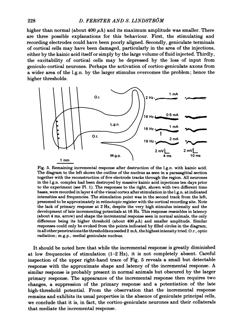
Evoked potentials were recorded in the visual cortex of the cat after electrical stimulation of the lateral geniculate nucleus (l.g.n.). The primary response, mediated by geniculo-cortical fibres, was depressed at stimulation frequencies above 7 Hz and replaced by a late potential, the incremental response, which gradually increased in amplitude with successive stimuli. The incremental response was a negative-positive potential in the depth of the cortex with the negative component having maximal amplitude in layer 4. The response reversed polarity in layer 1 to become a positive-negative potential at the surface. The latency of the negative component of the incremental response was about 3.5-4 ms in layer 4, compared to about 1.5 and 2.5 ms for the mono- and disynaptic components of the primary response. The incremental response could only be evoked from the l.g.n. and the optic radiation, not from the optic tract, superior colliculus or other surrounding structures. Within the l.g.n., the effect was only evoked from stimulation sites in approximate retinotopic register with the recording site in the cortex. Low threshold points were found in the A laminae, completely overlapping with the low threshold points for the primary response. Thresholds increased steeply when the stimulation electrode was lowered into the C laminae. The incremental response could still be evoked ten days after the destruction of all cells in the l.g.n. complex by kainic acid. It is concluded that the described incremental response is identical to the augmenting response of Dempsey & Morison (1943) and is mediated by intracortical axon collaterals of antidromically activated cortico-geniculate neurones.
Full text
PDF


Images in this article
Selected References
These references are in PubMed. This may not be the complete list of references from this article.
- Ahlsen G., Grant K., Lindström S. Monosynaptic excitation of principal cells in the lateral geniculate nucleus by corticofugal fibers. Brain Res. 1982 Feb 25;234(2):454–458. doi: 10.1016/0006-8993(82)90886-1. [DOI] [PubMed] [Google Scholar]
- Ferster D., Lindström S. An intracellular analysis of geniculo-cortical connectivity in area 17 of the cat. J Physiol. 1983 Sep;342:181–215. doi: 10.1113/jphysiol.1983.sp014846. [DOI] [PMC free article] [PubMed] [Google Scholar]
- Ferster D., Lindström S. Synaptic excitation of neurones in area 17 of the cat by intracortical axon collaterals of cortico-geniculate cells. J Physiol. 1985 Oct;367:233–252. doi: 10.1113/jphysiol.1985.sp015822. [DOI] [PMC free article] [PubMed] [Google Scholar]
- Gilbert C. D., Kelly J. P. The projections of cells in different layers of the cat's visual cortex. J Comp Neurol. 1975 Sep;163(1):81–105. doi: 10.1002/cne.901630106. [DOI] [PubMed] [Google Scholar]
- Gilbert C. D., Wiesel T. N. Morphology and intracortical projections of functionally characterised neurones in the cat visual cortex. Nature. 1979 Jul 12;280(5718):120–125. doi: 10.1038/280120a0. [DOI] [PubMed] [Google Scholar]
- HUBEL D. H., WIESEL T. N. Receptive fields, binocular interaction and functional architecture in the cat's visual cortex. J Physiol. 1962 Jan;160:106–154. doi: 10.1113/jphysiol.1962.sp006837. [DOI] [PMC free article] [PubMed] [Google Scholar]
- Matsuda Y., Sasaki K., Mizuno N. Examination of responses evoked in the sensory cortex by thalamic stimulation. Jpn J Physiol. 1972 Dec;22(6):651–666. doi: 10.2170/jjphysiol.22.651. [DOI] [PubMed] [Google Scholar]
- Mitzdorf U., Singer W. Prominent excitatory pathways in the cat visual cortex (A 17 and A 18): a current source density analysis of electrically evoked potentials. Exp Brain Res. 1978 Nov 15;33(3-4):371–394. doi: 10.1007/BF00235560. [DOI] [PubMed] [Google Scholar]
- Morin D., Steriade M. Development from primary to augmenting responses in the somatosensory system. Brain Res. 1981 Jan 26;205(1):49–66. doi: 10.1016/0006-8993(81)90719-8. [DOI] [PubMed] [Google Scholar]
- PURPURA D. P., SHOFER R. J., MUSGRAVE F. S. CORTICAL INTRACELLULAR POTENTIALS DURING AUGMENTING AND RECRUITING RESPONSES. II. PATTERNS OF SYNAPTIC ACTIVITIES IN PYRAMIDAL AND NONPYRAMIDAL TRACT NEURONS. J Neurophysiol. 1964 Mar;27:133–151. doi: 10.1152/jn.1964.27.2.133. [DOI] [PubMed] [Google Scholar]
- Sasaki K., Staunton H. P., Dieckmann G. Characteristic features of augmenting and recruiting responses in the cerebral cortex. Exp Neurol. 1970 Feb;26(2):369–392. doi: 10.1016/0014-4886(70)90133-0. [DOI] [PubMed] [Google Scholar]
- Schwarcz R., Coyle J. T. Striatal lesions with kainic acid: neurochemical characteristics. Brain Res. 1977 May 27;127(2):235–249. doi: 10.1016/0006-8993(77)90538-8. [DOI] [PubMed] [Google Scholar]
- Tusa R. J., Palmer L. A., Rosenquist A. C. The retinotopic organization of area 17 (striate cortex) in the cat. J Comp Neurol. 1978 Jan 15;177(2):213–235. doi: 10.1002/cne.901770204. [DOI] [PubMed] [Google Scholar]
- Wiitanen J. T. Selective silver impregnation of degenerating axons and axon terminals in the central nervous system of the monkey (Macaca mulatta). Brain Res. 1969 Jul;14(2):546–548. doi: 10.1016/0006-8993(69)90136-x. [DOI] [PubMed] [Google Scholar]
- Woodward W. R., Coull B. M. Studies of effects of kainic acid lesions in the dorsal lateral geniculate nucleus of rat. J Comp Neurol. 1982 Oct 10;211(1):93–103. doi: 10.1002/cne.902110109. [DOI] [PubMed] [Google Scholar]


