Abstract
In early 2004, an H5N2 avian influenza virus (AIV) that met the molecular criteria for classification as a highly pathogenic AIV was isolated from chickens in the state of Texas in the United States. However, clinical manifestations in the affected flock were consistent with avian influenza caused by a low-pathogenicity AIV and the representative virus (A/chicken/Texas/298313/04 [TX/04]) was not virulent for experimentally inoculated chickens. The hemagglutinin (HA) gene of the TX/04 isolate was similar in sequence to A/chicken/Texas/167280-4/02 (TX/02), a low-pathogenicity AIV isolate recovered from chickens in Texas in 2002. However, the TX/04 isolate had one additional basic amino acid at the HA cleavage site, which could be attributed to a single point mutation. The TX/04 isolate was similar in sequence to TX/02 isolate in several internal genes (NP, M, and NS), but some genes (PA, PB1, and PB2) had sequence of a clearly different origin. The TX/04 isolate also had a stalk deletion in the NA gene, characteristic of a chicken-adapted AIV. By analyzing viruses constructed by in vitro mutagenesis followed by reverse genetics, we found that the pathogenicity of the TX/04 virus could be increased in vitro and in vivo by the insertion of an additional basic amino acid at the HA cleavage site and not by the loss of a glycosylation site near the cleavage site. Our study provides the genetic and biologic characteristics of the TX/04 isolate, which highlight the complexity of the polygenic nature of the virulence of influenza viruses.
Influenza A viruses, which include all avian influenza viruses (AIVs), can be divided into subtypes based on their surface proteins, the hemagglutinin (HA) and neuraminidase (NA). There are 16 known HA subtypes (H1 through H16) and nine known NA subtypes (N1 through N9) in type A influenza viruses (5). Although AIVs possessing most HA subtypes have been recovered from poultry, particular emphasis is placed on the H5 and H7 subtypes of AIVs because they are the only ones associated with the high-pathogenicity phenotype (39).
At the First International Symposium on Avian Influenza held in 1981, it was resolved to define highly pathogenic AIV on the basis of the ability to produce not less than 75% mortality within 8 days in at least eight susceptible 4- to 8-week-old chickens inoculated by the intramuscular, intravenous, or caudal air sac route (1). However, the viruses responsible for the 1983 Pennsylvania outbreak elicited a problem with the previous definition of highly pathogenic AIV (11). The viruses first isolated in the outbreak were low pathogenic in laboratory tests, although they had four basic amino acids at the HA cleavage site. However, some viruses isolated later in the outbreak contained a single point mutation that eliminated a glycosylation site conformationally close to the HA cleavage site and they were highly pathogenic in animal studies. For this reason, the current definition of highly pathogenic AIV includes such potentially pathogenic viruses.
A recent change to the Office International des Epizooties (OIE) definition of highly pathogenic notifiable avian influenza is that they have an intravenous pathogenicity index in 6-week-old chickens greater than 1.2 or, as an alternative, cause at least 75% mortality in 4- to 8-week-old chickens infected intravenously. H5 and H7 viruses which do not have an intravenous pathogenicity index of greater than 1.2 or cause less than 75% mortality in an intravenous lethality test should be sequenced to determine whether multiple basic amino acids are present at the cleavage site of the HA molecule (HA0): if the amino acid motif is similar to that observed for other highly pathogenic notifiable AIV isolates, the isolate being tested should be considered a highly pathogenic notifiable AIV.
On 17 February 2004, the state of Texas reported the detection of a type A influenza virus from chickens. The mild clinical signs observed in the affected flock were consistent with an infection caused by a low pathogenicity AIV. However, the presence of multiple basic amino acids at the HA cleavage site, which was identical to the amino acid sequence in the highly pathogenic A/chicken/Scotland/59 (H5N1) virus, required that the United States Department of Agriculture (USDA) National Veterinary Services Laboratories report that the A/chicken/Texas/298313/04 (TX/04) (H5N2) isolate was classified as a highly pathogenic AIV on 23 February 2004. However, on 1 March 2004, National Veterinary Services Laboratories announced the results of the chicken pathogenicity test that showed the virus as being avirulent for chickens; the chickens remained healthy throughout the 10-day observation period. Consequently, the 2004 Texas H5N2 AIV meets the OIE molecular criterion for classification as highly pathogenic AIV, but it is not virulent for experimentally inoculated chickens.
Discordant results between the molecular classification, derived by sequencing the HA cleavage site, and virulence for experimentally infected chickens have been observed with several H5 and H7 subtype AIVs (11, 31; unpublished data). One good example is the A/chicken/PA/1/83 (H5N2) isolate. Although the virus contained multiple basic amino acids at the HA cleavage site that would be consistent with the classification as a highly pathogenic AIV, the virus initially was not virulent for experimentally inoculated chickens. Subsequently, this virus became highly pathogenic for chickens after a single point mutation (4, 11, 21).
Although we cannot predict which low-pathogenicity AIVs will remain low pathogenic and which will mutate to become highly pathogenic, it is acknowledged that low pathogenic H5 and H7 viruses, when allowed to circulate in poultry for an extended period of time, have sporadically undergone mutational changes that can result in the emergence of highly pathogenic AIV (11, 22, 31). In Texas, there was serologic and virologic evidence of H5 AIV infections during the previous 5 years. In 1999, H5N3 infections in commercial turkeys and live bird market ducks and H5N2 infections in live-bird market ducks were identified serologically (unpublished data). In 2002, a single outbreak of H5N3 avian influenza, confirmed by virus isolation as A/chicken/Texas/167280-4-/02 (TX/02) (H5N3), occurred in chickens being raised for the live-bird market and in the same year, H5N3 infections were identified serologically in a commercial layer flock (13). These incidences since 1999 provide evidence that AIVs possessing the H5 subtype have been sporadically introduced into poultry in Texas, particularly in association with the live-bird marketing system.
This study elucidates genetic and biologic characteristics of the 2004 H5N2 virus isolated from Texas, which is the first highly pathogenic strain reported in the United States in 20 years.
MATERIALS AND METHODS
Viruses.
The TX/04 isolate was obtained from the National Veterinary Services Laboratories in Ames, Iowa. The virus was received in allantoic fluid after a single passage in embryonating chicken eggs (ECE) (37). The virus was passaged one additional time at the Southeast Poultry Research Laboratory to make working stocks of the virus.
Sequence and phylogenetic analysis.
Viral RNA was extracted with Trizol LS reagent (Life Technologies, Rockville, Md.) from infectious allantoic fluid from ECE. The full-length protein coding region sequences of all eight viral RNA segments of the TX/04 virus were determined by cycle sequencing (PRISM Ready Reaction DyeDeoxy terminator cycle sequencing kit; Perine-Elmer, Foster City, Calif.), following amplification by standard reverse transcription-PCR using the QIAGEN one-step reverse transcription-PCR kit (QIAGEN, Valencia, Calif.). The sequences of the primers used for amplification and sequencing of the individual genes are available upon request. BLAST analyses (http://www.ncbi.nlm.nih.gov/BLAST) were conducted on each sequence to identify related reference viruses. Sequence comparisons to selected viruses were conducted by using the Megalign program using the Clustal V alignment algorithm (DNASTAR, Madison, Wisc.), and phylogenetic relationships were estimated by the method of maximum parsimony (PAUP software, version 4.0b10; Sinauer Associates, Inc, Sunderland, Mass.) using a bootstrap resampling method with a heuristic search algorithm.
Fourteen-day-old ECE passage system.
The TX/04 virus was passaged through 14-day-old ECEs that favor the emergence of highly pathogenic derivatives (38). Briefly, derivatives were obtained by allantoic sac inoculation of parent TX/04 virus stock into 641 14-day-old ECEs. Amnioallantoic fluid was harvested from each of the 233 ECEs that died during the 5-day observation period. Allantoic fluid from dead ECEs was diluted 10−3 and examined for increased production of cytopathic effect in trypsin-free chicken embryo fibroblast cultures compared to the parent virus. Ten of the allantoic fluid samples that showed increased cytopathic effect were selected and further examined for high plaquing efficiency on chicken embryo fibroblast cultures in the presence versus absence of trypsin. Because none of the derivatives showed increased plaquing efficiency, derivatives 2 and 10, which showed the most extensive cytopathic effect, were selected for further in vivo pathotyping studies.
Pathogenicity study of parent and 14-day ECE derivatives in chickens.
The parent TX/04 isolate and 14-day ECE derivative viruses 2 and 10 were pathotyped by intravenous inoculation in eight 4-week-old White Plymouth Rock (WPR) chickens as previously described (42) plus an additional two birds for virus isolation studies on day 3 postinoculation. The parent virus was examined for the ability to infect and cause disease following simulated natural exposure; i.e., intranasal inoculation of 106 50% egg infectious doses/0.1 ml of virus in eight 4-week-old WPR chickens plus two additional chickens for virus isolation studies. The birds were inspected daily for clinical signs. On day 3 postinoculation, two chickens per group were euthanized (100 mg sodium pentobarbital per kg of body weight), necropsied, and tissue samples were collected for histopathology and immunohistochemistry.
For immunohistochemistry, the previously described specific anti-avian influenza nucleoprotein antibody and staining method was used, and included as batch controls, AIV-positive and -negative tissue sections stained with and without the primary antibody (24). The tissues collected included: lung, thyroid, thymus, trachea, liver, spleen, heart, pancreas with segments of descending and ascending duodenum, small intestine at Meckel's diverticulum, cecal tonsils, cloacal bursa, kidney, adrenal gland, gonad, proventriculus, ventriculus, esophagus, comb, eyelid, brain, intranasal tissues, eye, and proximal tibiotarsal joint with the physis. These tissue samples were also collected from all chickens that died. Oropharyngeal and cloacal swabs were taken for virus isolation and titration from euthanized birds on day 3 postinoculation (26, 37). Individual swabs were suspended in 1.5 ml of brain heart infusion (Difco, Detroit, MI 48232-7059). In addition, chickens that died had virus isolation and titration performed on swabs and brain, heart, and spleen tissues. On day 10 postinoculation, all survivors were bled and then euthanized. All sera were tested for antibodies to type A influenza virus by the agar gel immunodiffusion test (37).
Plasmid construction and site-directed mutagenesis.
Eight transcription plasmids were generated by insertion of cDNA of eight viral gene segments into pHH21 vector between the promoter and terminator sequences of RNA polymerase I (14, 19). The HA and NA genes were derived from the TX/04 virus, the M, NS, PA, and PB2 genes from the A/chicken/Indonesia/7/03 (H5N1) virus, and the NP and PB1 genes from the A/duck/Anyang/AVL-1/01 (H5N1) virus (41), respectively. In addition, we cloned a HA gene of the A/chicken/Scotland/59 highly pathogenic AIV (44) into the pHH21 vector. We used the pHH21 plasmid that contained the HA gene of TX/04 isolate (pHH21-TX04-H5) as a template to introduce a series of mutations by using the Quick-Change site-directed mutagenesis kit (Stratagene, La Jolla, Calif.).
Generation of reverse-genetic reassortant viruses.
Reassortant viruses were generated by DNA transfection as described previously (14, 19). Briefly, 293T cells were transfected with 1 μg of each of the eight transcription plasmids and four expression plasmids with the use of Lipofectamine 2000 reagent (Invitrogen, San Diego, Calif.). After 48 h of incubation, supernatant was collected and subsequently inoculated into 10-day-old ECE. After 48 h of incubation, allantoic fluid containing reassortant virus was harvested and stored at −70 C for additional experiments. The identity of the reassortant virus was confirmed by partial sequencing of each viral segment.
In vitro and in vivo characterization of reverse-genetic viruses.
The growth of the rescued viruses was determined in 10-day-old ECE and chicken embryo fibroblasts. Plaque forming efficiency of the viruses was examined in chicken embryo fibroblasts with trypsin (0.4 μg/ml) and without trypsin as previously described (37). To calculate the mean death time (MDT) of the embryos, 104 50% egg infectious doses/0.2 ml dose of each virus was inoculated into 15 10-day-old ECEs and observed for 7 days for mortality.
Groups of 10 4-week-old SPF White Leghorn chickens were used in chicken intravenous pathogenicity tests with reverse-genetic reassortant viruses. One of the groups was inoculated with TX/04 parent virus for comparison. We also included four uninoculated birds as a negative control group in this experiment. Oropharyngeal and cloacal swabs were collected from three birds in each group on day 3 postinoculation. Morbidity and mortality were observed for 10 days and sera were collected from all birds at the end of the experiment to determine the antibody level to the HA by hemagglutination inhibition test (37).
GenBank accession numbers.
The sequences reported in this paper have been deposited in the GenBank database (accession nos. AY849782 to AY849793).
RESULTS
Epidemiology.
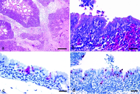
On 16 February 2004, six 10-week-old black and red broiler chickens were submitted to the Texas Veterinary Medical Diagnostic Laboratory, Poultry Diagnostic Laboratory in Gonzales, Texas. Chickens in the flock had a history of gasping and the chickens submitted to the laboratory for diagnostic investigation had easily heard moist rales. On necropsy all the chickens had mucopurulent to caseous exudate in the sinuses and increased mucus in the trachea. Most birds had fibrinous exudate in the thoracic airsacs and focal pulmonary edema. Histologically, birds had fibrinoheterophilic pneumonia with severe edema (Fig. 1a) and moderately severe lymphocytic tracheitis with epithelial necrosis.
FIG. 1.
Histological lesions in chickens infected with TX/04 virus. Photomicrographs of hematoxylin-and-eosin-stained tissue sections (a and b) or sections stained by immunohistochemistry to demonstrate avian influenza virus (c and d). (a) Fibrinoheterophilic pneumonia with severe edema in a chicken from the field. Bar = 200 μm. (b) Mild heterophilic bronchitis associated with bronchial associated lymphoid tissue from chicken intranasally inoculated with parent TX/04 virus and euthanized 2 days later. Bar = 25 μm. (c) Avian influenza viral antigen in respiratory epithelium of the trachea from chicken intranasally inoculated with parent TX/04 virus and euthanized 2 days later. Bar = 25 μm. (d) Avian influenza viral antigen in respiratory epithelium of the bronchus from a chicken intranasally inoculated with parent TX/04 virus and euthanized 2 days later. Bar = 50 μm.
Pooled sinus and tracheal exudates were tested immediately with the BD Directigen Flu A test (27) that can detect all type A influenza virus, and the results were positive. The same samples were also positive for the type A influenza matrix gene and an H5 HA gene by real-time reverse transcription-PCR analysis (29). Serum samples collected at necropsy were positive for antibodies to influenza A virus by the agar gel immunodiffusion test and positive for Mycoplasma gallisepticum by plate agglutination test (12). Subsequently, a H5N2 AIV was isolated at the National Veterinary Services Laboratories from the samples, the TX/04 virus.
The chickens at the affected farm were for sale to the Houston live-bird markets. The farm was a former commercial turkey operation and had four poultry houses, but only two were in use. House 2 contained a flock of 3,949 13-week-old colored broilers. This flock had no clinical signs and tested avian influenza negative 3 days before depopulation. House 4 contained 2,659 colored broilers in multiple age groups divided in six pens, including the 10-week-old chickens submitted to the laboratory. Chickens at this farm were vaccinated at 6 weeks of age with Newcastle disease virus, infectious bronchitis virus, and fowlpox vaccines. A total of 1,700 chickens had been sold to the live-bird markets in partial loads. The last group was sold on 9 February 2004, which is a week prior to submitting chickens to the laboratory. The index flock was depopulated on February 21 and a total of 6,608 chickens were destroyed.
In addition, epidemiological investigations conducted by the Texas Animal Health Commission identified two live bird markets in Houston with avian influenza-positive chickens. Birds in these two markets were depopulated along with birds in three additional markets that were voluntarily depopulated. On February 23, the USDA's National Veterinary Services Laboratories reported that the amino acid sequence of the HA cleavage site of the isolate was compatible with highly pathogenic AIV. A joint USDA-Texas Animal Health Commission surveillance program was conducted in order to regain highly pathogenic avian influenza-free status for Texas and the United States. A total of 2,938 serum samples were tested by AIV-specific agar gel immunodiffusion test and a total of 3,595 tracheal and cloacal swabs were tested by an AIV-specific real-time reverse transcription-PCR. No additional positive flocks were identified. On April 1, the outbreak was declared over.
Sequence and phylogenetic analysis.
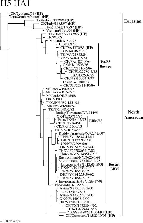
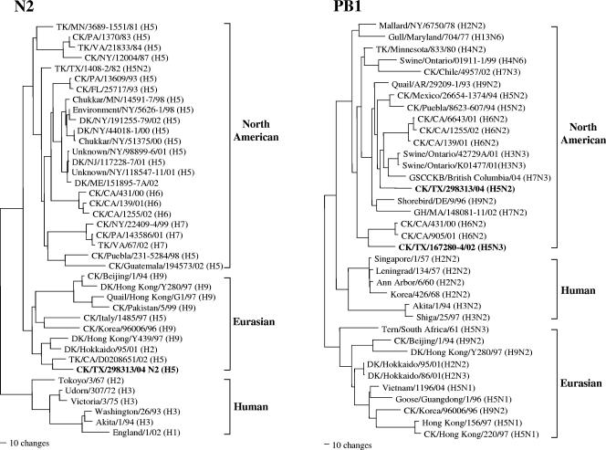
Sequence analysis demonstrated that each of the RNA segments, except the NA gene, of the TX/04 isolate was related to viruses of the North American avian lineage (Table 1). The NA gene belonged to the Eurasian phylogenetic lineage. The TX/04 isolate shared similar sequences with TX/02 isolate in four genes (HA, NS, M, and NP), while the other four genes showed various degree of similarity and were phylogenetically distinct (Table 1, Fig. 2 and 3).
TABLE 1.
Genetic similarity between eight gene segments of A/chicken/Texas/298313/04 (H5N2) and other influenza virus isolates
| Gene | Region of comparison (nucleotides) | % Nucleotide (amino acid) similarity with influenza virus:
|
Lineage | |
|---|---|---|---|---|
| TX/02 (H5N3)a | Virus with highest identity, excluding TX/02b | |||
| H5 HA | 29-723 | 97.8 (98.6) | DK/NY/44018-2/00 (H5N2)—91.6 (91.1) | North American |
| N2 NA | 20-1390 | TK/CA/D0208652-C/02 (H5N2)—95.8 (96.3) | Eurasian | |
| NS (NS1, NS2) | 27-864 | 98.7 (97.8, 99.1) | Pintail/Alberta/156/97 (H3N8)—95.3 (92.6, 98.2) | North American |
| M (M1, M2) | 26-1014 | 99.0 (99.2, 99.0) | Mallard/Alberta/17/91 (H9N2)—96.1 (96.3, 97.9) | North American |
| NP | 46-1542 | 99.1 (99.6) | Mallard/Alberta/226/98 (H2N3)—96.3 (98.4) | North American |
| PA | 25-2175 | 95.1 (97.5) | Shorebird/DE/9/96 (H9N2)—94.8 (98.9) | North American |
| PB1 | 25-2298 | 91.4 (97.6) | Swine/Ontario/K01477/01 (H3N3)—95.2 (98.4) | North American |
| PB2 | 28-2307 | 92.8 (96.8) | CK/Quaretaro/14588-19/95 (H5N2)—94.4 (97.6) | North American |
A/chicken/Texas/67280-4/02 (H5N3) isolate.
Abbreviations: CK, chicken; DK, duck; TK, turkey. Standard two-letter abbreviations are used for states in the United States.
FIG. 2.
Phylogenetic tree based on nucleotide sequences of the H5 HA gene. The tree was generated by the maximum parsimony method with the PAUP4.0b10 program with bootstrap replication (100 bootstraps) and a heuristic search method. The tree is rooted to A/chicken/Scotland/59. Previously identified highly pathogenic viruses are indicated as HP in parentheses. *, Ruddy Turnstone/NJ/2242/00 virus belongs to the recent live-bird market phylogenetic lineage, but the virus was not recovered from live-bird market. Abbreviations: CK, chicken; DK, duck; TK, turkey. Standard two-letter abbreviations are used for states in the United States.
FIG. 3.
Phylogenetic tree based on nucleotide sequences of the N2 NA and PB1 genes. Both trees are midpoint rooted.
The HA gene of the TX/04 isolate shared 98.6% amino acid identity with the TX/02 isolate. The potential glycosylation sites and amino acids at proposed receptor binding sites were conserved between the two isolates. However, the TX/04 virus had one additional basic amino acid (lysine at position 328 instead of a glutamic acid of the TX/02 isolate) at the HA cleavage site. The HA cleavage site sequence of different isolates were compared (Table 2) and the TX/04 isolate possessed the same cleavage site sequence as the A/chicken/Scotland/59, a known highly pathogenic AIV isolate. Phylogenetically, the TX/04 isolate was clearly distinguished from the Pennsylvania/83, Mexican, and Eurasian lineage highly pathogenic AIVs and formed a separate clade with the TX/02 isolate (Fig. 2).
TABLE 2.
Comparison of HA cleavage site and potential glycosylation site sequences of Texas isolates with other known highly pathogenic viruses
| Virusa | Glycosylation siteb
|
Cleavage site sequence
|
|||||||||||
|---|---|---|---|---|---|---|---|---|---|---|---|---|---|
| 11* | 12 | 13 | 321 | 322 | 323 | 324 | 325 | 326 | 327 | 328 | 329 | 330 | |
| CK/TX/167280-4/02 (H5N3) | N | S | T | P | Q | — | — | — | — | R | E | K | R |
| CK/TX/298313/04 (H5N2) | N | S | T | P | Q | — | — | — | — | R | K | K | R |
| CK/Scotland/59 (H5N1) | K | S | T | P | Q | — | — | — | — | R | K | K | R |
| CK/PA/1370/83 (H5N2) | N | S | K | P | Q | — | — | — | — | K | K | K | R |
| CK/Puebla/94 (H5N2) | N | S | T | P | Q | — | — | R | K | R | K | T | R |
| TK/Ireland/1378/83 (H5N8) | N | S | T | P | Q | — | — | R | K | R | K | K | R |
| TK/Ontario/7732/66 (H5N9) | N | S | T | P | Q | — | — | R | R | R | K | K | R |
| CK/Italy/1485/97 (H5N2) | N | S | T | P | Q | — | — | R | R | R | K | K | R |
| Tern/South Africa/61 (H5N3) | N | S | T | P | Q | R | E | T | R | R | Q | K | R |
| CK/Indonesia/7/03 (H5N1) | N | S | T | P | Q | R | E | R | R | R | K | K | R |
Abbreviations: CK, chicken; DK, duck; TK, turkey. Standard two-letter abbreviations are used for states in the United States.
* indicates the position where glycosylation occurs. Amino acids that affect the loss of glyocylation are in bold.
The TX/04 isolate had a unique N2 NA sequence and shared highest nucleotide sequence identity with the A/turkey/CA/D0208652-C/02 (H5N2) virus (95.8%). The virus had a 13 amino acid stalk deletion (at positions 51 to 63) in the NA gene, a characteristic of a chicken adapted AIV (17). The N2 sequence of TX/04 virus was not related to the N2 sequence of H7N2 subtype isolates from the Northeastern live-bird markets or that of the H6N2 subtype isolates from the recent California outbreak, the two most common subtypes currently found in the United States (30, 43) (Fig. 3).
The TX/04 isolate was compared with the H5N1 viruses currently circulating in Asia, which have been documented to cross the species barrier and infect humans, and no markers related to human infection were observed. Specifically, the TX/04 isolate did not have the five-amino-acid deletion in the NS1, thought to confer some resistance to the antiviral effect of the host, and no mutations in the M2 protein, which are commonly related to resistance to one class of antiviral drugs (amantadine) that are commonly used for influenza (28, 32). Also, the TX/04 isolate also had no E-to-K mutation at position 627 of the PB2 protein, which was responsible for the high virulence of A/Hong Kong/483/97 in mice (7).
Pathogenicity of the parent virus.
The parent virus replicated well in the respiratory tract of chickens as evidenced by mean virus titers of 5.0 and 6.1 log10 50% egg infectious doses/ml for oropharyngeal swabs collected on day 3 after intranasal and intravenous inoculation, respectively (Table 3). These titers are comparable to other chicken-adapted H5 and H7 subtype viruses (13, 15, 16). All surviving birds seroconverted as evidenced by the presence of antibodies to type A influenza virus by the agar gel immunodiffusion test. However, the parent virus did not produce clinical signs or death in intranasally or intravenously inoculated chickens. For the chickens inoculated intranasally with the parent virus and sampled on day 3 postinoculation, one chicken had a slightly enlarged spleen while the two intravenously inoculated birds posted had pulmonary congestion. One intravenously inoculated bird had a slightly enlarged spleen while the other had urates in the ureters (day 3 postinoculation).
TABLE 3.
Pathogenicity of parent (A/chicken/TX/298313/04) and 14-day-old ECE derivatives in chickens
| Virus | Inoculation route | Inoculation dose (EID50) | Morbiditya | Mortalityb | Virus titer (no. of positive samples)
|
Gel immunodiffusion testd | |
|---|---|---|---|---|---|---|---|
| Oropharyngeal | Cloacal | ||||||
| Parent | Intranasal | 106.0/0.1 | 0/8 | 0/8 | 6.1 ± 1.7 (2) | < 0.9 (2) | 8/8 |
| Intravenous | 108.1/0.2 | 0/8 | 0/8 | 5.0 ± 1.6 (2) | 4.7 ± 0.3 (2) | 8/8 | |
| 14-day derivative 2 | Intravenous | 107.6/0.2 | 1/8 | 1/8 | 5.4 ± 0.4 (3) | 4.8 ± 0.4 (3) | 7/7 |
| 14-day derivative 10 | Intravenous | 108.4/0.2 | 1/8 | 1/8 | 5.0 ± 0.1 (3) | 5.1 ± 0.3 (3) | 7/7 |
Number of birds that manifested clinical signs/number of birds inoculated.
Number of birds that died/number of birds inoculated.
The virus titer is express as the log10 50% egg infections dose (EID50) per milliliter ± standard deviation.
Number of positive sera/number of sera tested.
Histopathologically, intranasally inoculated chickens had mild focal areas of heterophilic-to-lymphocytic tracheitis and bronchitis (Fig. 1b). Avian influenza viral antigen was demonstrated in respiratory epithelial cells on the trachea and bronchi (Fig. 1c and d). The intravenously inoculated chickens had mild hyperplasia of macrophage-phagocytic system in the spleen, mild-to-moderate necrosis of pancreatic acini and severe lymphocytic-to-heterophilic interstitial nephritis with associated multifocal necrosis of tubule epithelium. avian influenza viral antigen was common in necrotic renal tubule epithelium and occasionally seen in the necrotic pancreatic acinar epithelium.
Pathogenicity of the 14-day-old ECE derivatives.
Of the 641 14-day ECEs inoculated, 233 died and the allantoic fluid from each dead embryo was screened for the ability to produce cytopathic effect without exogenous trypsin in chicken embryo fibroblast cells compared to the parent virus. Although none of the derivatives formed plaques without exogenous trypsin, some showed increased cytopathic effect compared to the parent virus and two of the derivatives (2 and 10), which showed the most extensive cytopathic effect, were selected for intravenous pathotyping in chickens. Both derivatives produced clinical signs of depression in one bird each, and each of these died; one on day 3 postinoculation and the other day 7 postinoculation (Table 3). Low titer of virus was detected from the brain (2.0 to 2.3 log10 50% egg infectious doses/gram) of both birds and also from the spleen (3.7 log10 50% egg infectious doses/gram) of the bird that was intravenously inoculated with derivative 2. Both birds that died had swollen kidneys filled with urates. The chickens euthanized and sampled on day 3 postinoculation had mild pulmonary congestion, moderately enlarged spleens, and mildly swollen kidneys.
Histopathologically, euthanized as well as naturally dead intravenously inoculated birds had consistent lesions in the kidney and pancreas. Kidney lesions ranged from mild multifocal lymphocytic interstitial nephritis with occasional tubule necrosis to severe diffuse renal tubule necrosis with edema and heterophilic inflammation. Avian influenza viral antigen was localized in the necrotic renal tubule epithelium. A few birds had necrosis of pancreatic acini with associated avian influenza viral antigen in the epithelial cells. In addition, the two birds that died had macrophage-phagocytic system hyperplasia in the spleen, and moderate apoptosis and lymphocyte depletion in thymus and cloacal bursa. Most interesting, one of the chickens inoculated with derivative 2 and sampled on day 3 postinoculation had avian influenza viral antigen in a few cardiac myocytes.
To determine the genetic changes during the passage of the parent virus in ECE and chickens, we sequenced the entire HA coding region of the 14-day ECE derivatives (2 and 10) and viruses reisolated from the brain and spleen tissues of the infected animals. We observed one to two amino acid changes at positions 158 (arginine to glutamine) and 181 (proline to serine) in the HA gene of those derivative viruses compared to the parent virus (data not shown). Those amino acid positions do not overlap with known amino acids sites that are related to HA cleavability or receptor binding.
In vitro and in vivo characterization of reverse-genetic viruses.
To predict possible changes that can increase the virulence of the TX/04 virus, we introduced a series of mutations or insertions in the HA gene by in vitro mutagenesis and rescued reassortant viruses that contained those mutated HA genes by reverse genetics. We were able to rescue the viruses that had the same HA sequence as the TX/04 isolate, HA sequences that had different mutations, and also the virus that had HA sequence of the highly pathogenic AIV, A/chicken/Scotland/59. All the reverse-genetic reassortant viruses replicated well in ECE (108.9 to 1010.9 50% egg infectious doses/ml) (Table 4).
TABLE 4.
In vitro and in vivo characteristics of the reverse-genetic viruses
| Virus | Glycosylation | Cleavage site sequence | ECEa
|
PFU/ml in CEF cells
|
Intravenous pathotyping in chickens
|
|||||
|---|---|---|---|---|---|---|---|---|---|---|
| EID50/ml | MDTb | Trypsin | No trypsin | Morbidityc | Mortalityd | Virus titere | Hi titerf | |||
| Parent | NST (+) | PQ——RKKR | 9.7 | >5 | 8 × 10⁹ | none | 1/10 | 0/0 | 5.8 ± 0.4 | 8.2 ± 0.6 |
| rg1 | NST (+) | PQ——RKKR | 8.9 | >5 | 1 × 10⁹ | none | 0/10 | 0/10 | 1.5 ± 2.5 | 4.7 ± 1.4 |
| rg2 | NSK (−) | PQ——RKKR | 9.2 | >5 | 3 × 10⁸ | none | NDg | ND | ND | ND |
| rg3 | KST (−) | PQ——RKKR | 10.5 | >5 | 4 × 10⁸ | none | 0/10 | 0/0 | <1.0 | 2.1 ± 1.8 |
| rg4 | NST (+) | PQ—RRKKR | 9.2 | 3.6 | 2 × 10⁹ | none | 0/10 | 0/0 | <1.0 | 5.2 ± 1.0 |
| rg5 | NST (+) | PQRKRKKR | 10.9 | 2.2 | 4 × 10⁷ | 1 × 10⁶ | 10/10 | 1/10 | 3.3 ± 1.4 | 6.8 ± 0.7 |
| rg6-CK/Scot/59 H5 | KST (−) | PQ——RKKR | 10.5 | 2.2 | 3 × 10⁷ | 3 × 10⁵ | 10/10 | 3/10 | 2.5 ± 0.1 | 6.0 ± 1.3 |
The challenge doses were adjusted to 104 EID50/0.2 ml and 106 EID50/0.2 ml for the pathogenicity test in ECE and chickens, respectively.
MDT, average number of days required to kill the inoculated eggs.
Number of birds that manifested clinical signs/number of birds inoculated.
Number of birds that died/number of birds inoculated.
The mean virus titer of three samples is expressed as the log10 EID50 per milliliter ± standard deviation.
Log2 hemagglutination inhibition titer (mean titer of sera collected from the surviving birds 10 days after challenge) ± standard deviation.
ND, test not done.
The reverse-genetic virus that had HA gene of the wild type virus (rg1) showed similar MDT in embryos as the wild type parent viruses. The rg2 and rg3 viruses, which had the potential glycosylation near the cleavage site removed so that the potential glycosylation site at position 11 resembles that of the known highly pathogenic AIVs A/chicken/Scotland/59, and A/chicken/PA/1370/83, respectively (Table 2), showed no increased virulence in embryos compared to the parent virus. The rg4 virus has one more basic amino acid insertion at the cleavage site and the rg5 virus has two additional basic amino acids inserted at the cleavage site. The cleavage site sequence of the rg5 is the same as the highly pathogenic AIV A/turkey/Ireland/1378/83 virus (Table 2). Both (rg4 and rg5) viruses showed increased virulence in ECE compared to the parent virus as evidenced by shortened MDT. The rg5 virus showed similar MDT as the reverse-genetic virus that had the A/chicken/Scotland/59 HA sequence (rg6).
The plaque-forming ability in the chicken embryo fibroblasts in the absence of trypsin is also known as one of the characteristics of highly pathogenic AIV (3). Only the rg5 and rg6 viruses were able to form plaques without addition of exogenous trypsin (Table 4).
We also evaluated the pathogenicity of the reverse-genetic virus in chickens by intravenous inoculation of 106 50% egg infectious doses/0.2 ml dose of each virus. The virus isolation and the postchallenge hemagglutination inhibition titer results indicated that all the reverse-engineered viruses had limited replication in chickens compared to the wild type virus (Table 4). However, rg5 and rg6 viruses showed increased virulence in chickens as evident by severe depression of all the infected birds in the early phase of infection (2 to 4 days postinoculation) and the marked decrease in food consumption. One and three chickens died within 10 days of observation period in groups of birds that were infected with rg5 and rg6 viruses, respectively.
DISCUSSION
Limited lesions and avian influenza viral antigen in the trachea and bronchi of chickens intranasally inoculated with the parent TX/04 virus are typical of low-pathogenicity AIVs, where infection is usually limited to the respiratory tract (18, 34, 40). The more severe lesions in the respiratory tract of naturally infected chickens suggests secondary bacterial infections or other cofactors contributed to a more severe clinical disease than experimentally infected birds in the laboratory (39). On intravenous inoculation in the standard pathotyping test, an aberrant route of inoculation, the parent virus was demonstrated in and produced lesions within the kidney tubular and pancreatic acinar epithelium, which supports the premise that replication of low-pathogenicity AIVs takes place in epithelial-type cells and not in mesenchymal-type cells. Similar viral replication and lesion distribution have been seen in other intravenous experiments using low-pathogenicity AIVs in chickens (33, 35, 36). By contrast, highly pathogenic AIVs on intranasal or intravenous inoculation typically produce systemic virus infection, including viremia, with virus replication and necrobiotic lesions in multiple visceral organs, brain, and skin (8, 10, 18, 25, 40).
Although pathogenicity relates to the viral strain's ability to cause disease and mortality in birds, the current OIE guideline considers H5 and H7 subtype viruses highly pathogenic AIVs if they meet the molecular criterion of cleavage site sequence that is compatible with other highly pathogenic AIVs regardless of their virulence in birds (20). The rationale behind this guideline is the potential for H5 and H7 strains to become highly pathogenic. Several low-pathogenicity AIVs of the H5 and H7 subtypes have been shown to undergo a virulence shift to highly pathogenic AIV either in the field or in laboratory test systems (2, 9, 11, 22, 31, 38). Among the H5 and H7 influenza viruses, however, we cannot predict which low-pathogenicity AIVs will remain low pathogenic and which will mutate to become highly pathogenic. Also, the type of mutation that is necessary to increase the virulence is not predictable. What we do know and have observed is that some H5 and H7 subtype AIVs, when allowed to circulate in poultry for an extended period of time, can undergo mutational changes that can have serious consequences.
Although it is difficult to estimate when the TX/04-like virus was first introduced into poultry, evidence would suggest that the virus has been circulating for an extended period of time. First, the TX/04 isolate shared 98.6% amino acid identity in the HA sequence with the TX/02 virus as well as high sequence identity with three other internal genes (13). Phylogenetic analysis of the HA and several internal genes (NP, M, and NS) further demonstrated the close relationship between these two isolates. However, four genes (NA, PA, PB1, and PB2) were clearly different, which indicates a reassortment event had occurred with unknown viruses. Second, the N2 gene of the TX/04 isolate had a stalk deletion, which is believed to be a marker of poultry adaptation of the virus (17). The virus replicated well in the respiratory tract of chickens, as has been observed with other poultry-adapted H5 and H7 viruses found in the United States (13, 15). Third, there has been sporadic serologic evidence of H5 infection in Texas since 1999, as explained in the Results section. However, these may be separate introductions of the H5 virus into poultry from the wild bird reservoir and evidence to demonstrate continued circulation of H5 virus in Texas or surrounding states is lacking. Furthermore, it is unlikely that the virus has circulated in the integrated poultry industry because of the ongoing passive and active surveillance programs in Texas. Although the virus could be present in backyard or gaming birds in Texas or surrounding states or countries, no evidence of H5 virus infection was seen in any of these poultry sectors during the enhanced surveillance that occurred in Texas during 2004.
To determine the pathogenic potential of the TX/04 virus, we conducted a 14-day-old ECE passage that is known to favor the emergence of highly pathogenic derivatives (22, 38). The change from low-pathogenicity AIV to highly pathogenic AIV in this system with some H5 and H7 subtype viruses had resulted in a change from local virus replication in respiratory and gastrointestinal tracts to wide spread systemic infection. In the current study, the low-pathogenic parent virus did not change in virulence following passage in the 14-day-old ECE passage model system, and all the resulting derivatives had biological characteristics of low-pathogenicity AIVs, including lack of plaque formation in the chicken embryo fibroblasts in the absence of added trypsin, absence of high mortality in intravenous pathotyping tests, and virus demonstration and lesions limited to epithelial cells. However, the presence of viral antigen in some cardiac myocytes of derivative 2-inoculated chickens is a pattern seen with highly pathogenic AIVs (8, 10, 18). This could represent an early stage in the transition from low- to high-pathogenicity AIV.
We also applied the reverse-genetic system to examine what amino acid changes might be necessary to increase the virulence of the virus. Because the cleavage site of the TX/04 isolate was the same as the highly pathogenic A/chicken/Scotland/59 isolate, and because it had a glycosylation site at position 11, we were interested to see the effect of the glycosylation in the virulence of the virus. It was previously postulated that the loss of this carbohydrate may permit access of ubiquitous proteases that recognizes the basic amino acid sequences and result in cleavage activation of the HA in the virulent virus (4, 21). However, removal of this glycosylation site in two different ways in this study had no effect on virulence of the virus (Table 4). Instead, insertion of basic amino acids at the cleavage site, which is more commonly observed in previous highly pathogenic AIVs, clearly increased the virulence of the virus in vitro.
The rg5 virus, which has two additional basic amino acid inserted at the HA cleavage site compared to the parent virus, showed virulence comparable to the reverse-genetic virus that had the HA gene of the A/chicken/Scotland/59 highly pathogenic AIV. Insertion of two additional basic amino acids at the cleavage site has been observed in many highly pathogenic AIVs and the possible mechanism of such insertion was postulated previously (6, 23). We also observed increased virulence of rg5 virus in vivo in terms of morbidity, but all the reverse-genetic viruses showed limited growth in chickens compared to wild-type parent virus, which likely protected the chickens from high mortality. Although all the internal genes of the reverse-genetic viruses were derived from highly pathogenic avian-origin viruses, we speculate the optimal combination of genes was not present for efficient replication in chickens, which reinforces the previous idea that the virulence of AIV is polygenic.
In conclusion, a molecularly defined highly pathogenic AIV with a low pathogenic phenotype was observed in Texas in 2004. This was the first time since 1983-1984 that a highly pathogenic AIV has been reported from the United States. The divergence of the proposed genetic and phenotypic markers of highly pathogenic AIV require us to reexamine what should be considered a reportable virus by OIE standards, since the declaration of highly pathogenic AIV results in severe effects on trade for the entire country.
It is important to note that the majority of the highly pathogenic AIVs of the H5 subtype and all the reported highly pathogenic AIVs of H7 subtype have at least two additional amino acids inserted at the HA cleavage site. A/chicken/Scotland/59 and A/chicken/Pennsylvania/1370/83 are the only reported highly pathogenic AIVs that do not have an additional basic amino acid insertion. It is likely that these two viruses have other HA structural features that allowed the virus to become highly pathogenic in the absence of the carbohydrate near the HA cleavage site. Thus, future research should focus on understanding the differences between the A/chicken/Scotland/59 highly pathogenic AIV and the TX/04 low-pathogenicity AIV. The understanding of structural differences between those two viruses will help the OIE define the highly pathogenic AIV more specifically than the broad definition currently used. Fortunately, because the outbreak was diagnosed quickly and state animal health officials reacted quickly to control the outbreak, the virus was not allowed to spread. Continued active and passive surveillance is critical to identify avian influenza outbreaks early and eradication plans should be developed before an outbreak occurs to ensure a rapid response.
Acknowledgments
This work was supported by USDA ARS CRIS project 6612-32000-039-00 D.
We thank Suzanne Deblois, Joan Beck, James Doster, and the SAA sequencing facility (Erica Spackman, Melissa Scott, and Joyce Bennett) for technical assistance and Roger Brock for animal care assistance.
REFERENCES
- 1.Bankowski, R. A. 1982. Proceedings of the First International Symposium on Avian Influenza, 1981. Carter Co., Richmond, Va.
- 2.Banks, J., E. S. Speidel, E. Moore, L. Plowright, A. Piccirillo, I. Capua, P. Cordioli, A. Fioretti, and D. J. Alexander. 2001. Changes in the haemagglutinin and the neuraminidase genes prior to the emergence of highly pathogenic H7N1 avian influenza viruses in Italy. Arch. Virol. 146:963-973. [DOI] [PubMed] [Google Scholar]
- 3.Bosch, F. X., M. Orlich, H. D. Klenk, and R. Rott. 1979. The structure of the hemagglutinin, a determinant for the pathogenicity of influenza viruses. Virology 95:197-207. [DOI] [PubMed] [Google Scholar]
- 4.Deshpande, K. L., V. A. Fried, M. Ando, and R. G. Webster. 1987. Glycosylation affects cleavage of an H5N2 influenza virus hemagglutinin and regulates virulence. Proc. Natl. Acad. Sci. USA 84:36-40. [DOI] [PMC free article] [PubMed] [Google Scholar]
- 5.Fouchier, R. A., V. Munster, A. Wallensten, T. M. Bestebroer, S. Herfst, D. Smith, G. F. Rimmelzwaan, B. Olsen, and A. D. Osterhaus. 2005. Characterization of a novel influenza A virus hemagglutinin subtype (H16) obtained from black-headed gulls. J. Virol. 79:2814-2822. [DOI] [PMC free article] [PubMed] [Google Scholar]
- 6.Garcia, M., J. M. Crawford, J. W. Latimer, E. Rivera-Cruz, and M. L. Perdue. 1996. Heterogeneity in the haemagglutinin gene and emergence of the highly pathogenic phenotype among recent H5N2 avian influenza viruses from Mexico. J. Gen. Virol. 77:1493-1504. [DOI] [PubMed] [Google Scholar]
- 7.Hatta, M., P. Gao, P. Halfmann, and Y. Kawaoka. 2001. Molecular basis for high virulence of Hong Kong H5N1 influenza A viruses. Science 293:1840-1842. [DOI] [PubMed] [Google Scholar]
- 8.Hooper, P., and P. Selleck. 1998. Pathology of low and high virulent influenza virus infections. p. 134-141. In D.E. Swayne and R. D. Slemons (ed.). Proceedings of the Fourth International Symposium on Avian Influenza. U.S. Animal Health Association, Richmond, Va.
- 9.Horimoto, T., and Y. Kawaoka. 1995. Molecular changes in virulent mutants arising from avirulent avian influenza viruses during replication in 14-day-old embryonated eggs. Virology 206:755-759. [DOI] [PubMed] [Google Scholar]
- 10.Jones, Y. L., and D. E. Swayne. 2004. Comparative pathobiology of low and high pathogenicity H7N3 Chilean avian influenza viruses in chickens. Avian Dis. 48:119-128. [DOI] [PubMed] [Google Scholar]
- 11.Kawaoka, Y., C. W. Naeve, and R. G. Webster. 1984. Is virulence of H5N2 influenza viruses in chickens associated with loss of carbohydrate from the hemagglutinin? Virology. 139:303-316. [DOI] [PubMed] [Google Scholar]
- 12.Kleven, S. H. 1998. Mycoplasmosis, p. 74-80. In D. E. Swayne (ed.), A laboratory manual for the isolation and identification of avian pathogens. American Association of Avian Pathologists, Kennett Square, Pa.
- 13.Lee, C. W., D. A. Senne, J. A. Linares, P. R. Woolcock, D. E. Stallknecht, E. Spackman, D. E. Swayne, and D. L. Suarez. 2004. Characterization of recent H5 subtype avian influenza viruses from US poultry. Avian Pathol. 33:288-297. [DOI] [PubMed] [Google Scholar]
- 14.Lee, C. W., D. A. Senne, and D. L. Suarez. 2004. Generation of reassortant influenza vaccines by reverse genetics that allows utilization of a DIVA (Differentiating Infected from Vaccinated Animals) strategy for the control of avian influenza. Vaccine 22:3175-3181. [DOI] [PubMed] [Google Scholar]
- 15.Lee, C. W., and D. L. Suarez. 2004. Application of real-time reverse transcription-PCR for the quantitation and competitive replication study of H5 and H7 subtype avian influenza virus. J. Virol. Methods 119:151-158. [DOI] [PubMed] [Google Scholar]
- 16.Lee, C. W., D. A. Senne, and D. L. Suarez. 2004. Effect of vaccine use in the evolution of Mexican lineage H5N2 avian influenza virus. J. Virol. 78:8372-8381. [DOI] [PMC free article] [PubMed] [Google Scholar]
- 17.Matrosovich, M., N. Zhou, Y. Kawaoka, and R. Webster. 1999. The surface glycoproteins of H5 influenza viruses isolated from humans, chickens, and wild aquatic birds have distinguishable properties. J. Virol. 73:1146-1155. [DOI] [PMC free article] [PubMed] [Google Scholar]
- 18.Mo, I. P., M. Brugh, O. J. Fletcher, G. N. Rowland, and D. E. Swayne. 1997. Comparative pathology of chickens experimentally inoculated with avian influenza viruses of low and high pathogenicity. Avian Dis. 41:125-136. [PubMed] [Google Scholar]
- 19.Neumann, G., T. Watanabe, H. Ito, S. Watanabe, H. Goto, P. Gao, M. Hughes, D. R. Perez, R. Donis, E. Hoffmann, G. Hobom, and Y. Kawaoka. 1999. Generation of influenza A viruses entirely from cloned cDNAs. Proc. Natl. Acad. Sci. USA 96:9345-9350. [DOI] [PMC free article] [PubMed] [Google Scholar]
- 20.Office International des Epizooties. 2004. Manual of diagnostic tests and vaccines for terrestrial animal, 5th ed. Office International des Epizooties, Paris, France.
- 21.Ohuchi, M., M, Orlich, R. Ohuchi, B. E. Simpson, W. Garten, H. D. Klenk, and R. Rott. 1989. Mutations at the cleavage site of the hemagglutinin after the pathogenicity of influenza virus A/chick/Penn/83 (H5N2). Virology 168:274-280. [DOI] [PubMed] [Google Scholar]
- 22.Perdue, M. L., M. Garcia, J. Beck, M. Brugh, and D. Swayne. 1996. An Arg-Lys insertion at the hemagglutinin cleavage site of an H5N2 avian influenza isolate. Virus Genes 12:77-84. [DOI] [PubMed] [Google Scholar]
- 23.Perdue, M. L., and D. L. Suarez. 2000. Structural features of the avian influenza virus hemagglutinin that influence virulence. Vet. Microbiol. 74:77-86. [DOI] [PubMed] [Google Scholar]
- 24.Perkins, L. E., and D. E. Swayne. 2001. Pathobiology of A/Chicken/Hong Kong/220/97 (H5N1) avian influenza virus in seven gallinaceous species. Vet. Pathol. 38:149-164. [DOI] [PubMed] [Google Scholar]
- 25.Perkins, L. E. L., and D. E. Swayne. 2003. Comparative susceptibility of selected avian and mammalian species to a Hong Kong-origin H5N1 high-pathogenicity avian influenza virus. Avian Dis. 47:956-967. [DOI] [PubMed] [Google Scholar]
- 26.Reed, L. J., and H. Muench. 1938. A simple method for estimating fifty percent endpoints. Am. J. Hyg. 27:493-497. [Google Scholar]
- 27.Ryan-Poirier, K. A., J. M. Katz, R. G. Webster, and Y. Kawaoka. 1992. Application of Directigen FLU-A for the detection of influenza A virus in human and nonhuman specimens. J. Clin. Microbiol. 30:1072-1075. [DOI] [PMC free article] [PubMed] [Google Scholar]
- 28.Seo, S. H., E. Hoffmann, and R. G. Webster. 2002. Lethal H5N1 influenza viruses escape host anti-viral cytokine responses. Nat. Med. 8:950-954. [DOI] [PubMed] [Google Scholar]
- 29.Spackman, E., D. A. Senne, T. J. Myers, L. L. Bulaga, L. P. Garber, M. L. Perdue, K. Lohman, L. T. Daum, and D. L. Suarez. 2002. Development of a real-time reverse transcriptase PCR assay for type A influenza virus and the avian H5 and H7 hemagglutinin subtypes. J. Clin. Microbiol. 40:3256-3260. [DOI] [PMC free article] [PubMed] [Google Scholar]
- 30.Spackman, E., D. A. Senne, S. Davison, and D. L. Suarez. 2003. Sequence analysis of recent H7 avian influenza viruses associated with three different outbreaks in commercial poultry in the United States. J. Virol. 77:13399-13402. [DOI] [PMC free article] [PubMed] [Google Scholar]
- 31.Suarez, D. L., D. A. Senne, J. Banks, I. H. Brown, S. C. Essen, C. W. Lee, R. J. Manvell, C. Mathieu-Benson, V. Mareno, J. Pedersen, B. Panigrahy, H. Rojas, E. Spackman, and D. J. Alexander. 2004. Recombination resulting in virulence shift in avian influenza outbreak, Chile. Emerg. Infect. Dis. 10:693-699. [DOI] [PMC free article] [PubMed] [Google Scholar]
- 32.Suzuki, H., R. Saito, H. Masuda, H. Oshitani, M. Sato, and I. Sato. 2003. Emergence of amantadine-resistant influenza A viruses: epidemiological study. J. Infect. Chemother. 9:195-200. [DOI] [PubMed] [Google Scholar]
- 33.Swayne, D. E., and R. D. Slemons. 1990. Renal pathology in specific-pathogen-free chickens inoculated with a waterfowl-origin type A influenza virus. Avian Dis. 34:285-294. [PubMed] [Google Scholar]
- 34.Swayne, D. E., and R. D. Slemons. 1994. Comparative pathology of a chicken-origin and two duck-origin influenza virus isolates in chickens: The effect of route of inoculation. Vet. Pathol. 31:237-245. [DOI] [PubMed] [Google Scholar]
- 35.Swayne, D. E., and D. J. Alexander. 1994. Confirmation of nephrotropism and nephropathogenicity of three low-pathogenic chicken-origin influenza viruses for chickens. Avian Pathol. 23:345-352. [DOI] [PubMed] [Google Scholar]
- 36.Swayne, D. E., and R. D. Slemons. 1995. Comparative pathology of intravenously inoculated wild duck- and turkey-origin type A influenza virus in chickens. Avian Dis. 39:74-84. [PubMed] [Google Scholar]
- 37.Swayne, D. E., D. A. Senne, and C. W. Beard. 1998. Avian influenza, p. 150-155. In D. E. Swayne (ed.), A laboratory manual for the isolation and identification of avian pathogens. American Association of Avian Pathologists, Kennett Square, Pa.
- 38.Swayne, D. E., J. R. Beck, M. Garcia, M. L. Perdue, and M. Brugh. 1998. Pathogenicity shifts in experimental avian influenza virus infections in chickens, p. 171-181. In D.E. Swayne and R. D. Slemons (ed.), Proceedings of the Fourth International Symposium on Avian Influenza. U.S. Animal Health Association, Richmond, Va.
- 39.Swayne, D. E., and D. A. Halvorson. 2003. Influenza, p. 135-160. In Y. M. Saif, H. J. Barnes, J. R. Glisson, A. M. Fadly, L. R. McDougald, and D. E. Swayne (ed.), Diseases of poultry. Iowa State University Press, Ames, Ia.
- 40.Swayne, D. E., and J. R. Beck. 2005. Experimental study to determine if low pathogenicity and high pathogenicity avian influenza viruses can be present in chicken breast and thigh meat following intranasal virus inoculation. Avian Dis. 49:81-85. [DOI] [PubMed] [Google Scholar]
- 41.Tumpey, T. M., D. L. Suarez, L. E. Perkins, D. A. Senne, J. G. Lee, Y. J. Lee, I. P. Mo, H. W. Sung, and D. E. Swayne. 2002. Characterization of a highly pathogenic H5N1 avian influenza A virus isolated from duck meat. J. Virol. 76:6344-6355. [DOI] [PMC free article] [PubMed] [Google Scholar]
- 42.U.S. Animal Health Association. 1994. Report of the Committee on Transmissible Diseases of Poultry and Other Avian Species. Criteria for determining that an avian influenza virus isolation causing an outbreak must be considered for eradication. p. 522. In Proceedings of the 98th Annual Meeting of the U.S. Animal Health Association. U.S. Animal Health Association, Grand Rapids, Mich.
- 43.Webby, R. J., P. R. Woolcock, S. L. Krauss, and R. G. Webster. 2002. Reassortment and interspecies transmission of North American H6N2 influenza viruses. Virology 295:44-53. [DOI] [PubMed] [Google Scholar]
- 44.Wood, G. W., J. W. McCauley, J. B. Bashiruddin, and D. J. Alexander. 1993. Deduced amino acid sequences at the haemagglutinin cleavage site of avian influenza A viruses of H5 and H7 subtypes. Arch. Virol. 130:209-217. [DOI] [PubMed] [Google Scholar]


