



Abstract
During the unprecedented COVID-19 outbreak, new-onset or relapsing glomerulonephritis, such as ANCA-associated glomerulonephritis and Immunoglobulin A (IgA) nephropathy, following COVID-19 has been reported. However, to date, the association of COVID-19 with preexistent IgA vasculitis (IgAV) remains unclear. Here, we present the case of a 20-something old Japanese woman with preexistent IgAV who newly developed glomerulonephritis following COVID-19. At the diagnosis of IgAV, she had cutaneous purpura, joint pains, and gastrointestinal symptoms, but no signs of kidney involvement. Three months ago, she was tested positive for COVID-19 and subsequently developed hematuria and proteinuria. She was then admitted to our hospital and renal biopsy showed glomerular mesangial expansion and hypercellularity and cellular and fibrocellular crescents, accompanied by diffuse IgA and C3 deposits. With the diagnosis of de novo IgAV nephritis, the patient was treated with intravenous methylprednisolone followed by oral prednisolone. She had favorable responses to this treatment and has achieved and maintained the remission of hematuria and proteinuria after initiation of glucocorticoid therapy. Our case highlights that immune response to SARS-CoV-2 infection could trigger the onset of glomerulonephritis in the IgAV patients who have no renal involvement.
Keywords: IgA vasculitis, Glomerulonephritis, COVID-19, Glucocorticoid therapy
Introduction
Immunoglobulin A (IgA) vasculitis (IgAV) is a leukocytoclastic, small-vessel vasculitis caused by deposits of the IgA1-dominant immune complexes [1]. IgAV is the most common form of vasculitis in children, but it can occur in adults with an incidence of 0.8–1.8 per 100,000 person-years [2]. The major clinical manifestations of IgAV include palpable skin purpura, arthritis, gastrointestinal symptoms, and nephritis. IgAV nephritis (IgAVN) is characterized by glomerular mesangial IgA deposits and it is generally more severe in adults than in children [1, 3]. Several studies have reported risk factors for the development of IgAVN in adult IgAV patients, such as age, smoking habit, generalized purpura, and elevated neutrophil-to-lymphocyte ratio [4–6]. Importantly, the severity of IgAVN is considered as a key prognostic factor in the adult IgAV patients [1, 7].
Previous studies have demonstrated the association between onset of IgAV and the history of bacterial infection, viral infection, and vaccination. During the unprecedented COVID-19 outbreak, several case reports have presented patients who newly developed IgAV and glomerulonephritis after SARS-CoV-2 infection or vaccination [8–11]. In addition, there are limited number of cases with recurrence of IgAV following COVID-19 in the patients with preexistent IgAV [12]. Herein, we report the first case of biopsy-proven, new-onset IgAVN following COVID-19 in a patient with preexistent IgAV who had have no signs and symptoms of kidney involvement.
Case report
We report a 20-something old woman who had been diagnosed with IgAV 6 months earlier, but had no other medical history of interest. At diagnosis of IgAV, the patient had cutaneous purpura in both lower limbs and bilateral joint pains following development of gastrointestinal symptoms. Skin biopsy confirmed leukocytoclastic vasculitis and she was diagnosed IgAV. At that time, she had normal renal function (estimated Glomerular Filtration Rate [eGFR] = 87.0 ml/min/1.73m2). In addition, she had no urinary abnormalities, such as hematuria (1–4 red blood cells [RBCs]/high power field [HPF]), proteinuria (urine protein/creatinine [Cr] ratio [UPCR] < 0.1 g/gCr), epithelial cell casts, red blood cell casts, and increased concentrations of β2-microglobulin and N-acetyl-beta-glucosaminidase. Taken together, these findings strongly suggest that the patient had no kidney involvement at the time of IgAV diagnosis. Tests for proteinase 3- and myeloperoxidase-anti-neutrophil cytoplasm antibodies (PR3-ANCA, MPO-ANCA) antibodies and cryoglobulin were negative. The patient thereafter required only symptomatic treatment with acetaminophen.
Three months ago, the patient presented mild symptoms suggestive of COVID-19 including fever and sore throat and she was then tested positive for COVID-19 by nicking enzyme amplification reaction (NEAR) method. She had no history of previous SARS-CoV-2 infection or vaccination. Subsequently, she developed microhematuria (10–19 RBCs/HPF) and proteinuria (UPCR = 0.53 g/gCr) with the flaring of cutaneous purpura and she was admitted to our hospital for a renal biopsy. Physical examination revealed cutaneous purpura in both lower limbs (Fig. 1). Vital signs were unremarkable. Her eGFR value remained normal (94.6 ml/min/1.73m2). Urinalysis showed worsening microhematuria (30–49 RBCs/HPF) and proteinuria (UPCR = 0.62 g/gCr). C3/4 levels were within normal reference ranges. IgA was also within the reference range (231 mg/dl). Tests for hepatitis B surface antigen and antibodies to hepatitis C virus were negative tests for anti-nuclear antibodies and other autoantibodies were negative. The test for the salivary IgG against SARS-CoV-2 was negative.
Fig. 1.
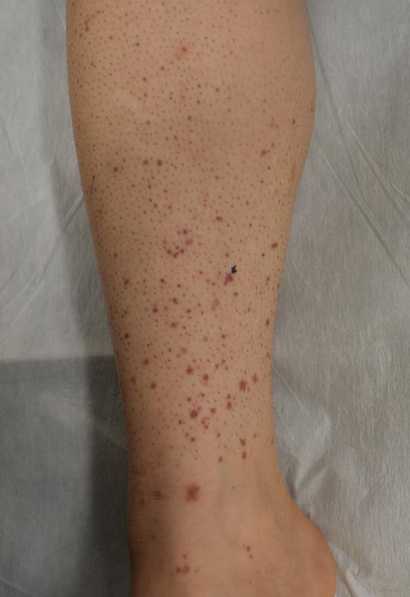
Cutaneous purpura in the lower limb
In the specimens from the kidney, 33 glomeruli were identified. Eight glomeruli showed mesangial area expansion, six glomeruli showed mesangial cell proliferation, and five glomeruli showed cellular or fibrocellular crescents (Fig. 2). Immunofluorescent staining showed diffuse IgA and C3 deposition in the mesangial area, while IgM and C1q are negative (Fig. 3). In addition, an electron microscopy examination revealed electron dense mesangial deposits with partial foot process effacement (Fig. 4).
Fig. 2.

Light microscope images of renal biopsy specimens. Glomerulus with mesangial expansion and hypercellularity that shows the formation of cellular and fibrocellular crescents. A Periodic acid Schiff staining. B Periodic acid-methenamine silver staining
Fig. 3.

Immunofluorescent staining. Anti-Immunoglobulin A (IgA) (A) and anti-C3 staining (B) are positive in the glomerular mesangial areas
Fig. 4.

Electron microscope images of glomeruli. The cell proliferation, the matrix expansion, and the electron-dense deposits are present in the glomerular mesangial area
A further worsening of proteinuria was observed after renal biopsy (UPCR = 1.42 g/gCr). With the diagnosis of IgAVN (International Study of Kidney Diseases in Children [ISKDC] Classification IIIa), the patient was given intravenous methylprednisolone 1000 mg/day for three consecutive days, followed by oral prednisolone 1 mg/kg/day. Prednisolone therapy immediately improved IgAV-associated clinical manifestation, such as cutaneous purpura, proteinuria, and hematuria (Fig. 5). At 8 months after initiation of prednisolone therapy, urinalysis showed remission of hematuria (1–4 RBCs/HPF) and proteinuria (UPCR = 0.17 g/gCr) (Fig. 5). The oral glucocorticoid (prednisolone) during tapering was reduced to 8 mg/day at that point.
Fig. 5.

Clinical course and responses to glucocorticoid treatment. IgAV IgA vasculitis, mPSL methylprednisolone; PSL prednisolone, UPCR urine protein/creatinine [Cr] ratio, U-Bld urine blood, RBC red blood cell, HPF high power field
Discussion
Since the beginning of the COVID-19 pandemic, new-onset or relapsing glomerulonephritis, such as ANCA-associated glomerulonephritis, IgA nephropathy (IgAN), and membranous nephropathy, following SARS-CoV-2 infection or vaccination has been well documented [13–15]. Although there are several case reports that have presented patients who newly developed IgAVN following COVID-19 diagnosis [8–11, 16], none of these patients were diagnosed IgAV before COVID-19. To our best knowledge, this is the first report showing the development of de novo glomerulonephritis following COVID-19 in a patient with preexistent IgAV who had have no signs and symptoms of kidney involvement.
As far as we are aware, there was only one paper that reported two cases with preexistent IgAV who developed a renal flare of vasculitis after COVID-19 diagnosis [12]. One patient achieved remission of IgAVN and experienced a recurrence of hematuria and renal dysfunction 4 months after mild COVID-19. The other patient had no history of renal involvement at the initial diagnosis of IgAV and developed new-onset ANCA-associated vasculitis 3 months after asymptomatic COVID-19. Both patients showed favorable responses after treatment with glucocorticoid and immunosuppressive agent such as azathioprine or rituximab. The time to onset of kidney involvement after COVID-19 diagnosis, presence of hematuria, and successful treatment response to immunosuppressive agent(s) were overall similar between previous and present cases. However, our patient presented proteinuria and hematuria relatively late after COVID-19 diagnosis compared to the previous cases with other types of glomerulonephritis associated with COVID-19 [13]. The mechanism explaining this discrepancy remains unclear, but could involve the distinct immune responses to SARS-CoV-2 infection in patients with preexistent IgAV. In addition, we cannot exclude the possibility that IgAVN coincidentally occurred following COVID-19.
There are several case reports describing the development of de novo IgAVN following COVID-19 vaccination [9]. In addition, we are aware of one reported case with a history of cutaneous leukocytoclastic vasculitis but not nephritis who presented biopsy-proven IgANV after receiving the messenger RNA (mRNA) COVID-19 vaccination [17]. Taken together, these findings suggest that COVID-19 vaccination-induced immune responses could induce de novo and relapsing glomerulonephritis in patients with IgAV. However, the risk for such adverse events is very rare and the clinical course of glomerulonephritis following COVID-19 vaccine is overall mild and transient [18, 19]. Thus, the benefits COVID-19 vaccine could preponderate over the potential risks and it is generally recommended that patients with immune-mediated glomerulonephritis should receive COVID-19 vaccine [18].
The pathogenic mechanism of IgAVN associated with COVID-19 remains unclear. However, recent accumulating evidence has supported several possible mechanisms mediated by immune responses to SARS-CoV-2, such as direct renal infection through angiotensin converting enzyme-2 (ACE-2) expressed in podocytes, proximal epithelial cells, and endothelial cells, autoantibody generation, and cytokine release syndrome [20]. Suzuki et al. recently found that IgAV patients with nephritis have elevated serum concentration and cellular production of galactose-deficient IgA1 (Gd-IgA1) and Gd-IgA1-specific IgG than those without nephritis or healthy control people [21]. These findings suggest that generation of Gd-IgA1-specific autoantibodies and subsequent Gd-IgA1-containing immune complexes formation are involved in the development of IgAVN [22]. Indeed, it was recently reported that plasma Gd-IgA1 IgG concentration was increased in the patient who newly developed IgAN following the first dose of the mRNA-1273 COVID-19 vaccine [23]. Taken together, these findings indicate that the immune response to SARS-CoV-2 could trigger production of Gd-IgA1 and Gd-IgA1 autoantibodies, leading to the development of de novo IgAVN, although unfortunately serum Gd-IgA1 and Gd-IgA1 IgG concentrations were not determined in our case. Given that glomerulonephritis was accompanied with worsening of cutaneous purpura following COVID-19 in our case, it is also possible that SARS-CoV-2 infection could induce the flare of systemic vasculitis in patients with preexistent IgAV, contributing to the new-onset of IgAVN as a ‘second hit’.
Data obtained from the retrospective study conducted in 250 adult patients with biopsy-proven IgAVN found that the risks of progression to end-stage renal disease (ESRD) and severe renal failure (CCr < 30 ml/min) are 11% and 13%, respectively [24]. Previous studies identified several risk factors associated with ESRD in adult IgAV patients, such as baseline renal insufficiency, baseline proteinuria (> 1 or 1.5 g/day), proteinuria (> 1 g/day) during the follow-up, hypertension, macroscopic hematuria, and degree of interstitial fibrosis, sclerotic glomeruli, and fibrinoid necrosis [24–26]. Our patient met the criteria for only baseline proteinuria (UPCR = 1.42 g/gCr) but not for other clinical or histological parameters, and thus, the risk of progression to ESRD is considered relatively low. Nonetheless, the clinical course and prognostic factors could vary between COVID-19-related and -unrelated IgAVN. Although our patient has achieved and maintained the remission of hematuria and proteinuria under glucocorticoid treatment, long-term careful monitoring of disease activity would be required.
In summary, we present a case of the IgAV patient who newly displayed hematuria and proteinuria with glomerular mesangial IgA deposits following COVID-19. Although we cannot definitely rule out the possibility that glomerulonephritis is unrelated to COVID-19 in our case, this report highlights that immune response to SARS-CoV-2 could trigger the onset of glomerulonephritis in the IgAV patients who have no renal involvement.
Declarations
Conflicts of interest
All the authors have declared no competing interest.
Informed consent
Informed written consent was obtained from the patient for publication of this case report and all accompanying images.
Footnotes
Publisher's Note
Springer Nature remains neutral with regard to jurisdictional claims in published maps and institutional affiliations.
Daigo Kobayashi and Jun Yoshino authors have equally contributed to this work.
References
- 1.Davin JC. Henoch-Schonlein purpura nephritis: pathophysiology, treatment, and future strategy. Clin J Am Soc Nephrol. 2011;6(3):679–89. 10.2215/CJN.06710810. [DOI] [PubMed] [Google Scholar]
- 2.Piram M, Mahr A. Epidemiology of immunoglobulin A vasculitis (Henoch-Schonlein): current state of knowledge. Curr Opin Rheumatol. 2013;25(2):171–8. 10.1097/BOR.0b013e32835d8e2a. [DOI] [PubMed] [Google Scholar]
- 3.Komatsu H, Fujimoto S, Yoshikawa N, Kitamura H, Sugiyama H, Yokoyama H. Clinical manifestations of Henoch-Schonlein purpura nephritis and IgA nephropathy: comparative analysis of data from the Japan Renal Biopsy Registry (J-RBR). Clin Exp Nephrol. 2016;20(4):552–60. 10.1007/s10157-015-1177-0. [DOI] [PMC free article] [PubMed] [Google Scholar]
- 4.Hocevar A, Tomsic M, Jurcic V, Perdan Pirkmajer K, Rotar Z. Predicting gastrointestinal and renal involvement in adult IgA vasculitis. Arthritis Res Ther. 2019;21(1):302. 10.1186/s13075-019-2089-2. [DOI] [PMC free article] [PubMed] [Google Scholar]
- 5.Cao R, Lau S, Tan V, Tey HL. Adult Henoch-Schonlein purpura: Clinical and histopathological predictors of systemic disease and profound renal disease. Indian J Dermatol Venereol Leprol. 2017;83(5):577–82. 10.4103/ijdvl.IJDVL_571_16. [DOI] [PubMed] [Google Scholar]
- 6.Nagy GR, Kemeny L, Bata-Csorgo Z. Neutrophil-to-lymphocyte ratio: a biomarker for predicting systemic involvement in adult IgA vasculitis patients. J Eur Acad Dermatol Venereol. 2017;31(6):1033–7. 10.1111/jdv.14176. [DOI] [PubMed] [Google Scholar]
- 7.Pillebout E, Sunderkotter C. IgA vasculitis. Semin Immunopathol. 2021;43(5):729–38. 10.1007/s00281-021-00874-9. [DOI] [PubMed] [Google Scholar]
- 8.Messova A, Pivina L, Muzdubayeva Z, Sanbayev D, Urazalina Z, Adams A. COVID-19 and new onset IgA Vasculitis: a systematic review of case reports. J Emerg Nurs. 2022;48(4):348–65. 10.1016/j.jen.2022.05.002. [DOI] [PMC free article] [PubMed] [Google Scholar]
- 9.Sugita K, Kaneko S, Hisada R, Harano M, Anno E, Hagiwara S, et al. Development of IgA vasculitis with severe glomerulonephritis after COVID-19 vaccination: a case report and literature review. CEN Case Rep. 2022;11(4):436–41. 10.1007/s13730-022-00695-1. [DOI] [PMC free article] [PubMed] [Google Scholar]
- 10.Morioka F, Nakatani S, Tsuda A, Mori K, Emoto M. Successful alternative vaccination with BNT162b2 mRNA COVID-19 vaccine for new-onset IgA vasculitis after receiving mRNA-1273-case report. CEN Case Rep. 2022;11(4):511–2. 10.1007/s13730-022-00735-w. [DOI] [PMC free article] [PubMed] [Google Scholar]
- 11.Nakatani S, Mori K, Morioka F, Hirata C, Tsuda A, Uedono H, et al. New-onset kidney biopsy-proven IgA vasculitis after receiving mRNA-1273 COVID-19 vaccine: case report. CEN Case Rep. 2022;11(3):358–62. 10.1007/s13730-021-00677-9. [DOI] [PMC free article] [PubMed] [Google Scholar]
- 12.Valero C, Baldivieso-Acha JP, Uriarte M, Vicente-Rabaneda EF, Castaneda S, Garcia-Vicuna R. Vasculitis flare after COVID-19: report of two cases in patients with preexistent controlled IgA vasculitis and review of the literature. Rheumatol Int. 2022;42(9):1643–52. 10.1007/s00296-022-05153-w. [DOI] [PMC free article] [PubMed] [Google Scholar]
- 13.Klomjit N, Zand L, Cornell LD, Alexander MP. COVID-19 and glomerular diseases. Kidney Int Rep. 2023;8(6):1137–50. 10.1016/j.ekir.2023.03.016. [DOI] [PMC free article] [PubMed] [Google Scholar]
- 14.Ota Y, Kuroki R, Iwata M, Taira H, Matsuo S, Kamijo M, et al. Association between COVID-19 vaccination and relapse of glomerulonephritis. Clin Exp Nephrol. 2023;27(3):236–42. 10.1007/s10157-022-02299-6. [DOI] [PMC free article] [PubMed] [Google Scholar]
- 15.Li H, Li Z, Wu Z, Wang F, Xing Y, Liu Y, et al. Clinical and pathological findings of IgA nephropathy following SARS-CoV-2 infection. Clin Exp Med. 2024;24(1):43. 10.1007/s10238-023-01271-2. [DOI] [PMC free article] [PubMed] [Google Scholar]
- 16.Ramdani Y, Galempoix JM, Augusto JF, Dekmeer E, Perard L, Ferreira N, et al. Immunoglobulin A vasculitis following COVID-19: a french multicenter case series. J Rheumatol. 2022;49(12):1390–4. 10.3899/jrheum.220503. [DOI] [PubMed] [Google Scholar]
- 17.Cho I, Kim JK, Kim SG. IgA vasculitis presenting as nephrotic syndrome following COVID-19 vaccination: a case report. BMC Nephrol. 2022;23(1):403. 10.1186/s12882-022-03028-7. [DOI] [PMC free article] [PubMed] [Google Scholar]
- 18.Stevens KI, Frangou E, Shin JIL, Anders HJ, Bruchfeld A, Schonermarck U, et al. Perspective on COVID-19 vaccination in patients with immune-mediated kidney diseases: consensus statements from the ERA-IWG and EUVAS. Nephrol Dial Transplant. 2022;37(8):1400–10. 10.1093/ndt/gfac052. [DOI] [PMC free article] [PubMed] [Google Scholar]
- 19.Waldman M, Sinaii N, Lerma EV, Kurien AA, Jhaveri KD, Uppal NN, et al. COVID-19 vaccination and new onset glomerular disease: results from the irocgn2 international registry. Kidney. 2023. 10.34067/KID.0006832022. [DOI] [PMC free article] [PubMed] [Google Scholar]
- 20.Ahmadian E, Hosseiniyan Khatibi SM, Razi Soofiyani S, Abediazar S, Shoja MM, Ardalan M, et al. Covid-19 and kidney injury: Pathophysiology and molecular mechanisms. Rev Med Virol. 2021;31(3): e2176. 10.1002/rmv.2176. [DOI] [PMC free article] [PubMed] [Google Scholar]
- 21.Suzuki H, Moldoveanu Z, Julian BA, Wyatt RJ, Novak J. Autoantibodies specific for galactose-deficient IgA1 in IgA vasculitis with nephritis. Kidney Int Rep. 2019;4(12):1717–24. 10.1016/j.ekir.2019.08.015. [DOI] [PMC free article] [PubMed] [Google Scholar]
- 22.Suzuki H, Yasutake J, Makita Y, Tanbo Y, Yamasaki K, Sofue T, et al. IgA nephropathy and IgA vasculitis with nephritis have a shared feature involving galactose-deficient IgA1-oriented pathogenesis. Kidney Int. 2018;93(3):700–5. 10.1016/j.kint.2017.10.019. [DOI] [PubMed] [Google Scholar]
- 23.Chen YS, Yang CW, Tsai CC, Ang MD, Chou SF, Chiang WC, et al. Newly-diagnosed immunoglobulin A nephropathy with increased plasma galactose-deficient-IgA(1) antibody associated with mRNA COVID-19 vaccination: a case report. J Int Med Res. 2022;50(10):3000605221129674. 10.1177/03000605221129674. [DOI] [PMC free article] [PubMed] [Google Scholar]
- 24.Pillebout E, Thervet E, Hill G, Alberti C, Vanhille P, Nochy D. Henoch-Schonlein Purpura in adults: outcome and prognostic factors. J Am Soc Nephrol. 2002;13(5):1271–8. 10.1097/01.asn.0000013883.99976.22. [DOI] [PubMed] [Google Scholar]
- 25.Coppo R, Mazzucco G, Cagnoli L, Lupo A, Schena FP. Long-term prognosis of Henoch-schonlein nephritis in adults and children. Nephrol Dial Transplant. 1997. 10.1093/ndt/12.11.2277. [DOI] [PubMed] [Google Scholar]
- 26.Shrestha S, Sumingan N, Tan J, Alhous H, McWilliam L, Ballardie F. Henoch Schonlein purpura with nephritis in adults: adverse prognostic indicators in a UK population. QJM. 2006;99(4):253–65. 10.1093/qjmed/hcl034. [DOI] [PubMed] [Google Scholar]