



Abstract
Purpose
HEV is an emerging pathogen in Europe and was previously shown to be hyperendemic in areas of Abruzzo and Lazio, Central Italy. No systematic analysis of the HEV strains responsible for human infections over several years in Central Italy has previously been reported. Aim of the study was the molecular characterization of HEV from autochthonous hepatitis E cases occurred in Abruzzo and Lazio between 2015 and 2023.
Methods
Samples from 118 cases collected as part of virological surveillance in Abruzzo and Lazio from 2015 to 2023 were subjected to HEV sequencing and phylogenetic analysis.
Results
The main observed subtype was 3f, followed by 3c and 3e. The annual subtype distribution was quite stable over the observation period, but 3f cases tended to concentrate in winter/early spring whereas 3e cases in summer. Phylogenetic clusters of highly related sequences (a) highlighted unrecognized “point source outbreaks”, (b) provided molecular support to temporally and/or geographically linked cases and (c) provided evidence for transmission of identical/highly related strains up to months/years following their first detection.
Conclusions
The data provide an overview of the HEV strains responsible for human infections over eight years in Central Italy. The observed subtype distribution appears to agree better with the subtype distribution reported in Italy in pigs rather than in geographically matched wild boars, suggesting pig and its derivate food was a more frequent source of infection than wild boar in Abruzzo and Lazio. Molecular characterization is essential to recognize “point source outbreaks” and to monitor HEV circulation.
Supplementary Information
The online version contains supplementary material available at 10.1007/s15010-024-02348-0.
Keywords: Hepatitis E Virus, Foodborne diseases, Zoonoses, Phylogenetic analysis
Introduction
Hepatitis E Virus (HEV) belongs to the species Paslahepevirus balayani (Family Hepeviridae, Subfamily Orthohepevirinae, Genus Paslahepevirus) [1]. Based on phylogenetic analysis, HEV is classified into eight genotypes (1 to 8) [2]. Genotypes 1 to 4 are the most widespread among humans. Genotypes 1 and 2 infect exclusively humans; they are the cause of HEV outbreaks in developing countries and are mainly transmitted by contaminated water. In contrast, genotypes 3 and 4 are zoonoses [3–5]. Genotypes 5 and 6 had been isolated so far only in swine and wild boar [4, 5]. Genotypes 7 and 8 had been detected in camels (Gt7) and in Bactrian camels (Gt8) [6, 7]. Recently, human HEV acute hepatitis has also been linked to Rocahepevirus ratti (previously classified as Orthohepevirus C), commonly denominated rat HEV, another member of the Family Hepeviridae. Rodents and mustelids are natural reservoirs of rat HEV, while human infections were described in Spain and in France [8, 9].
Genotype 3 (Gt3) is the main genotype diffused in Europe [10]. Even though swine (Suis scrofa domesticus) and wild boars (Suis scrofa) represent the main reservoir, Gt3 had been isolated in several other mammals, including deer, rabbit, camels and dolphins [11, 12]. According to the last updated international proposal for subtype reference sequences of HEV, 14 subtypes have been recognised within genotype 3 (3a, 3b, 3c, 3d, 3e, 3f, 3 g, 3 h, 3i, 3j, 3k, 3 L, 3 m, 3ra) [2]. They are grouped in three main clades: clade 1, including 3e, 3f and 3 g subtypes, clade 2, including 3a, 3b, 3c, 3 h, 3i, 3j, 3k, 3 L, 3 m subtypes and clade 3 with 3ra subtype only. Currently, 3ra seems to have a specific tropism for rabbit, indeed it has been isolated only in rabbit and not in other commonly HEV Gt3 reservoirs like pigs or wild boars. In Italy, different prevalence of Gt3 subtypes has been found in analysed animal reservoirs (pigs and wild boars). Processed products (liver, meat, blood) of pigs and wild boars are believed to represent the main sources of human infections.
The HEV prevalence in wild boar livers is very similar in different Italian geographical areas: the highest prevalence was observed in Calabrian region (26.7%) [13], followed by Central Italy (16.3%) [14], North Italy (14.3%) [15] and South Italy (13.7%) [16]. Several studies were also carried out in pigs. For example, in Piedmont region (North Italy), the anti-HEV IgG in adult swine was 50.3% among intensive pig farming, while the HEV RNA positivity was 10.9% [17] and 13.4% in another recent study [18]. Both pigs and wild boars were infected with 3c, 3e and 3f subtypes, although with different relative proportions, and wild boars were also infected with 3a subtype. Among humans, HEV infections represent a consistent percentage of acute hepatitis: 30.4% in Marche, 29.1% in Abruzzo and 17% in Toscana regions [19]. The main Gt3 subtypes identified in humans were 3f, 3e, 3c, 3a; moreover, some strains could not be assigned to any subtypes by phylogenetic analysis [20]. These different subtypes were associated to different clinical manifestations and they were also linked to outbreaks [21, 22], suggesting the possibility that some HEV strains may possess high virulence and infectivity capacity.
Previous national studies in blood donors showed high prevalence of anti-HEV IgG in some Italian Regions, providing indirect evidence for frequent HEV infection in the human population living in those areas. Analysis at the Province sub-level showed areas with very high prevalence in some Regions, such as Abruzzo and Lazio, two bordering Regions in Central Italy [23, 24] (see a map of Lazio and Abruzzo in Italy in Supplementary Fig. 1).
Aim of the present study was to provide an overview of the HEV genotypes/subtypes and strains responsible for autochthonous HEV infection in humans in Abruzzo and Lazio and to investigate the commonality of infection sources. Through a collaboration between the Italian National Institute of Health (Istituto Superiore di Sanità, ISS) and the Reference Regional Laboratory of Lazio (INMI Spallanzani), the viruses from sera collected between 2015 and 2023 from HEV positive cases were molecularly characterized by sequencing a fragment from the ORF2 region of the HEV genome and phylogenetic analysis. In addition to providing a molecular epidemiology update, data were evaluated to highlight any temporal changes in the distribution of HEV subtypes as well as to assess whether these changes could be related to subtypes circulating in pigs and wild boars.
Methods
Study population
Overall, 118 cases of autochthonous HEV infection were investigated (66 cases from the INMI laboratory and 52 cases from the ISS laboratory). Almost all the patients included in the study were anti-HEV IgM/IgG positive. Anti-HEV IgM and IgG antibodies were detected by DIAPRO ELISA assays (DIA.PRO, Milan, Italy) at INMI “L. Spallanzani” and by Wantai HEV-IgM and Wantai HEV-IgG ELISA assays (Beijing WANTAI Biological Pharmacy Enterprise Co. Ltd., Beijing, China) at ISS, according to manufacturer’s instructions.
The two collaborating laboratories of the present study (INMI lab and ISS lab) receive samples in different settings and workflows. The INMI laboratory (the Regional Reference Laboratory of Lazio) receives samples for diagnosis and Regional surveillance from suspected viral hepatitis cases hospitalized in Lazio. Thus, each sample is routinely tested at INMI laboratory for markers of hepatitis A, B, C and E infection. Overall, 1068 samples were tested for these markers in the period 2015–2023; a positive anti-HEV IgM result was obtained in 93 of them. A PCR fragment suitable for sequencing (ORF2 region, 411 nt) was amplified from 72/93 samples. Upon sequence analysis, 6/72 cases had HEV Gt1, consistent with case history of travel abroad and, thus, were excluded from the present study, whose focus was on autochthonous HEV Gt3 infections; the remaining 66 cases had HEV Gt3 and no history of travel abroad (autochthonous cases) and were included in the present study.
The ISS laboratory (the Italian National Institute of Health laboratory) receives samples for HEV diagnosis and National surveillance from hospitalized non-A/non-B/non-C hepatitis cases from Regions with no established Regional reference laboratory, such as Abruzzo, i.e. one of the two regions involved in the present study. In contrast with the INMI laboratory, the ISS laboratory receives samples already tested (and with negative result) for markers of hepatitis A, B, and C infection. Overall, 215 samples were tested for anti-HEV IgM in the period 2015–2023; a positive anti-HEV IgM result was obtained in 71 of them and a PCR fragment for sequencing (ORF2 region, 493 nt) was amplified from 48/71 samples. In addition, four autochthonous asymptomatic cases from one blood donor and three transplanted patients, identified in the period 2015–2023 in the frame of other studies carried out in Abruzzo, were also included in the study. All 52 cases had HEV Gt3 and no history of travel abroad (all autochthonous cases) and were included in the present study.
Summarising the above information, 1,283 samples were tested for anti-HEV IgM/IgG, 164 samples were anti-HEV IgM positive and a PCR product from autochthonous hepatitis E cases was obtained from 118 cases.
A medical history was taken from the cases, including the collection of general data, such as age, sex, symptoms, travel history before the onset of illness, history of blood transfusion or ingestion of raw meat or undercooked liver/viscera from HEV animal reservoirs. The association between known risk factors and HEV infection in the studied population was evaluated by Fisher’s test, comparing through a contingency table the frequency of each risk factor in the HEV positive group vs. the frequency in a matched HEV negative group (this latter consisting of non-A/non-B/non-C hepatitis cases, whose samples had been received for HEV testing in the same period of the HEV positive group and had a negative result for all tested HEV markers, i.e. anti-HEV IgM, anti-HEV IgG, HEV-RNA); a p < 0.05 was considered statistically significant. In laboratory examination, the blood levels of total bilirubin (T-bil), alanine aminotransferase (ALT), aspartate aminotransferase (AST) were determined. Possible differences of T-bil, ALT and AST levels among the different HEV Gt3 subtypes were evaluated by Student’s T-test, by comparing observed values in paired subtypes; a p < 0.05 was considered statistically significant.
HEV characterization by sequencing
The two laboratories (INMI and ISS) involved in the present study routinely use two different HEV sequencing protocols, that produce partially overlapping sequences corresponding to the following nucleotide positions in the reference sequence Acc.N. M73218: nt 5945–6355 (sequence by the INMI protocol) and nt 5986–6478 (sequence by the ISS protocol). Their sizes are 411 nt and 493 nt, respectively; the size of the overlapping region is 370 nt.
Procedure at INMI laboratory: HEV-RNA was extracted from 0.6 mL of human serum using QIASYMPHONY automated instruments (Qiagen, Hilden, Germany). Faeces were pre-treated with AL lysis buffer (100 mgr in 5 ml of AL), vortexed for 10 s, centrifuged at 3.500xg for 10 min, and 0.6 mL of supernatant were extracted. The RNA was reverse transcribed using one step RT-PCR until 2018 or Superscript IV (Thermo Fisher Scientific) after 2018; nested PCR was subsequently carried out using methods previously described [21, 22]. Specific primers for a well conserved region of ORF2 were used [25]. TaqGold polymerase was used for PCR amplification. Cycling profile included the following setting: 15 min at 95 °C, 35 cycles for 30 s at 95 °C, 30 s at 56 °C, and 45 s at 72 °C, and at 72 °C for 7 min as final extension. The PCR products from the second round amplification (457 nt ORF2 region, nucleotide position 5922–6378:M73218) were subsequently sequenced using methods previously described [20]. After removal of primer sequence from both ends, the final sequence (size: 411 nt) was used for phylogenetic analysis. Sequencing was successful in 66 samples; the sequences were deposited in GenBank, with the following Accession Numbers: MN432489; MN444828 to MN444844; MN444848 to MN444850; MN444852; MN444853; MN537875 to MN537879; MN604404; MN737482 to MN737484; MT263983 to MT263985; MT769323 to MT769325; MT786652; MT786653; MT990345; MW013049 to MW013052; OK216750; OK310498 to OK310501; OR795721 to OR795729.
Procedure at ISS laboratory: HEV-RNA was extracted from 200 µL serum by the QIAmp MinElute Virus Spin kit (Qiagen, Hilden, Germany). Five µL extracted RNA was reverse transcribed by the SuperScript IV (Thermo Fisher Scientific) with a specific reverse primer and amplified by nested PCR as previously described [26]. For the first PCR step, PCR cycling conditions were as follows: 35 cycles of 95 °C 30 s; 42 °C 30 s; 60 °C 45 s. For the second (nested) PCR, PCR cycling conditions were as follows: initial denaturation 95 °C 6 min, then 40 cycles of 95 °C 30 s; 60 °C 20 s; 72 °C 15 s. The PCR products from the second round amplification (566 nt ORF2 region, nucleotide position 5948–6513 in the reference sequence Accession Number M73218) were subsequently sequenced as previously described [26]. After removal of primer sequence from both ends, the final sequence (size: 493 nt) was used for phylogenetic analysis. Sequencing was successful in 52 samples; the sequences were deposited in GenBank, with the following Accession Numbers: MZ274229 to MZ274264, MZ274266 to to MZ274271 and PP898058 to PP898067.
Phylogenetic analysis
The ISS and INMI laboratories sequencing protocols produce 493 nt and 411 nt sequences, respectively, from the ORF2 region. The sequences share a 370 nt overlapping region. To include in the analysis as much as possible genetic information, the sequences obtained by the two different protocols were subtyped separately. To search for possible clusters, the 370 nt overlapping region was used for phylogenetic analysis.
The sequences were aligned by ClustalW and the alignment inspected for optimal alignment. The best evolutionary model for the dataset under study was evaluated by the Models tool in MEGA [27]. Then, a Maximum Likelihood approach was used to build a phylogenetic tree, using the best evolutionary model (GTR + G + I). Significance of the tree nodes was evaluated by bootstrap analysis; bootstrap values > 70% were considered significant.
Results
Overall, 118 cases of HEV infection from Abruzzo and Lazio from 2015 to 2023 were included in the present study. While the vast majority of analyzed samples were from acute hepatitis cases admitted to hospital because of hepatitis symptoms (n = 114), four cases were asymptomatic (one blood donor and three transplanted individuals), with infection detected either in the frame of a prevalence study in blood donors or in the frame of a project of active surveillance of transplanted patients in Abruzzo.
Table 1 summarizes the demographic features of the study population. Most cases were males (100/117, 85.5%; data not available in one case). The mean age was 58.3 years (± 13.2 years), with no significant difference between males and females.
Table 1.
Demographic features of the study population (n = 118)
| n | % | ||
|---|---|---|---|
| Gender * | Males | 100 | 85.5% |
| Females | 17 | 14.5% | |
| Mean age (± sd) | overall | 58.3 (± 13.2) | |
| in males | 58.8 (± 13.1) | ||
| in females | 55.9 (± 14.2) | ||
| Residence | Abruzzo^ | 42 | 35.6% |
| Lazio | 76 | 64.4% |
* data not available in one case
^ one case attending an Abruzzo hospital had residence in the nearby Molise region and was included in the Abruzzo group
Figure 1A shows the monthly distribution of HEV infection cases of the present study, collected in the period 2015–2023; the distribution is based on the date of symptom onset (or the date of sample collection, as a proxy of the onset date). Only three months (July, November and December) showed less than 8 cases, with a quite uniform distribution of cases over the year. However, while the considered population mostly includes sporadic cases, some of them (represented by the orange part of the bars in Fig. 1B) were part of an outbreak occurred in a single year (2019) [22]: thus, in this specific analysis the exclusion of these exceptional cases may more accurately represent the actual monthly distribution of “background” sporadic cases in the period under study (2015–2023). In this latter case, i.e. by considering only the blue bars in Fig. 1B, it is evident a seasonality, with higher frequency in February to April than in the remaining months.
Fig. 1.
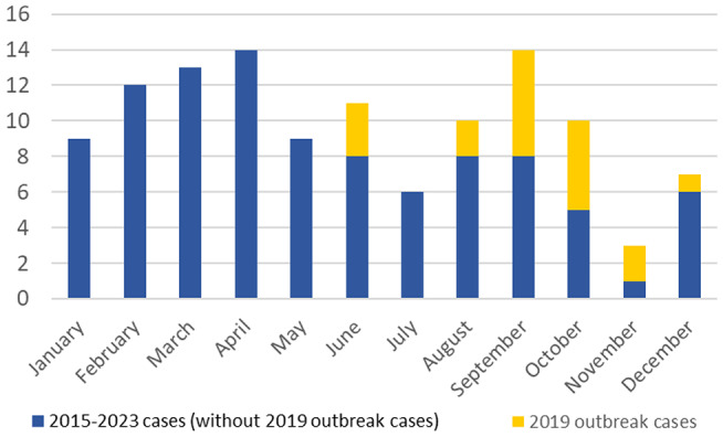
Monthly distribution of cases observed in the period 2015–2023, in which cases from a HEV outbreak occurred in Central Italy in 2019 are highlighted (orange end of the bars)
Genotyping/subtyping of viral sequences: overall, annual and monthly distribution of subtypes
Genotyping by phylogenetic analysis of the 118 HEV sequences together with references of the known eight genotypes showed clearly that all autochthonous HEV infection cases in Abruzzo and Lazio during 2015–2023 had been caused by HEV genotype 3 (data not shown). During this period, we had also observed five cases caused by HEV genotype 1, all from foreign people from India, Pakistan, and Bangladesh. All of them manifested acute hepatitis symptoms when came back from their country of origin. Those sequences were omitted from the study because they were not autochthonous strains.
Figure 2 shows the subtyping results of the 118 sequences by phylogenetic analysis. The ISS and INMI laboratories sequencing protocols produce 493 nt and 411 nt sequences, respectively, with a 370 nt overlapping region. To include in the analysis as much as possible genetic information, the sequences obtained by the two different protocols were subtyped separately, reserving phylogenetic analysis of the 370 nt overlapping region only to the search for possible clusters (see below). Figure 2a and b report the phylogenetic trees obtained from the 493 nt and 411 nt sequences, respectively.
Fig. 2.

Phylogenetic trees for HEV subtyping, obtained by the Maximum Likelihood approach. (A) Analysis of n = 52 sequences, size 493 nt. (B) Analysis of n = 66 sequences, size 411 nt. Both trees include 39 Reference sequences representing subtypes 3a to 3 m, as reported in Smith et al., 2020 [2]. The meaning of the red and black dots is reported in the insert in the lower right corner of the figure
A p-distance analysis of the sequences was also carried out. Supplementary Table 1 reports the individual p-distance values calculated between each of the 118 sequences and each of the same 39 reference sequences used for phylogenetic analysis. The average p-distances of the sequences, grouped by subtype according to the results of phylogenetic analysis, are reported in Supplementary Table 2. The 3f, 3e, 3a and 3c strains correctly showed the lowest average p-distance only when compared with reference sequences of the same subtype; one only exception was a sequence that could not be unambiguously assigned to a specific subtype by phylogenetic analysis (INMI_1909/Dec.2019/ in Fig. 2B) and that, in fact, showed low p-distance both with 3c and 3i references (Supplementary Table 2, column “3chilm strain”).
Figure 3A summarizes the distribution of subtypes according to the results of phylogenetic and p-distance analysis. One half cases was subtype 3f (61/118, 51.7%), the great majority of remaining cases were split between 3e (30/118, 25.4%) and 3c (25/118, 21.2%); one case observed in 2017 was 3a; finally, in one case occurred in 2019 (the above mentioned INMI_1909/Dec.2019 in Fig. 2B) phylogenetic analysis places its sequence in the 3chilm clade (i.e. in the clade including 3c, 3 h, 3i, 3 L, 3 m reference sequences) and it appears most similar to the 3i reference, but without significant boostrap support; p-distance analysis also did not provide any further elements for subtype assigment: so it was classified as a member of the 3chilm clade.
Fig. 3.

(A) Subtype distribution, HEV infection cases (n = 118) from Abruzzo and Lazio, 2015–2023. (B) Annual trend of subtype distribution of 3f, 3e and 3c cases from Abruzzo and Lazio, 2015–2023. The annual number of cases in the years 2015, 2020, 2021, 2022 was not large enough (n = 3, n = 7, n = 7 and n = 3, respectively) to give rise to a reliable annual distribution. Thus, cases from 2015 were joined to 2016, cases from 2020 were joined to 2021 and cases from 2022 were joined to 2023. (C) Monthly distribution of 3f, 3e and 3c subtype cases cumulatively observed in the period 2015–2023 in Abruzzo and Lazio, including 3f and 3e cases from the 2019 outbreak. (D) Monthly distribution of 3f, 3e and 3c subtype cases after exclusion of the 3f and 3e cases from the 2019 outbreak
Figure 3B shows the annual trend of subtype distribution of 3f, 3e and 3c cases. It is evident that year 2019 contributed heavily to the total number of 3e and 3f sequences in the overall study period, because of the above-mentioned outbreak occurred in that year. Except in 2019, the 3f subtype appears to be the most frequent over the studied period, followed by 3c and 3e; this latter subtype was rarely detected before and after the 2019 outbreak. Careful analysis of residence data of individual cases showed that in Abruzzo the few 3e cases detected in 2015–2018 were from the coastal areas (Pescara and Chieti provinces); in the internal areas (L’Aquila province) the 3e subtype had never been detected previously and was first observed upon the 2019 outbreak, that was sustained mainly by 3e strains, possibly introduced by pork products or live animals imported from outside Abruzzo [22].
Figure 3C shows the monthly distribution of 3f, 3e and 3c subtypes in the period 2015–2023 in Abruzzo and Lazio. A seasonal trend can be recognized: the winter/early spring months show an excess of 3f subtype while summer months include most subtype 3e cases observed in 2015–2023. Figure 3D shows that this trend holds even after having excluded the 3f and 3e outbreak cases occurred in summer/fall 2019.
Subtypes vs. demographic and clinical variables
No differences in the distribution of 3f, 3e and 3c subtypes were observed between males and females or in different age groups (data not shown).
No significant differences of ALT and AST levels could be observed among 3f, 3e and 3c, however total bilirubin levels were significantly higher in 3f (6.53 mg/dL ± 7.93) than in 3c (2.82 mg/dL ± 3.08) (t-Student’s, p = 0.01).
Analysis of molecular clusters
The sequences obtained by the different ISS and INMI sequencing protocols were aligned and the shared overlapping region (size: 370 nt) was subjected to phylogenetic analysis by a Maximum Likelihood approach.
Figure 4 shows the resulting phylogenetic tree. It was preliminary controlled if the “cut” 370 nt sequences had been assigned to the same subtypes as in the two phylogenetic trees obtained from the “uncut” 493 nt and 411 nt sequences (Fig. 2A and B, see above). Careful comparison of the trees demonstrates that the “cut” 370 nt sequences were correctly grouped in the same subtype clades as in the trees obtained from the “uncut” sequences.
Fig. 4.

Phylogenetic tree resulting from analysis of a 370 nt region shared by the HEV sequences obtained with different protocols (n = 118). A Maximum Likelihood approach was applied, with a GTR + G + I evolutionary model preliminarily estimated by the Model tool in MEGA. The tree includes reference sequences (n = 39) representing subtypes 3a to 3 m, as reported in in Smith et al., 2020 [2]. The meaning of red, blue and black circles is described in the box. Clusters are delimited by red lines and marked by alphabetical letters, A to N
Several sequence clusters could be identified according to the following criteria: (a) the cluster had to be statistically supported (bootstrap value ≥ 70) and (b) sequences in the cluster had to be identical or closely related (≤ 3 nt differences over 370 nt). Identified clusters were then evaluated for any possible temporal and/or geographical link among the cases from which the sequences were obtained.
Seven clusters (A to G) were identified in the 3f clade (Fig. 4). Cluster A includes sequences from the above mentioned HEV outbreak that occurred in 2019, that was sustained by multiple 3f and 3e HEV strains [22]. Cluster B includes five sequences, four of them identical each other and the fifth with 1 nt difference; they show geographical link (all occurred in Lazio) and they also show a temporal link (onset between mid-February and mid-March 2017). Cluster C includes two closely related sequences (1 nt difference) showing geographical as well as temporal link (both cases from Lazio in April 2017, onset three days apart). Cluster D includes five sequences (three identical, the two others with 1 nt and 2 nt differences); the cases occurred in a narrow timeframe (between mid-April and the end of May 2018). Three cases were from Lazio, but one of them reported a dinner in Abruzzo with consumption of pork products about three weeks before onset of symptoms; the two other cases were from Abruzzo. Cluster E includes two identical sequences from cases occurred in Lazio in January 2016, with onset on two consecutive days, admitted in two different hospitals. Cluster F includes two identical sequences from two cases occurred in Lazio in May 2021, with onset two days apart, admitted to the same hospital. Cluster G includes two identical sequences from two cases occurred in Abruzzo in February 2018, with onset four days apart.
Three clusters (H to L) were identified in the 3e clade (Fig. 4). Clusters H and L mostly include 3e sequences from the above-mentioned outbreak occurred in 2019, in which two distinct 3e clusters had been recognized [22]. However, cluster L also includes two further sequences detected one-and-a-half and two years later, respectively: a closely related sequence (ISS_ID_366, 1 nt difference) detected in January 2021 from a case in Abruzzo and an identical sequence (INMI_2109) detected in August 2021 from a case in Lazio. Cluster I includes two identical sequences from two cases occurred in Lazio in February 2016, with onset one week apart, admitted to the same hospital.
Two clusters (M and N) were identified in the 3c clade (Fig. 4). Cluster M includes two closely related sequences (1 nt difference) from two cases occurred in Lazio in 2023, with onset two months apart (mid-June and mid-August). Cluster N includes five sequences (two identical and three closely related sequences with 1–3 nt difference) occurred in cases from both Lazio and Abruzzo between February 2017 and April 2018. However, even though the cases formally occurred in two different regions, actually they are geographically linked: in fact, the cases have their residence in towns/villages quite close to each other, in an area at the border of Lazio and Abruzzo (maximum straight-line distance between those towns/villages: 25 Km).
Analysis of risk factors
Information about risk factors was available for 48 HEV positive cases; they were compared with risk factors available from 99 non-A/non-B/non-C hepatitis cases whose samples had been received for HEV testing during 2015–2023 and had proved to be negative for anti-HEV IgM and IgG as well as for HEV-RNA. No significant differences were observed in the frequency of consumption of raw or undercooked meat (from pig/wild boar/dear) and in consumption of wild boar sausages, while consumption of both pig sausages and liver sausages was significantly more frequent in the HEV positive group than in the HEV negative one (pig sausages: 40/46, 87% vs. 59/92, 64%, Fisher’s test: p < 0.005; liver sausages: 28/45, 62% vs. 21/90, 23%, Fisher’s test: p < 0.0001).
Discussion
The present study provides an overview of the HEV subtypes responsible for hepatitis E cases occurred in Abruzzo and Lazio, Central Italy, in 2015–2023. The monthly distribution of sporadic HEV cases in the observation period showed some degree of seasonality, with higher frequency in January to April than in the remaining months. According to the risk factors for HEV genotype 3 infection reported in the literature, confirmed in the present case series, a possible hypothesis to explain the finding is that the observed annual distribution pattern might be the result of more frequent consumption of home-made pork products in winter months, particularly in the first months of the year. In particular, seasonality might be related to the widespread habit of people living in rural areas to raise one or a few pigs for familial consumption and distribution to relatives and friends; this habit is especially frequent in the internal areas and small towns of Abruzzo and Lazio. These pigs are mostly slaughtered in December: some pork products are consumed fresh or in the days immediately following cutting, while uncooked cured sausages (both meat and liver sausages), cold cuts and other cured pork products are consumed starting from January, after a few weeks (4 to 8 weeks) of ripening. Considering the variable incubation period before onset of symptoms, this timing is compatible with the peak of hepatitis E cases observed in January to April.
Overall, in 2015–2023 the predominant subtype was 3f (about one-half cases), followed by 3c and 3e with roughly similar proportion. However, examination of the annual subtype distribution (Fig. 3B) showed that the overall number of 3e cases in the studied period was highly affected by the excess of 3e cases occurred in a single year (2019) because of an outbreak (mostly involving 3e subtype and, to a less extent, 3f): thus, with the only exception of 2019, 3e cases were actually less frequent than 3c and the annual distribution of subtypes appeared to be quite stable through the years under study. Even excluding 3f cases involved in the 2019 outbreak, 3f still continued to be the most frequently observed subtype, as reported in France [28] and particularly in Spain, where 3f reaches a prevalence of 88.3% [29]. However, this contrasts with Germany and Belgium, where 3c is the predominant subtype, representing 67.3% and 50.3% of cases, respectively [30, 31].
The subtype prevalence pattern observed among our cases differs from observations in native wild boars in Central Italy and Southern Regions, where 3c appears to be the most frequent subtype. In a study in Abruzzo about HEV prevalence in wild boar samples collected between October 2015 and January 2016, the viral sequences obtained from nine samples proved to be subtype 3c [16]; although 3f subtype was detected in 2017 in six wild boar liver samples collected in a small area [32], samples collected in the following hunting season 2018–2019 in a large area confirmed 3c to be the most frequent subtype in wild boar in Abruzzo (9/12, 75%) [33]. A study in wild boars in 2011–2014 in Lazio (Viterbo province) showed that one-half sequences belonged to an unclassified subtype (12/26) whereas the others were 3c (4/26), 3f (5/26), 3a (3/26) and 3 L (2/26) [14]. A subsequent study in wild boars in 2016–2017 in the same area (Lazio, Viterbo province) confirmed a consistent fraction of unclassified subtype(s) (11/18), whereas the remaining strains were 3c (5/18) and 3f (2/18) [34]. In the nearby Marche region in Central Italy, Ascoli Piceno province, bordering the North of Abruzzo, all sequences (55/55, 100%) from wild boar liver samples collected between October 2020 and January 2021 harboured HEV subtype 3c and the strains showed high nucleotide identity with two subtype 3c strains identified in the previous year in the same area [35]. In the Calabria region (South Italy), 19/23 (83%) HEV sequences from samples collected between October 2019 and January 2020 showed subtype 3c [13]. In Apulia and Basilicata regions (South-East Italy), 5/13 (38%) strains were 3c, with most remaining strains being of undetermined subtypes [36].
Although further phylogenetic studies are needed, the proportion of the 3c subtype in humans in Abruzzo and Lazio, much lower than in wild boars from roughly the same or nearby geographical areas, as well as the absence of unclassified subtypes as observed in wild boars, suggest wild boar was not the main source for human HEV infection in Central Italy in 2015 to 2023. The high prevalence of 3f, 3e, and 3c subtypes in our cases appears to agree better with the subtype distribution overall observed in pigs in Italy [18, 37, 38], suggesting consumption of pork and its derivate food (swine meat products, e.g. under-cooked sausages and liver meat) may be the main source of HEV transmission. This is in line with the fact that human consumption of pig products by far exceeds the consumption of wild boar derived food: therefore, at population level, it is expected that the overall exposure to HEV subtypes circulating in pigs is significantly greater than exposure to subtypes circulating in wild boars.
Analysis of the monthly subtype distribution showed that 3c cases were quite distributed throughout the year, whereas 3f cases were mostly detected in January to May, in contrast with 3e cases that mostly occurred in August and September (a pattern that holds even excluding the 3f and 3e cases of the 2019 outbreak, Fig. 3D). The underlying reasons of this distribution remain, at present, unknown and deserve further investigations in the future.
No conclusions can be done regarding the greater or lesser pathogenicity of Gt3 subtypes. In the present study, 3f subtype appears to be associated with more severe cases (higher total bilirubine level) than 3c, apparently contradicting a previous study where subtype 3c seemed to be the most pathogenic [21]. Contradictory results were also reported from other European countries, such as Belgium, where 3c subtype appears more pathogenic than subtype 3f [39], while in France 3f subtype is described with greater pathogenicity [40] and in Germany severe disease was more often associated with the group of 3e, 3f, and 3 g subtypes than with the group including all other subtypes. These apparent discrepancies may be due to bias in patient analysis/enrolment in each study. In our study, the amount of cases with 3e and 3f subtypes was significantly higher than that of subtype 3c and also included samples from the 2019 outbreak [22].
Phylogenetic analysis showed that clusters of identical or closely related sequences were detected throughout the period under study. Three clusters included the strains involved in a previously described HEV outbreak occurred in 2019 (Fig. 4, clusters A, H and L) [22]. The outbreak was sustained by three strains (one 3f and two 3e), appearing as three distinct clusters upon phylogenetic analysis; based on the onset dates, the cases from the three clusters showed partially overlapped temporal distribution. Although the specific source of infection could not be identified, epidemiological data suggested consumption of pork products in Abruzzo as the most likely source of the outbreak.
In addition to the clusters due to the 2019 outbreak, several other small clusters were observed. They provide evidence that, in addition to sporadic isolated cases and 2019 outbreak cases, small “point source outbreaks” had occurred; however, in most cases they went unrecognised: in fact, individual cases appeared as sporadic unrelated cases until evidence of a molecular link, obtained by viral sequence comparison. Early identification of these “point source outbreaks” would require more timely molecular characterization, a goal that will require the concerted effort of all the involved professionals, from the hospital admission to the final sequence comparison.
Some clusters of molecularly and temporally linked sequences can also include sequences detected months/years later, such as cluster L that, in addition to the strains involved in the 2019 outbreak, also includes two identical/closely related strains detected 16–24 months later (Fig. 4, cluster L). Cluster L provides evidence for the circulation of identical or evolutionarily related strains for a long time following the first detection. This may be due to prolonged viral circulation in the host animal populations or to consumption of infected products after long term storage. A possible limit of this observation is that only a small fragment of viral genome was compared: two viruses may appear to be identical, but possible differences in the remaining regions of the genome get undetected. Future sequencing of the full viral genome by Next Generation Sequencing technologies will provide a more accurate picture of the genetic relatedness of the viral strains and a more accurate reconstruction of HEV molecular epidemiology.
Electronic supplementary material
Below is the link to the electronic supplementary material.
Acknowledgements
The authors are very grateful to the many physicians and employees of the Italian National Health Service for their silent but valuable work that allowed the present study. The authors also thank the Secretariat Staff of the Department of Infectious Diseases at Istituto Superiore di Sanità for valuable support.
Author contributions
R.B., A.R.G., A.G., A.R.C. designed the study. G.P., J.D.B., M.D.A., A.R., K.C., B.C., E.M. identified cases, collected clinical and epidemiological data, collected specimens. U.V., C.M., M.E., S.P. carried out laboratory investigation. U.V., C.M., M.E., S.P., A.C., E.M. collected laboratory data. R.B., C.M., A.R.G. carried out phylogenetic analysis: A.R.G., G.K., R.B. analysed data. R.B., A.R.G., A.R.C. interpreted data. R.B. prepared tables/figures. A.R.G., R.B., A.G., R.M., A.R.C. supervised the study. R.B. and A.R.G. wrote the main manuscript text. All authors made substantial contributions to revising the manuscript.
Funding
This study was partly supported by project n.7S06 from the Ministry of Health and by institutional funds of Istituto Superiore di Sanità and received funding by “Ricerca corrente Linea 3 Epatiti” from the Ministry of Health, Italy.
Data availability
Sequence data that support the findings of this study were deposited in GenBank and Accession Numbers are reported in the Methods section.
Declarations
Ethical approval
This study was approved by INMI L Spallanzani Istituto di ricovero e cura a carattere scientifico (IRCCS) Hospital Ethics Committee (agreement 29/2013, approved on 05-21-2013).
Competing interests
The authors declare no competing interests.
References
- 1.Purdy MA, Drexler JF, Meng XJ, Norder H, Okamoto H, Van der Poel WHM, et al. ICTV Virus Taxonomy Profile: Hepeviridae 2022. J Gen Virol. 2022;103:001778. 10.1099/jgv.0.001778. [DOI] [PMC free article] [PubMed] [Google Scholar]
- 2.Smith DB, Izopet J, Nicot F, Simmonds P, Jameel S, Meng XJ, et al. Update: proposed reference sequences for subtypes of hepatitis E virus (species Orthohepevirus A). J Gen Virol. 2020;101(7):692–8. 10.1099/jgv.0.001435. [DOI] [PMC free article] [PubMed] [Google Scholar]
- 3.Doceul V, Bagdassarian E, Demange A, Pavio N, Zoonotic Hepatitis E, Virus. Classification, Animal reservoirs and Transmission routes. Viruses. 2016;8(10):270. 10.3390/v8100270. [DOI] [PMC free article] [PubMed] [Google Scholar]
- 4.Si F, Widén F, Dong S, Li Z. Hepatitis E as a zoonosis. Adv Exp Med Biol. 2023;1417:49–58. 10.1007/978-981-99-1304-6_4.PMID. [DOI] [PubMed] [Google Scholar]
- 5.EFSA BIOHAZ Panel (EFSA Panel on Biological Hazards), Ricci A, Allende A, Bolton D, Chemaly M, Davies R, Fernandez Escamez PS, Herman L, Koutsoumanis K, Lindqvist R, Nørrung B, Robertson L, Ru G, Sanaa M, Simmons M, Skandamis P, Snary E, Speybroeck N, Ter Kuile B, Threlfall J, Wahlstr€om H, Di Bartolo I, Johne R, Pavio N, Rutjes S, van der Poel W, Vasickova P, Hempen M, Messens W, Rizzi V, Latronico F, Girones R. 2017. Scientific Opinion on the public health risks associated with hepatitis E virus (HEV) as a food-borne pathogen. EFSA Journal 2017;15(7):4886, 89 pp. 10.2903/j.efsa.2017.4886. [DOI] [PMC free article] [PubMed]
- 6.Lee GH, Tan BH, Teo EC, Lim SG, Dan YY, Wee A, et al. Chronic infection with camelid hepatitis E virus in a liver transplant recipient who regularly consumes camel meat and milk. Gastroenterology. 2016;150:355–e73. 10.1053/j.gastro.2015.10.048. [DOI] [PubMed] [Google Scholar]
- 7.Woo PC, Lau SK, Teng JL, Cao KY, Wernery U, Schountz T, et al. New Hepatitis E virus genotype in bactrian camels, Xinjiang, China, 2013. Emerg Infect Dis. 2016;22(12):2219–21. 10.3201/eid2212.160979. [DOI] [PMC free article] [PubMed] [Google Scholar]
- 8.Rivero-Juarez A, Frias M, Perez AB, Pineda JA, Reina G, Fuentes-Lopez A, et al. Orthohepevirus C infection as an emerging cause of acute hepatitis in Spain: first report in Europe. J Hepatol. 2022;77(2):326–31. 10.1016/j.jhep.2022.01.028. [DOI] [PubMed] [Google Scholar]
- 9.Rodriguez C, Marchand S, Sessa A, Cappy P, Pawlotsky JM. Orthohepevirus C Hepatitis, an underdiagnosed disease? J Hepatol. 2023;79(1):e39–41. 10.1016/j.jhep.2023.02.008. [DOI] [PubMed] [Google Scholar]
- 10.Aspinall EJ, Couturier E, Faber M, Said B, Ijaz S, Tavoschi L, et al. Hepatitis E virus infection in Europe: surveillance and descriptive epidemiology of confirmed cases, 2005 to 2015. Euro Surveill. 2017;22(26):30561. 10.2807/1560-7917.ES.2017.22.26.30561. [DOI] [PMC free article] [PubMed] [Google Scholar]
- 11.Zahmanova G, Takova K, Tonova V, Koynarski T, Lukov LL, Minkov I, et al. The re-emergence of Hepatitis E Virus in Europe and Vaccine Development. Viruses. 2023;15(7):1558. 10.3390/v15071558. [DOI] [PMC free article] [PubMed] [Google Scholar]
- 12.Kenney SP. The current host range of Hepatitis E viruses. Viruses. 2019;11(5):452. 10.3390/v11050452. Published 2019 May 17. [DOI] [PMC free article] [PubMed] [Google Scholar]
- 13.Lorusso P, Bonerba E, Pandiscia A, Mottola A, Di Pinto A, Piredda R, et al. Occurrence of hepatitis E virus (HEV) in calabrian wild boars. Int J Food Microbiol. 2022;371:109671. 10.1016/j.ijfoodmicro.2022.109671. [DOI] [PubMed] [Google Scholar]
- 14.Di Pasquale S, De Santis P, La Rosa G, Di Domenico K, Iaconelli M, Micarelli G, et al. Quantification and genetic diversity of Hepatitis E virus in wild boar (Sus scrofa) hunted for domestic consumption in Central Italy. Food Microbiol. 2019;82:194–201. 10.1016/j.fm.2019.02.005. [DOI] [PubMed] [Google Scholar]
- 15.Martinelli N, Pavoni E, Filogari D, Ferrari N, Chiari M, Canelli E, et al. Hepatitis E virus in wild boar in the central northern part of Italy. Transbound Emerg Dis. 2015;62(2):217–22. 10.1111/tbed.12118. [DOI] [PubMed] [Google Scholar]
- 16.Aprea G, Amoroso MG, Di Bartolo I, D’Alessio N, Di Sabatino D, Boni A, et al. Molecular detection and phylogenetic analysis of hepatitis E virus strains circulating in wild boars in south-central Italy. Transbound Emerg Dis. 2018;65(1):e25–31. 10.1111/tbed.12661. [DOI] [PubMed] [Google Scholar]
- 17.Caruso C, Peletto S, Rosamilia A, Modesto P, Chiavacci L, Sona B, et al. Hepatitis E Virus: a cross-sectional Serological and Virological Study in pigs and humans at zoonotic risk within a high-density Pig Farming Area. Transbound Emerg Dis. 2017;64(5):1443–53. 10.1111/tbed.12533. [DOI] [PubMed] [Google Scholar]
- 18.Carella E, Oberto F, Romano A, Peletto S, Vitale N, Costa A, et al. Molecular and serological investigation of Hepatitis E virus in pigs slaughtered in Northwestern Italy. BMC Vet Res. 2023;19(1):21. 10.1186/s12917-023-03578-4. [DOI] [PMC free article] [PubMed] [Google Scholar]
- 19.Seieva. 2021. The Integrated Epidemiological surveillance system for acute hepatitis E. Available online: from https://www.epicentro.iss.it/epatite/epatite-e (accessed on 10 October 2023).
- 20.De Sabato L, Di Bartolo I, Lapa D, Capobianchi MR, Garbuglia AR. Molecular characterization of HEV Genotype 3 in Italy at Human/Animal Interface. Front Microbiol. 2020;11:137. 10.3389/fmicb.2020.00137. [DOI] [PMC free article] [PubMed] [Google Scholar]
- 21.Minosse C, Biliotti E, Lapa D, Rianda A, Marchili M, Luzzitelli I, et al. Clinical characteristics of Acute Hepatitis E and their correlation with HEV Genotype 3 subtypes in Italy. Pathogens. 2020;9(10):832. 10.3390/pathogens9100832. Published 2020 Oct 11. [DOI] [PMC free article] [PubMed] [Google Scholar]
- 22.Garbuglia AR, Bruni R, Villano U, Vairo F, Lapa D, Madonna E, et al. Hepatitis E outbreak in the Central Part of Italy sustained by multiple HEV genotype 3 strains, June-December 2019. Viruses. 2021;13(6):1159. 10.3390/v13061159. Published 2021 Jun 17. [DOI] [PMC free article] [PubMed] [Google Scholar]
- 23.Spada E, Pupella S, Pisani G, Bruni R, Chionne P, Madonna E, et al. A nationwide retrospective study on prevalence of hepatitis E virus infection in Italian blood donors. Blood Transfus. 2018;16(5):413–21. 10.2450/2018.0033-18. [DOI] [PMC free article] [PubMed] [Google Scholar]
- 24.Spada E, Simeoni M, Martina A, Pati I, Villano U, Adriani D, et al. Prevalence and risk factors for hepatitis E virus infection in blood donors: a nationwide survey in Italy, 2017 to 2019. Euro Surveill. 2022;27(22):2100516. 10.2807/1560-7917.ES.2022.27.22.2100516. [DOI] [PMC free article] [PubMed] [Google Scholar]
- 25.Mizuo H, Suzuki K, Takikawa Y, Sugai Y, Tokita H, Akahane Y, et al. Polyphyletic strains of hepatitis E virus are responsible for sporadic cases of acute hepatitis in Japan. J Clin Microbiol. 2002;40(9):3209–18. 10.1128/JCM.40.9.3209-3218.2002. [DOI] [PMC free article] [PubMed] [Google Scholar]
- 26.Boxman ILA, Jansen CCC, Hägele G, Zwartkruis-Nahuis A, Cremer J, Vennema H, Tijsma ASL. Porcine blood used as ingredient in meat productions may serve as a vehicle for hepatitis E virus transmission. Int J Food Microbiol. 2017;257:225–31. 10.1016/j.ijfoodmicro.2017.06.029. [DOI] [PubMed] [Google Scholar]
- 27.Tamura K, Stecher G, Kumar S. Mol Biol Evol. 2021;38(7):3022–27. 10.1093/molbev/msab120. MEGA11: Molecular Evolutionary Genetics Analysis Version 11. [DOI] [PMC free article] [PubMed]
- 28.Izopet J, Tremeaux P, Marion O, Migueres M, Capelli N, Chapuy-Regaud S, et al. Hepatitis E virus infections in Europe. J Clin Virol. 2019;120:20–6. 10.1016/j.jcv.2019.09.004. [DOI] [PubMed] [Google Scholar]
- 29.Muñoz-Chimeno M, Bartúren S, García-Lugo MA, Morago L, Rodríguez Á, Galán JC, et al. Hepatitis E virus genotype 3 microbiological surveillance by the Spanish reference laboratory: geographic distribution and phylogenetic analysis of subtypes from 2009 to 2019. Euro Surveill. 2022;27(23):2100542. 10.2807/1560-7917.ES.2022.27.23.2100542. [DOI] [PMC free article] [PubMed] [Google Scholar]
- 30.Schemmerer M, Wenzel JJ, Stark K, Faber M. Molecular epidemiology and genotype-specific disease severity of hepatitis E virus infections in Germany, 2010–2019. Emerg Microbes Infect. 2022;11(1):1754–63. 10.1080/22221751.2022.2091479. [DOI] [PMC free article] [PubMed] [Google Scholar]
- 31.Suin V, Klamer SE, Hutse V, Wautier M, Jacques M, Abady M, et al. Epidemiology and genotype 3 subtype dynamics of hepatitis E virus in Belgium, 2010 to 2017. Euro Surveill. 2019;24(10):1800141. 10.2807/1560-7917.ES.2019.24.10.1800141. [DOI] [PMC free article] [PubMed] [Google Scholar]
- 32.Lo Presti A, Bruni R, Villano U, Marcantonio C, Equestre M, Ciuffetelli M, Grimaldi A, Suffredini E, Di Pasquale S, De Medici D, Ciccaglione AR. Phylogenetic analysis and epidemiological history of Hepatitis E virus 3f and 3c in swine and wild boar. Italy Heliyon. 2020;6(10):e05110. 10.1016/j.heliyon.2020.e05110. [DOI] [PMC free article] [PubMed] [Google Scholar]
- 33.Aprea G, Scattolini S, D’Angelantonio D, Chiaverini A, Di Lollo V, Olivieri S, Marcacci M, Mangone I, Salucci S, Antoci S, Cammà C, Di Pasquale A, Migliorati G, Pomilio F. Whole genome sequencing characterization of HEV3-e and HEV3-f subtypes among the Wild Boar Population in the Abruzzo Region, Italy: First Report. Microorganisms. 2020;8(9):1393. 10.3390/microorganisms8091393. [DOI] [PMC free article] [PubMed] [Google Scholar]
- 34.De Sabato L, Ostanello F, De Grossi L, Marcario A, Franzetti B, Monini M, Di Bartolo I. Molecular survey of HEV infection in wild boar population in Italy. Transbound Emerg Dis. 2018;65(6):1749–56. 10.1111/tbed.12948. [DOI] [PubMed] [Google Scholar]
- 35.Ferri G, Giantomassi G, Piccinini A, Olivastri A, Vergara A. Hepatitis E Virus RNA detection from hunted wild boars in Central Italy: an Epidemiological Investigation. Food Environ Virol. 2023;15(2):158–66. 10.1007/s12560-023-09554-3. [DOI] [PMC free article] [PubMed] [Google Scholar]
- 36.La Bella G, Basanisi MG, Nobili G, Coppola R, Damato AM, Donatiello A, et al. Evidence of circulation and phylogenetic analysis of Hepatitis E Virus (HEV) in wild boar in South-East Italy. Viruses. 2023;15(10):2021. 10.3390/v15102021. [DOI] [PMC free article] [PubMed] [Google Scholar]
- 37.Pavoni E, Barbieri I, Bertasi B, Lombardi G, Giangrosso G, Cordioli P, et al. Detection and Molecular Characterisation of Swine Hepatitis E Virus in Brescia Province, Italy. Ital J Food Saf. 2015;4(2):4587. 10.4081/ijfs.2015.4587. [DOI] [PMC free article] [PubMed] [Google Scholar]
- 38.Di Bartolo I, Angeloni G, Monini M, Maione E, Marrone R, Ostanello F, et al. Complete genome sequencing of a genotype 3 hepatitis E virus strain identified in a swine farm in Italy. Virus Res. 2016;211:89–95. 10.1016/j.virusres.2015.09.014. [DOI] [PubMed] [Google Scholar]
- 39.Peeters M, Schenk J, De Somer T, Roskams T, Locus T, Klamer S, et al. Viral clade is associated with severity of symptomatic genotype 3 hepatitis E virus infections in Belgium, 2010–2018. J Hepatol. 2023;78(1):67–77. 10.1016/j.jhep.2022.08.033. [DOI] [PubMed] [Google Scholar]
- 40.Abravanel F, Dimeglio C, Castanier M, Péron JM, Kamar N, Lhomme S, et al. Does HEV-3 subtype play a role in the severity of acute hepatitis E? Liver Int. 2020;40(2):333–7. 10.1111/liv.14329. [DOI] [PubMed] [Google Scholar]
Associated Data
This section collects any data citations, data availability statements, or supplementary materials included in this article.
Data Citations
- Tamura K, Stecher G, Kumar S. Mol Biol Evol. 2021;38(7):3022–27. 10.1093/molbev/msab120. MEGA11: Molecular Evolutionary Genetics Analysis Version 11. [DOI] [PMC free article] [PubMed]
Supplementary Materials
Data Availability Statement
Sequence data that support the findings of this study were deposited in GenBank and Accession Numbers are reported in the Methods section.