

Abstract
Introduction and importance
Second branchial cleft anomalies are common congenital malformations in children, presenting as cysts, fistulas, or cervical masses. Early detection and timely management are crucial to prevent complications such as superinfection. This study evaluates the clinical presentation, surgical treatment, and long-term outcomes of second branchial cleft anomalies in pediatric patients.
Case series presentation
We retrospectively reviewed 16 children treated for lateral cervical cysts and fistulas at our ENT department between January 2005 and December 2020. Of these, 10 had second branchial cleft anomalies, with a mean age of 8 years and a female predominance (80 %). Common presenting symptoms included cervical swelling, fistulas, and superinfection. Diagnostic approaches included ultrasound, CT, and auditory evoked potentials. Surgical excision of the cyst and fistula was performed in all cases.
Clinical discussion
Ultrasound was the primary diagnostic tool, followed by CT for detailed anatomical assessment. Surgical excision, including complete removal of the cyst and fistula, was performed. High ligation of the fistula, rather than ipsilateral tonsillectomy, was sufficient to prevent recurrence, with no cases of recurrence observed during the follow-up period.
Conclusion
Second branchial cleft anomalies in children require early diagnosis and surgical excision to avoid complications. High ligation of the fistula is an effective treatment to prevent recurrence, supporting the approach of complete excision without the need for tonsillectomy.
Keywords: Branchial cleft anomalies, Second branchial cleft cyst, Fistula, Surgery, Recurrence prevention, Case series
Highlights
-
•
Surgical management, involving excision of the cyst and fistulous tract, remains the treatment of choice for second branchial cleft anomalies.
-
•
Our study supports the view that high ligation of the fistula alone is effective in preventing recurrence, making ipsilateral tonsillectomy unnecessary.
-
•
Second branchial cleft anomalies typically present as cervical cysts, sinuses, or fistulas along the anterior border of the sternocleidomastoid muscle.
-
•
Diagnosis is often confirmed by ultrasound or CT/MRI imaging.
1. Introduction
Branchial cleft anomalies are rare congenital otorhinolaryngological malformations resulting from the persistence of embryonic structures (branchial sinuses) that should have been resorbed during the second month of embryogenesis [1,2]. Among these malformations, second branchial cleft anomalies are the most common in children, following thyroglossal duct cysts. They can be detected at birth or later, typically during a superinfection. These anomalies can present in isolation or be associated with other manifestations in the context of specific syndromes such as branchio-oto-renal (BOR) syndrome or oto-facial-cervical syndrome [2].
The majority of these lesions manifest in childhood as a fistulous opening, although they can also present as cysts or cervical masses. Treatment often requires surgical excision, which must be complete to prevent recurrence [[2], [3], [4]].
Our study aims to examine the epidemiological and clinical characteristics of second branchial cleft anomalies and detail their therapeutic management.
2. Methods
Our study was a retrospective analysis of children treated for branchial origin lateral cervical cysts and fistulas at our ENT and cervicofacial surgery department, covering a period of 16 years, from January 2005 to December 2020. The inclusion criteria comprised children diagnosed with lateral cervical cysts or fistulas of branchial origin, with epidemiological, clinical, therapeutic, and follow-up data supporting a diagnosis of a second branchial cleft cyst. Exclusion criteria included children with other congenital malformations affecting the lateral cervical region or the midline.
The work has been reported in line with the PROCESS criteria [5].
3. Results
Our study included 16 children treated for branchial origin lateral cervical cysts and fistulas. Of these, 10 had a second branchial cleft anomaly. The mean age at the time of the first consultation was 8 years, ranging from 3 to 16 years. A clear female predominance was noted (80 %), with a sex ratio of 0.2. The main reason for consultation in our study was the presence of a cervical swelling. This swelling was isolated in 6 cases and associated with an external fistulous opening in 4 cases. Two children presented with an isolated cervical fistula since birth (Fig. 1).
Fig. 1.

A - Isolated non-productive right latero-cervical fistula in a child with a second branchial cleft anomaly (blue arrow).
B - Low right latero-cervical swelling in a patient with a non-infected second branchial cleft cyst (red arrow).
C - 2 cm basi-cervical swelling with purulent discharge through a fistulous opening in a patient with a complete second branchial cleft anomaly (orange arrow).
A superinfection was reported in 3 patients in the form of a basicervical abscess, all of whom had a second branchial cleft anomaly. On physical examination, cervical swelling was found in 8 patients: 6 with a high jugulocarotid localization and 2 with a supraclavicular localization. For second branchial arch anomalies, a fistula, present since birth, was found in 4 patients with basicervical localization. In two of these cases, the fistula was isolated, without palpable cervical masses.
Cervical lymphadenopathy was present in 3 patients, bilateral, sub-centimetric, and inflammatory in appearance in all cases. A prehelical fistula was found in two children. Otoscopic examination was normal for all patients. Examination of the tonsillar fossa revealed no internal fistulous opening in any of the patients. The diagnosis of branchial arch anomalies is primarily based on clinical data. Paraclinical exams aimed to provide additional information to support the diagnosis and rule out certain differential diagnoses. Ultrasound (Fig. 2) was performed as the first-line examination for all patients. For second branchial cleft anomalies, it revealed a cystic formation in 8 cases and adenomegaly in 2 cases. Cervical CT was requested for one of our patients. It showed a cystic formation in contact with the anterior border of the sternocleidomastoid muscle, fistulized to the skin, suggesting a second branchial cleft cyst. A renal ultrasound was performed on two patients with second branchial arch malformations associated with prehelical fistulas, with normal results. Auditory evoked potentials were conducted in two of whom had branchial cysts associated with prehelical fistulas. Conductive hearing loss was detected in one patient, who also had concomitant serous otitis media. All of our patients underwent surgical excision. Antibiotic therapy was prescribed in three cases of infected cysts prior to surgery. All patients underwent complete excision of the cyst and fistulous tract (Fig. 3, Fig. 4). These malformations were classified according to Bailey's classification. In our series, of the 10 cases studied, 7 were classified as stage II, 2 as stage III, and 1 as stage I. No cases of malignant degeneration were identified in second branchial cleft cysts. Postoperative recovery was generally uncomplicated. The average length of hospital stay was 3 days [2–4 days]. All patients received regular follow-up in outpatient consultations, with an average follow-up period of 24 months. No cases of recurrence were observed in our study after an average follow-up of 24 months.
Fig. 2.
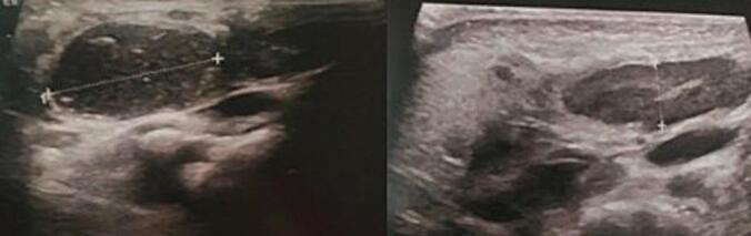
A cervical ultrasound showing a right lateral cervical cystic formation consistent with a second branchial cleft cyst.
Fig. 3.
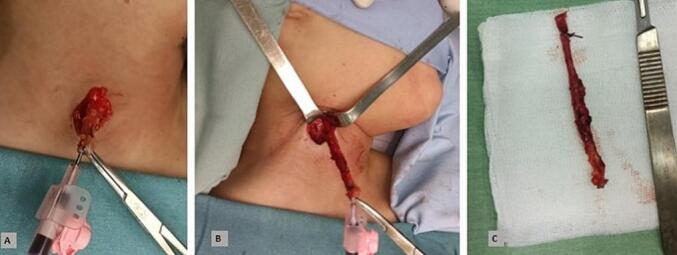
Surgical excision of a second branchial cleft fistula.
a - Orange peel incision encircling the fistulous opening.
b - Step-by-step dissection of the fistula, which ascends towards the tonsillar region.
c - Cervical fistula after resection.
Fig. 4.

Surgical excision of a second branchial cleft cyst en bloc with its tract.
a - Step-by-step dissection of the cyst.
b - Step-by-step dissection of the cyst after identification of its tract, which ascends towards the tonsillar region (white arrow).
c - Surgical specimen.
The Table 1 below presents a comprehensive overview of the clinical features, imaging findings, surgical approaches, and outcomes of the patients treated for second branchial cleft anomalies in this case series.
Table 1.
Clinical characteristics, management, and outcomes of the 10 pediatric cases.
| Case | Age (yrs) | Gender | Chief complaint | Delay | Neck Examination | Fistula opening location | Ultrasound findings | Management | Bailey stage | Outcome |
|---|---|---|---|---|---|---|---|---|---|---|
| 1 | 16 | F | Lateral neck swelling | 3 months | High left jugulocarotid swelling | Absent | Left jugulocarotid cystic lesion contacting middle cervical fascia | Surgical excision of cyst and fistula tract | II | No complications or recurrence |
| 2 | 14 | F | Lateral neck swelling | 8 months | High left jugulocarotid swelling | Absent | Inflammatory sub-digastric lymphadenopathy | Surgical excision | II | No complications or recurrence |
| 3 | 3 | F | Lateral neck swelling | 4 months | High left jugulocarotid swelling | Absent | Partially fistulized cystic lesion to skin | Surgical excision | III | No complications or recurrence |
| 4 | 11 | M | Lateral neck swelling | 3 months | High left jugulocarotid swelling | Absent | Well-defined cystic lesion | Surgical excision | II | No complications or recurrence |
| 5 | 16 | F | Lateral neck swelling | 2 months | High left jugulocarotid swelling | Absent | Partially necrotic lymphadenopathy in level II | Surgical excision | II | No complications or recurrence |
| 6 | 3 | F | Lateral neck swelling | 1 year | Low cervical swelling | Absent | Well-defined, non-fistulized cystic lesion | Surgical excision | II | No complications or recurrence |
| 7 | 7 | F | Cervical swelling with external fistula | Since birth | Right paramedian low cervical swelling | Low cervical, medial to SCM | Subcutaneous cystic lesion medial to SCM | Surgical excision | I | No complications or recurrence |
| 8 | 4 | F | Lateral neck swelling | Since birth | Right paramedian low cervical swelling | Right lateral cervical | Right lateral cervical collection with echogenic content | Surgical excision | II | No complications or recurrence |
| 9 | 5 | F | Cervical fistula | Since birth | No palpable mass | Low cervical, medial to SCM | Deep lesion with fluid content, fistulized to skin | Surgical excision | III | No complications or recurrence |
| 10 | 9 | M | Lateral neck swelling | Since birth | No palpable mass | Medial to SCM | Deep jugulocarotid cystic mass | Surgical excision | II | No complications or recurrence |
4. Discussion
These congenital anomalies of the branchial arches are the most frequent, accounting for 85 % to 95 % of all branchial anomalies [6,7]. In this study, anomalies of the second branchial cleft represented 62.5 % of all lateral cervical branchial anomalies.
The presence of bilateral or multiple branchial cysts or fistulas should prompt the search for a genetic anomaly, particularly branchio-oto-renal syndrome (BOR), an autosomal dominant syndrome with variable penetrance, characterized by the association of branchial anomalies, external, middle, or inner ear malformations, and renal malformations [8].
Although our series showed a clear female predominance, some earlier studies have not observed a gender predominance [9]. However, other researchers have noted that lesions were slightly more frequent in males [6,10].
Congenital anomalies of the second branchial cleft may present as a true fistula, sinus tract, or isolated cysts, with cysts being the most frequently observed [11].
Clinically, cystic lesions typically manifest as painless, solitary, slow-growing, fluctuating masses located in the lateral neck region [[11], [12], [13]]. However, in some cases, a rapid increase in cyst size can occur following upper respiratory tract infection, and pain or tenderness may develop if the cysts become superinfected [14,15]. This complication was observed in three patients presenting with a basicervical abscess.
A sinus or external fistula of the second branchial cleft opens to the outside, typically between the middle and lower third of the anterior border of the sternocleidomastoid (SCM) muscle. Although often observed at birth, this sinus or fistula typically becomes evident in early childhood, with recurrent mucopurulent infections [6,14].
Type II of Bailey's classification is the most common, while type IV is the rarest [15,16]. This is consistent with our results showing a predominance of type II.
Preoperative radiological investigations include ultrasound, computed tomography (CT), and magnetic resonance imaging (MRI), which are essential for confirming the clinical diagnosis and planning surgical management [2,9,12]. Ultrasound is typically the first examination performed due to its non-invasive nature, the absence of sedation requirements, and its ability to differentiate between solid masses and cysts. However, it has certain limitations in evaluating extensions and relationships with adjacent structures. Ultrasound can detect the presence of a cyst, whether homogeneous, heterogeneous, or dense. It is crucial to check for the absence of intracystic vegetation, which could signal an intracystic metastasis from a papillary thyroid carcinoma [9].
To study the localization, extent of the lesion, and its relationships with adjacent anatomical structures, CT is the examination of choice in clinical practice. It typically shows a well-defined, homogeneous, hypodense cystic mass with a thin, smooth wall. MRI generally shows a cystic image with hypointensity on T1-weighted imaging and hyperintensity on T2-weighted imaging [2,11,12].
Treatment consists of surgical excision due to the risk of superinfection [10,13,17]. Some authors have suggested performing not only excision of the fistula or sinus but also ipsilateral tonsillectomy, as the tract may end in the tonsillar fossa [17]. In this current study, we opted for surgical treatment for all our patients, including excision of the cyst and fistulous tract, but did not perform ipsilateral tonsillectomy.
Recent studies have shown that recurrence is not influenced by unilateral tonsillectomy, but that high ligation of the fistula is sufficient to prevent recurrence [2,6,10].
We did not observe any cases of recurrence after 48 months of follow-up for all in this case series.
5. Conclusion
This study highlights the clinical and epidemiological characteristics of second branchial cleft anomalies in children, underlining the importance of early diagnosis and complete surgical excision for effective management. The predominance of female patients observed in this current study aligns with some previous studies, while others have reported no significant gender bias. The presence of superinfection in a subset of patients further emphasizes the importance of timely intervention. This study confirms that second branchial cleft anomalies most commonly present as cysts, and surgical excision remains the gold standard treatment. With appropriate management, recurrence is rare, as evidenced by our findings of no recurrences after a mean follow-up of 24 months.
Author contribution
Jihene Houas, Monia Ghammam: conceptualisation, and writing original draft-editing.
Taissir Ben Arfi, Imen Boukattaya: data collection and writing the article.
Wassim Kermani: supervision and validation.
Mohamed Abdelkefi: supervision and validation.
The final version of the manuscript has been approved by all the authors.
Consent
Written informed consent was obtained from the patients for publication of this case report and accompanying images. A copy of the written consent is available for review by the Editor-in-Chief of this journal on request.
Ethical approval
Our institution Faculty of medicine of Sousse's Ethics Committee does not require ethical approval for reporting individual cases or case series.
(We maintained a high level of respect for both anonymity and confidentiality when presenting the patient in our case series.)
Guarantor
Monia Ghammam.
Research registration number
Not applicable.
Funding
This research did not receive any specific grant from funding agencies in the public, commercial or not-for-profit sectors.
Conflict of interest statement
None.
References
- 1.Goff C.J., Allred C., Glade R.S. Current management of congenital branchial cleft cysts, sinuses, and fistulae. Curr. Opin. Otolaryngol. Head Neck Surg. déc 2012;20(6):533–539. doi: 10.1097/MOO.0b013e32835873fb. [DOI] [PubMed] [Google Scholar]
- 2.Li W., Xu H., Zhao L., Li X. Branchial anomalies in children: a report of 105 surgical cases. Int. J. Pediatr. Otorhinolaryngol. janv 2018;104:14–18. doi: 10.1016/j.ijporl.2017.10.035. [DOI] [PubMed] [Google Scholar]
- 3.Paul I, Mohiyuddin SMA, A S, Mohammadi K, Babu P. The outcome of treatment in second branchial cleft anomalies: a case series. Cureus. 15(6):e40164. [DOI] [PMC free article] [PubMed]
- 4.Spinelli C., Rossi L., Strambi S., Piscioneri J., Natale G., Bertocchini A., et al. Branchial cleft and pouch anomalies in childhood: a report of 50 surgical cases. J. Endocrinol. Investig. mai 2016;39(5):529–535. doi: 10.1007/s40618-015-0390-8. [DOI] [PubMed] [Google Scholar]
- 5.Mathew G., Sohrabi C., Franchi T., Nicola M., Kerwan A., Agha R., et al. Preferred reporting of case series in surgery (PROCESS) 2023 guidelines. Int. J. Surg. Lond. Engl. 21 nov 2023;109(12):3760–3769. doi: 10.1097/JS9.0000000000000940. [DOI] [PMC free article] [PubMed] [Google Scholar]
- 6.Reddy A., Valika T., Maddalozzo J. Definitive surgical management for second branchial cleft fistula: a case series. J. Otolaryngol. - Head Neck Surg. J. Oto-Rhino-Laryngol. Chir. Cervico-Faciale. 5 août 2020;49(1):55. doi: 10.1186/s40463-020-00453-2. [DOI] [PMC free article] [PubMed] [Google Scholar]
- 7.Xing M.H., Mundi N., Govindan A., Khorsandi A., Urken M.L. Unusual location of a second branchial cleft cyst presenting in the suprasternal notch. Head Neck. avr 2021;43(4):E27–E29. doi: 10.1002/hed.26629. [DOI] [PubMed] [Google Scholar]
- 8.Can I.H., Doğan S., Dönmez M., Doğan M., Samim E.E. A different type of branchial fistula as part of a branchiootorenal syndrome. J. Pediatr. Surg. févr 2012;47(2):404–407. doi: 10.1016/j.jpedsurg.2011.11.045. [DOI] [PubMed] [Google Scholar]
- 9.Prosser J.D., Myer C.M. Branchial cleft anomalies and thymic cysts. Otolaryngol. Clin. N. Am. févr 2015;48(1):1–14. doi: 10.1016/j.otc.2014.09.002. [DOI] [PubMed] [Google Scholar]
- 10.Kajosaari L., Mäkitie A., Salminen P., Klockars T. Second branchial cleft fistulae: patient characteristics and surgical outcome. Int. J. Pediatr. Otorhinolaryngol. sept 2014;78(9):1503–1507. doi: 10.1016/j.ijporl.2014.06.020. [DOI] [PubMed] [Google Scholar]
- 11.Lee D.H., Yoon T.M., Lee J.K., Lim S.C. Clinical study of second branchial cleft anomalies. J. Craniofac. Surg. sept 2018;29(6):e557–e560. doi: 10.1097/SCS.0000000000004540. [DOI] [PubMed] [Google Scholar]
- 12.Adams A., Mankad K., Offiah C., Childs L. Branchial cleft anomalies: a pictorial review of embryological development and spectrum of imaging findings. Insights Imaging. févr 2016;7(1):69–76. doi: 10.1007/s13244-015-0454-5. [DOI] [PMC free article] [PubMed] [Google Scholar]
- 13.Magdy E.A., Hamza A., Youssef A., Yoneis A. Second branchial cleft fistula/sinus tract endoscopy: a novel intraoperative technique assisting complete surgical resection. Eur. Arch. Oto-Rhino-Laryngol Off. J. Eur. Fed. Oto-Rhino-Laryngol. Soc. EUFOS Affil. Ger. Soc. Oto-Rhino-Laryngol. - Head Neck Surg. mars 2021;278(3):833–838. doi: 10.1007/s00405-020-06158-6. [DOI] [PubMed] [Google Scholar]
- 14.Teng S.E., Paul B.C., Brumm J.D., Fritz M., Fang Y., Myssiorek D. Endoscope-assisted approach to excision of branchial cleft cysts. Laryngoscope. juin 2016;126(6):1339–1342. doi: 10.1002/lary.25711. [DOI] [PubMed] [Google Scholar]
- 15.Gao S., Xu Q., Yi Q. Endoscopically assisted transoral resection of a Bailey type IV second branchial cleft cyst: a case report. Medicine (Baltimore) 22 janv 2021;100(3) doi: 10.1097/MD.0000000000024375. [DOI] [PMC free article] [PubMed] [Google Scholar]
- 16.Gaillard F. Radiopaedia. [cité 16 févr 2025]. Bailey classification of second branchial cleft cysts | Radiology Reference Article | Radiopaedia.org. Disponible sur: https://radiopaedia.org/articles/bailey-classification-of-second-branchial-cleft-cysts.
- 17.Maddalozzo J., Rastatter J.C., Dreyfuss H.F., Jaffar R., Bhushan B. The second branchial cleft fistula. Int. J. Pediatr. Otorhinolaryngol. juill 2012;76(7):1042–1045. doi: 10.1016/j.ijporl.2012.04.002. [DOI] [PubMed] [Google Scholar]