Abstract
1. Quantitative ultrastructural examinations of axon terminals synapsing with normal α-motoneurones in segment T9 of cat spinal cord provided estimates of their numbers, sizes and synaptic structure. One synapse, the C type, derived from short-axon propriospinal segmental interneurones, was studied in detail.
2. The numbers, sizes and post-synaptic structure of normal C-type synapses at T9 were compared with similar estimates from material provided by cats subjected to partial central deafferentation by double spinal hemisection at T5 and T10 between 7 days and 2 years previously.
3. The proportion of C-type synapses present progessively increased from 1% in normal cats to 8·8% 200 days following hemisection, and had still attained a level of 3·1% by 2 years; these increases imply that the absolute number of C-type synapses underwent increase.
4. Mean sizes of C-type synapses increased from 4·0 μm (normal) to 5·8 μm (200 days) and retained their enlarged sizes up to 2 years (5·9 μm). Furthermore, while 84% of C-type synapses were under 6 μm in length in normal motoneurones, 48% were over 6 μm long 200 days post-operatively.
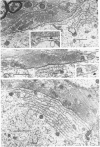
5. The unique post-synaptic structure of C-type synapses also proliferated following partial central deafferentation of the motoneurones. The elongated cistern, increased numbers and individual lengths of lamellae of the associated underlying rough endoplasmic reticulum indicated a trophic interaction between the presynaptic C terminal and its post-synaptic motoneurone.
6. Counts of ribosomes `bound' to lamellae of the subsynaptic rough endoplasmic reticulum, and of the lamellae-associated polyribosomes interposed between individual lamellae for normal and 200 day post-operative C-type synapses indicated an over-all post-operative increase in capacity for local subsynaptic protein synthesis topographically directed towards this type of axon terminal.
7. The observed greater increase in frequency of ribosomes `bound' to the rough endoplasmic reticulum, together with an over-all proliferation of this structure, specificially indicated an increased capacity for synthesis of protein for utilization in sites remote from those of synthesis (e.g. a trans-synaptic passage of protein).
8. A hypothesis is advanced on the basis of the above results relating both pre- and post-synaptic changes in structure to an increased functional activation of the segmental short-axon propriospinal interneurones forming the C-type synapses, as a compensatory response to partial central deafferentation of spinal motoneurones.
Full text
PDF


Images in this article
Selected References
These references are in PubMed. This may not be the complete list of references from this article.
- Beattie M. S., Bresnahan J. C., King J. S. Ultrastructural identification of dorsal root primary afferent terminals after anterograde filling with horseradish peroxidase. Brain Res. 1978 Sep 15;153(1):127–134. doi: 10.1016/0006-8993(78)91134-4. [DOI] [PubMed] [Google Scholar]
- Bernstein J. J., Bernstein M. E. Ventral horn synaptology in the rat. J Neurocytol. 1976 Feb;5(1):109–123. doi: 10.1007/BF01176185. [DOI] [PubMed] [Google Scholar]
- Bernstein M. E., Bernstein J. J. Synaptic frequency alteration on rat ventral horn neurons in the first segment proximal to spinal cord hemisection: an ultrastructural statistical study of regenerative capacity. J Neurocytol. 1977 Feb;6(1):85–102. doi: 10.1007/BF01175416. [DOI] [PubMed] [Google Scholar]
- Blobel G., Dobberstein B. Transfer of proteins across membranes. I. Presence of proteolytically processed and unprocessed nascent immunoglobulin light chains on membrane-bound ribosomes of murine myeloma. J Cell Biol. 1975 Dec;67(3):835–851. doi: 10.1083/jcb.67.3.835. [DOI] [PMC free article] [PubMed] [Google Scholar]
- Bodian D. Origin of specific synaptic types in the motoneuron neuropil of the monkey. J Comp Neurol. 1975 Jan 15;159(2):225–243. doi: 10.1002/cne.901590205. [DOI] [PubMed] [Google Scholar]
- Conradi S., Kellerth J. O., Berthold C. H., Hammarberg C. Electron microscopic studies of serially sectioned cat spinal alpha-motoneurons. IV. Motoneurons innervating slow-twitch (type S) units of the soleus muscle. J Comp Neurol. 1979 Apr 15;184(4):769–782. doi: 10.1002/cne.901840409. [DOI] [PubMed] [Google Scholar]
- Conradi S., Ronnevi L. O. Ultrastructure and synaptology of the initial axon segment of cat spinal motoneurons during early postnatal development. J Neurocytol. 1977 Apr;6(2):195–210. doi: 10.1007/BF01261505. [DOI] [PubMed] [Google Scholar]
- Conradi S., Skoglund S. Observations on the ultrastructure of the initial motor axon segment and dorsal root boutons on the motoneurons in the lumbosacral spinal cord of the cat during postnatal development. Acta Physiol Scand Suppl. 1969;333:53–76. [PubMed] [Google Scholar]
- Conradi S. Ultrastructure and distribution of neuronal and glial elements on the motoneuron surface in the lumbosacral spinal cord of the adult cat. Acta Physiol Scand Suppl. 1969;332:5–48. [PubMed] [Google Scholar]
- Hamos J. E., King J. S. The synaptic organization of the motor nucleus of the trigeminal nerve in the opossum. J Comp Neurol. 1980 Nov 15;194(2):441–463. doi: 10.1002/cne.901940210. [DOI] [PubMed] [Google Scholar]
- Kellerth J. O., Berthold C. H., Conradi S. Electron microscopic studies of serially sectioned cat spinal alpha-motoneurons. III. Motoneurons innervating fast-twitch (type FR) units of the gastrocnemius muscle. J Comp Neurol. 1979 Apr 15;184(4):755–767. doi: 10.1002/cne.901840408. [DOI] [PubMed] [Google Scholar]
- Kernell D., Peterson R. P. The effect of spike activity versus synaptic activation on the metabolism of ribonucleic acid in a molluscan giant neurone. J Neurochem. 1970 Jul;17(7):1087–1094. doi: 10.1111/j.1471-4159.1970.tb02262.x. [DOI] [PubMed] [Google Scholar]
- Lagerbäck P. A., Ronnevi L. O., Cullheim S., Kellerth J. O. An ultrastructural study of the synaptic contacts of alpha 1-motoneuron axon collaterals. II. Contacts in lamina VII. Brain Res. 1981 Oct 5;222(1):29–41. doi: 10.1016/0006-8993(81)90938-0. [DOI] [PubMed] [Google Scholar]
- Lagerbäck P. A., Ronnevi L. O., Cullheim S., Kellerth J. O. An ultrastructural study of the synaptic contacts of alpha-motoneurone axon collaterals. I. Contacts in lamina IX and with identified alpha-motoneurone dendrites in lamina VII. Brain Res. 1981 Mar 2;207(2):247–266. doi: 10.1016/0006-8993(81)90363-2. [DOI] [PubMed] [Google Scholar]
- Lenk R., Ransom L., Kaufmann Y., Penman S. A cytoskeletal structure with associated polyribosomes obtained from HeLa cells. Cell. 1977 Jan;10(1):67–78. doi: 10.1016/0092-8674(77)90141-6. [DOI] [PubMed] [Google Scholar]
- Mathers D. A. The influence of concanavalin A on glutamate-induced current fluctuations in locust muscle fibres. J Physiol. 1981 Mar;312:1–8. doi: 10.1113/jphysiol.1981.sp013611. [DOI] [PMC free article] [PubMed] [Google Scholar]
- Matsushita M., Ikeda M. Propriospinal fiber connections of the cervical motor nuclei in the cat: a light and electron microscope study. J Comp Neurol. 1973 Jul 1;150(1):1–32. doi: 10.1002/cne.901500102. [DOI] [PubMed] [Google Scholar]
- McLaughlin B. J. Dorsal root projections to the motor nuclei in the cat spinal cord. J Comp Neurol. 1972 Apr;144(4):461–474. doi: 10.1002/cne.901440405. [DOI] [PubMed] [Google Scholar]
- McLaughlin B. J. The fine structure of neurons and synapses in the motor nuclei of the cat spinal cord. J Comp Neurol. 1972 Apr;144(4):429–460. doi: 10.1002/cne.901440404. [DOI] [PubMed] [Google Scholar]
- PALADE G. E. A small particulate component of the cytoplasm. J Biophys Biochem Cytol. 1955 Jan;1(1):59–68. doi: 10.1083/jcb.1.1.59. [DOI] [PMC free article] [PubMed] [Google Scholar]
- PALAY S. L., PALADE G. E. The fine structure of neurons. J Biophys Biochem Cytol. 1955 Jan;1(1):69–88. doi: 10.1083/jcb.1.1.69. [DOI] [PMC free article] [PubMed] [Google Scholar]
- Pullen A. H., Sears T. A. Modification of "C" synapses following partial central deafferentation of thoracic motoneurones. Brain Res. 1978 Apr 21;145(1):141–146. doi: 10.1016/0006-8993(78)90802-8. [DOI] [PubMed] [Google Scholar]
- Raisman G., Field P. M. A quantitative investigation of the development of collateral reinnervation after partial deafferentation of the septal nuclei. Brain Res. 1973 Feb 28;50(2):241–264. doi: 10.1016/0006-8993(73)90729-4. [DOI] [PubMed] [Google Scholar]
- Ralston H. J., Ralston D. D. Identification of dorsal root synaptic terminals on monkey ventral horn cells by electron microscopic autoradiography. J Neurocytol. 1979 Apr;8(2):151–166. doi: 10.1007/BF01175558. [DOI] [PubMed] [Google Scholar]
- Ronnevi L. O. Spontaneous phagocytosis of C-type synaptic terminals by spinal alpha-motoneurons in newborn kittens. An electron microscopic study. Brain Res. 1979 Feb 23;162(2):189–199. doi: 10.1016/0006-8993(79)90283-x. [DOI] [PubMed] [Google Scholar]
- SIEKEVITZ P., PALADE G. E. A cytochemical study on the pancreas of the guinea pig. I. Isolation and enzymatic activities of cell fractions. J Biophys Biochem Cytol. 1958 Mar 25;4(2):203–218. doi: 10.1083/jcb.4.2.203. [DOI] [PMC free article] [PubMed] [Google Scholar]
- Schrøder H. D. Paramembranous densities of 'C' terminal-motoneuron synapses in the spinal cord of the rat. J Neurocytol. 1979 Feb;8(1):47–52. doi: 10.1007/BF01206457. [DOI] [PubMed] [Google Scholar]
- Watanabe H. Development of axosomatic synapses of the Xenopus spinal cord with special reference to subsurface cisterns and C-type synapses. J Comp Neurol. 1981 Aug 10;200(3):323–328. doi: 10.1002/cne.902000304. [DOI] [PubMed] [Google Scholar]
- Watanabe H., Yamamoto T. Y. Freeze fracture study on three types of synapses in the Xenopus spinal cord. J Comp Neurol. 1981 May 10;198(2):249–263. doi: 10.1002/cne.901980205. [DOI] [PubMed] [Google Scholar]
- Zwaagstra B., Kernell D. Sizes of soma and stem dendrites in intracellularly labelled alpha-motoneurones of the cat. Brain Res. 1981 Jan 12;204(2):295–309. doi: 10.1016/0006-8993(81)90590-4. [DOI] [PubMed] [Google Scholar]


