

Abstract
Purpose:
The purpose of this study was to detect the distribution of different lectin receptors in primary cancer cells as well as in the case of metastasis, as these biomolecules can potentially predict cancer development in certain tissues and systems.
Methods:
To detect lectin receptors in tumors, the authors used conjugates of lectins purified by affinity chromatography with peroxidase, and studied their localization in paraffin sections of 12 cases of primary cancer and 33 of its metastases.
Result:
In primary cancers and their metastases, there is a distinct mosaicity in the histotopography of individual lectins, especially peanut and soybean lectin. Mosaicity increases in metastases, which corresponds to the increase in malignancy of tumour cells. Detected cases of metastases with a decrease in mosaicity may be a sign of their monoclonality or a decrease in their malignancy. The study of lectins in the cells of cancer metastases and their comparison with the primary tumour and with each other suggests that in metastases, as a rule, not only signs of malignancy increase, but also the ability of cells to adhere and migrate. Thus, in the process of tumour growth and metastasis, there is a selection of clones of cells that are more prone to the development of new metastases.
Conclusions:
It has been revealed that the growth of a primary immature tumour from the epithelium and its metastases is accompanied by sialylation of the surface of tumour cells, which leads to the fact that tumour cells are not recognized by the system of mononuclear phagocytes and this, naturally, contributes to the progression of tumour growth.
Key Words: Oncology, tumour progression, sialylation, peroxidase, affinity chromatography
Introduction
Lectins are carbohydrate-binding proteins that have the ability to recognize and bind to specific sugar moieties found on glycoconjugates molecules consisting of proteins or lipids covalently bonded to carbohydrate chains on the surface of cells. These interactions occur at the molecular level, where lectins identify specific oligosaccharide structures, such as mannose, galactose, or fucose, present on glycoproteins and glycolipids. The carbohydrate-binding domains of lectins allow them to form non-covalent interactions with these sugars, which can include hydrogen bonds, van der Waals forces, and hydrophobic interactions. This precise molecular recognition enables lectins to mediate various biological processes, such as cell adhesion, signaling, and immune responses.
In the context of cancer, glycosylation patterns on the surface of tumor cells are often altered. These changes in the glycan structures of glycoconjugates can affect how lectins interact with cancer cells. For instance, sialylation the addition of sialic acid to glycan chains is frequently upregulated in cancer cells, which can shield the cell from immune detection and enhance its invasive properties. These altered interactions between lectins and glycoconjugates are critical in cancer progression because they influence key processes such as cell-cell communication, cell migration, and the ability of cancer cells to evade immune surveillance.
Changes in lectin-glycoconjugate interactions are closely linked to cancer aggressiveness due to their role in modulating cell behavior. For example, cancer cells with increased levels of specific lectin receptors, such as galectins or selectins, can exhibit enhanced adhesion to the extracellular matrix or to other cells, facilitating metastasis. Additionally, altered glycosylation can promote immune evasion by preventing recognition by immune cells, thereby allowing the tumor to grow and spread unchecked. As a result, the aggressiveness of cancer is often correlated with the degree to which these molecular interactions are altered, making lectin-glycoconjugate interactions a key factor in determining cancer malignancy and metastatic potential. Understanding these molecular mechanisms not only provides insights into cancer biology but also opens up potential avenues for therapeutic intervention by targeting aberrant glycosylation patterns [1, 2].
The unique recognition and binding of lectins to altered glycosylation patterns on cancer cells, which play a crucial role in the progression and metastasis of tumor diseases, may make them more successful than other markers or approaches for cancer prognosis and treatment. These interactions not only allow lectins to function as very sensitive biomarkers for detecting small changes in cancer cell behavior, but also provide valuable information about the aggressiveness and metastatic ability of tumors. In addition, some lectins demonstrate antiproliferative and cytotoxic properties, which opens up therapeutic opportunities beyond simple identification. The multimodal function of lectins both in predicting disease progression and functioning as therapeutic agents provides a significant advantage over traditional single-target oncological indicators [3, 4].
It has been noted by M.H. Wu et al. [5], that in colon, larynx, and larynx cancer patients, the correlation between poor prognosis and galectin-1 expression has been established. It has also been pointed out that the detection of galectin-1 in the tumour stroma is associated with poor prognosis. Glycoconjugates play an important role in the vital activity of individual cells and tissues, by influencing the molecular mechanism of intercellular recognition, the relationship of the cell with its microenvironment, regulation of elasticity and strength, shape-forming and other specialized functions of the extracellular matrix.
The terminal carbohydrate residues of glycoconjugates are particularly dynamic during changes in the functional state of the cell, as well as in the case of pathological changes, and specific reagents that allow identifying the terminal residues of oligosaccharide chains of carbohydrate-containing biopolymers and studying their redistribution in different morpho-functional states are lectins. According to A. Nobumoto et al. [6], lectins embedded in cell membranes are important in the organotroph of metastases, and inactivation of lectins allows blocking the process of metastasis. Thus, lectins can be used to predict the metastatic potential, which is of certain scientific interest in the study of the process of cancer metastasis.
According to H. Suzuki et al. [7], podoplanin (PDPN) is a cell surface glycoprotein that binds to the C-type lectin receptor (CLEC-2) on the surface of platelets and triggers their aggregation. This glycoprotein modulates carcinogenesis through mechanisms of tumour cell activation, proliferation, migration, invasion, and differentiation. As a result of joining PDPN to CLEC-2, immune cells become embolized and lose their protective function. At the same time, activated platelets release an excess of podoplanin and tumour growth factor beta (TGF-β). Podoplanin, in turn, stimulates the processes of division and increases the viability of cancer cells, and the growth factor initiates their migration and invasion, which ultimately determines the occurrence and spread of metastases [8, 9]. Therefore, PDPN and CLEC-2 are potential markers of cancer development.
The aim of this study was to detect the distribution of different lectin receptors in primary cancer cells as well as in case of metastasis. These biomolecules can predict potential cancer development in some tissues and systems. The objectives of the study are as follows:
- To study the distribution and expression patterns of different lectin receptors in primary cancer cells and their metastases.
- Determine the correlation between the expression of lectin receptors and the aggressiveness or metastatic potential of different types of cancer.
- To evaluate the relationship between specific lectin receptors and patient prognosis in different types of cancer.
- Investigate the role of lectins in the adhesion, migration and evasion of cancer cells from immunity.
- To evaluate the antiproliferative and cytotoxic effects of plant lectins on cancer cells.
Materials and Methods
The authors investigated the localization of lectin receptors in 12 observations of primary cancer and 33 of its metastases, including 6 cases of stomach cancer, 3 cases of lung cancer, 2 cases of pancreas cancer, 1 case of kidney cancer. To detect lectin receptors in tumours, conjugates of lectins with peroxidase purified by affinity chromatography were used (lentil seeds, soybeans, wheat ovules, peanuts, leguminous beans, grape snail), produced by the scientific and production cooperative “Lectinotest” in Lviv.
In this study, the preparative affinity chromatography of lectins was performed using the AKTA Pure 25 L chromatograph system (GE Healthcare, USA), a high-precision instrument designed for protein purification with an accuracy of ±0.01 mL in flow rate and pressure control. The system operated under specific parameters tailored to lectin binding, utilizing a Tricorn 5/50 column (Cytiva, USA) packed with agarose-bound resin from Vector Laboratories, USA. The column was equilibrated with 10 mM HEPES-NaOH buffer, pH 7.5, with a flow rate maintained at 8 mL/min. Detection of lectins was conducted at a wavelength of 230 nm, ensuring accurate identification of target proteins. For elution, a 20 mM sugar solution specific to each lectin in 10 mM HEPES-NaOH, pH 7.5, was employed, which effectively released the bound lectins from the column. Additional purification was achieved using reverse-phase HPLC on a C18 Mightysil RP-18GP column (2x50 mm, Kanto Chemical, Japan), pre-equilibrated with 0.1% trifluoroacetic acid (TFA). The system’s capability to maintain stable pH levels and precise flow rates, coupled with accurate wavelength detection, ensured the reliability and reproducibility of the results, thereby supporting the overall accuracy of the study’s findings [10].
The collected material was fixed in 4 % solution of neutral formalin (Sigma-Aldrich, Germany) according to the appropriate method. After embedding in paraffin, paraffin sections were made, which were then stained with the appropriate lectin-peroxidase conjugate. The receptors to lectin bounding the conjugate were detected by the determination of peroxidase activity. This enzyme catalyses a redox reaction between hydrogen peroxide and various substrates. As a result, a coloured product is formed and it is detected at a wavelength of 570 nm (extinction length 535 nm and emission length 587 nm, respectively) [11]. The study of the preparations was carried out under a light microscope (Kern OBE 134, Germany).
Results
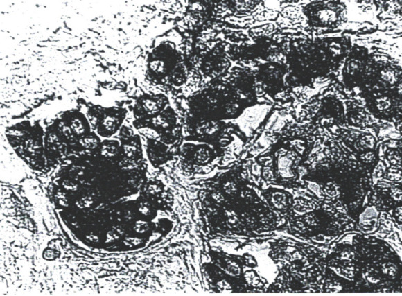
By studying the nature of distribution of peanut lectin in tumour cells of primary cancer and its metastases, it has been found out that the intensity of the peroxidase-peanut lectin reaction with tumour cells and other tissue structures is quite high (up to +3), but not uniform. In primary cancer, separate zones of tumour cells with the level of accumulation of peanut lectin in them up to +3, as well as zones with low concentration, are determined. Cells with peanut lectin receptors in the primary site are mainly located in the expected areas of tumour growth (along its edge, near small vessels) (Figure 1). Minimal staining was observed in the area of tumour cell necrosis.
Figure 1.

Low-Differentiated Stomach Cancer. Mosaic staining of tumour cells. Treatment with peanut peroxidase-lectin conjugate. x400
In metastatic nodes, the intensity of reaction in tumour cells peanut peroxidase-lectin was also uneven. In individual metastatic nodes, this response was lower than in the primary node and other daughter metastases. In a number of metastatic nodes, it was similar or higher than in the primary tumour, and with a rather significant mosaicity (Figure 2). Desialylation of tumour cells slightly enhanced the reaction with peanut lectin, but in the case where peanut lectin receptors were not detected in the metastasis before desialylation, lectin receptors were not revealed in tumour cells, which confirms their absence.
Figure 2.
Metastasis in the Liver. Treatment with peanut peroxidase-lectin conjugate. increased number of peanut lectin receptors in tumour cells. x400
Mosaicity in the histochemical reaction in tumour nodes when the peanut lectin receptor is detected is due to the fact that both the primary tumour and its metastases are a collection of polymorphic cells.
The study of the content of lectins in tumour cells before and after their desialylation shows that the desialylation of tumour cells was accompanied by an increase in the reaction with lectins, especially peanut, that is, in the studied tumours there is a distinct process of sialylation of the surface of tumour cells. It has been experimentally proven that sialylation of O-galactose residues is associated with an increase in the migratory abilities of cells. In particular, the correlation of O-galactose/sialic acid lying on the surface of cells can be a regulator of a balanced adhesion-migration system, the lower this correlation, the higher the migratory abilities of cells. The histochemically detected sialylation of tumour cells is evidence of an increase in their migratory abilities, while the expressiveness of this ability is uneven in different cells of both the primary tumour and metastases. Mannose-containing conjugates – lentil lectin receptors play the main role in the specific recognition of its targets by the cell. The role of mannose conjugates in increasing intercellular adhesion has been proven, in particular, to ensure strong contacts of epidermal cells.
The histochemical picture of the detection of lentil lectin is characterized by the fact that the primary centre of tumour growth has a locally quite pronounced (up to +3), but focal reaction. In most metastases, it is lower compared to the primary tumour, and in some places, it is equal to “0”. Attention is drawn to the observation that in metastases of lung cancer to the skeletal muscle, the level of reaction in tumour cells with lentil lectin was quite high, compared to other metastases, with a simultaneously low (close to “0”) level of reaction with peanut lectin, with positive reactions in other metastases. So, it has been revealed that in metastases cell clones with a much smaller number of lentil lectin receptors predominate, that is, intercellular contacts are reduced in metastases. Cases of metastases with an increased content of lentil lectin and a parallel decrease in the number of peanut lectin receptors were found out, which is confirmation of the heterogeneity of “daughter metastases”, as well as the fact that the degree of differentiation of cells in metastases can increase. In addition, the appearance of some receptors and the internalization or masking of others, according to the literature, correlates with the degree of cell (tissue) differentiation.
The histochemical picture of detected soy lectin receptors in tumour cells of primary cancer and its metastases is similar to the reaction with peanut lectin. Obviously, it is due to the fact that both of these lectins have an affinity for O-galactose. The localization of soybean lectin receptors in the primary and metastatic node is mosaic. The number of cells with a high level of reaction (up to +3) in the primary tumour is greater than in its metastases. The majority of cells with positive reaction to soy lectin were localized at the edge of the tumour node, which is growing, but there were metastases with an almost equal “zero” level of reaction, at the same time, in normal cells of the recipient organ, the reaction was up to +2.
Desialylation of histological preparations enhanced the reaction with soy lectin. In the necrosis zone, the reaction level remained equal to “0”. The authors detection of soybean lectin and peanut lectin receptors proved that glycoconjugates were mainly localized diffusely in the cytoplasm of tumour cells. Some researchers regard this as a sign of distinct cell malignancy. It has been revealed that the nature of distribution and degree of accumulation of soy and peanut lectin receptors in cells of primary cancer and its metastases are most often correlated with each other, and their number is higher in metastases cells compared to the primary tumour. Thus, it can be assumed that the accumulation of O-galactoconjugates in cells and sialylation contribute to the development of the metastatic process in cancer.
One of the leading factors in the pathogenesis of metastasis is the ability of tumour cells to adhere. A number of experimental studies revealed that during fertilization, the leading role in adhesion belongs to fucose-containing and sialic-containing glycoconjugates, including wheat ovary lectin receptors. There is information in the literature that the migration of tumour cells correlates with the redistribution of receptors for concovalin A and wheat lectin. The authors histochemical determination of lectin receptors in the wheat ovary revealed uneven localization of them in tumour cells of primary and metastatic cancer with a predominance of the latter (Figure 3). At the same time, desialylation of histological preparations actually did not change the nature of the histochemical reaction for the detection of lectin receptors in the wheat ovary.
Figure 3.

Lung Metastasis of Undifferentiated Pancreatic Cancer. Positive reaction of tumour cells with wheat ovule lectin. Treatment of wheat ovaries with peroxidase-lectin conjugate. x400
The results of wheat ovule lectin receptors detection indicate that tumour cells of primary cancer and especially its metastases have signs that indicate the ability of tumour cells to adhere and migrate. Since these properties are due to the presence of wheat germ lectin receptors, especially in the cells of metastatic nodes, it can be assumed that according to the growth of the tumour (during its progression), the ability of tumour cells to migrate and adhere increases. Histochemical detection of grape snail lectin receptors proved the presence of a minimal number of them in tumour cells as a primary cancer (0, +1) and its metastases (0, +1) with a relatively high concentration of peanut lectin receptors in tumour cells. At the same time, peanut lectin receptors were not detected in the lymph nodes.
The presence of separately located peroxidase-positive cells containing grape snail lectin receptors was noted in the loose fibrous connective tissue on the periphery of the tumour and in the normal tissue surrounding the tumour, as well as in regional lymph nodes (Figure 4). According to the literature, these cells are histiocytes-macrophages. The number of these cells among tumours of both primary and metastatic nodes is minimal, compared to normal tissues, which is a manifestation of low macrophage activity of cells in the area of tumour growth. According to the literature, the detection of histiocytes-macrophages with cytoplasmic granularity in lymphomas in lymph nodes, which intensively bind castor lectins and are reactive with peanut lectin in combination with tumour cells, the reaction of cytoplasmic glycoconjugates of which was directly opposite (RNA+/RCA-), correlated with a highly malignant form of the course of the disease, and with the opposite relationship, the prognosis is more favourable. It can be assumed that the authors’ detection of histiocytes-macrophages in the tumour, which intensively bind grape snail lectin and are reactive with peanut lectin, should be considered as an indicator of an unfavourable prognosis.
Figure 4.

Regional Pre-Cancerous Lymph Node. Macrophages with a clearly positive reaction in the cytoplasm with grape snail lectin. Treatment with grape snail peroxidase-lectin conjugate. x400
The number of lectin receptors of the leguminous legume in the cells of the primary metastatic nodes was insignificant (0+1). But in one of the metastases, after desialylation, the intensity of the reaction in the tumour cells increased to +2 in parallel with the strengthening of the reaction with wheat ovule lectin. It is known that leguminous lectin has an affinity for L-fucose, the role of which has been proven in cell adhesion, that is, leguminous lectin receptors are one of the factors of adhesion, which in this observation did not correlate with other factors that contribute to the adhesion of tumour cells (lectin wheat).
Desialylation is the phenomenon of eliminating sialic acid residues from glycoproteins or glycolipids located on the cellular surface. Terminal positions of glycan chains are commonly occupied by sialic acids, which can exert a substantial impact on the behaviour and characteristics of cells, particularly those in tumours. Within cancer cells, sialylation frequently facilitates immune evasion, cell-cell interactions, and signalling pathways that contribute to the development and spread of tumours. The removal of sialic acid residues by desialylation can result in substantial alterations in the functionality of lectin receptors on the cancer cell surface. Carbahydrate-binding proteins known as lectins selectively bind to glycan structures, and their binding affinity can be modified by the presence or absence of sialic acid. Desialylation reveals the underlying sugar residues, including galactose or N-acetylgalactosamine, in a manner that enhances their recognition by particular lectins. Enhanced exposure of lectin-binding sites resulting from desialylation can augment the binding affinity of lectins such as peanut or soybean lectin, therefore facilitating more robust interactions between these lectins and the tumour cells.
The comparison of the number and distribution of lectin receptors in tumour cells of metastasis with parenchymatous cells and other tissue structures of the recipient’s organs was made. It has been found out that the type of lectin receptors, the number and nature of their distribution in tumour cells and tumour stroma do not correlate with the recipient organ of metastasis. There are cases when the number of snail lectin receptors in the tumour cells of the metastasis is minimal (0+1), in single cells they are diffusely located, and in the epithelial cells of the renal tubules they are located in the apical part and their number is much larger. The stroma of metastases, as a rule, contains a smaller number of receptors of the studied lectins compared to the stroma of the organ. The obtained data allow to conclude that the selectivity of metastasis does not depend on the correspondence of the nature and number of receptors of the studied lectins in tumour cells that are implanted and cells of the target organ.
Discussion
In the course of the conducted research, no correlation of the certain type lectin receptor’s location with the organ or tissue in which metastases sprout was observed. Instead, according to T. Li et al. [12], the presence of C-type lectin receptors (CLRs) correlates with bladder cancer. As of today, one of the most effective ways to treat bladder cancer is adjuvant immunotherapy, which consists in key immune points blocking. In the case of bladder oncology, dectin receptors 1-3 as well as calcium-dependent lectin receptors of the C-type (Mincle) contribute to the colonization of the tumour by immunocompetent cells with specific antitumour activity. At the same time, this type of receptors is associated with pathogenic fungal microflora, which can weaken the anticancer immune response. The antifungal immune response is mainly implemented through C-type lectin receptors that recognize endogenous or surface fungal ligands (for example, dectin-3 is the main immune barrier against Candida tropicalis pathogens) [13-16]. Therefore, the microbiota of the urinary tract is one of the most important factors in cancer pathogenesis. The CLR-dependent antifungal immune response is considered as a promising target for immunotherapy of the progression of bladder malignancies [12, 17].
One of the “most dangerous” C-type lectin receptors in terms of carcinogenesis is the Mincle subtype, which is expressed by macrophages, neutrophils, B-lymphocytes and dendritic cells. In the tissue microenvironment of the tumour, these receptors trigger the process of immunosuppression mediated by macrophages. As a result, tumour cells are not affected by the body’s immune system and therefore progress (necrosome-induced oncogenesis) [18-20]. One of the ligands of these signalling systems is fucose. As revealed in the current study, fucose receptors specific to wheat lectin dominate in metastases of lung tissue compared to primary cancer cells and correlate with the rate of tumour cell migration. They probably belong to type C. According to the data of R.M. Perrotta et al. [21], beta-galactoside-binding proteins (galectins) are actively synthesized in the sites of metastasis and in the tissue microenvironment around tumours. The class of these compounds includes peanut lectins, as their marker is also galactose. On the other hand, according to the results of this study, a greater number of peanut lectin receptors was found not in metastases, but in primary tumours, in particular, in the peripheral area. Cells of metastases, which do not contain these receptors, are more differentiated, incapable to implantation.
According to S. Kremsreiter et al. [22], the main ligands of galactose-binding lectins are O-glycans (T- and Tn-antigens, tumour antigen MUC1 of cancer cells), as well as their sialyated forms. The MUC1 antigen is expressed by cancer cells of the cervix, rectum, breast, etc. The structural feature of this biomolecule, in comparison with the analogue of normal cells, is hypoglycosylation. As a result, after the binding of this lectin to a branched glycan, the activity of T-killers and T-helpers decreases. Therefore, these immune agents lose their immunoprotective function aimed at cancer cells. The sialyl form of MUC1 inhibits the differentiation of dendritic cells and their ability to synthesize interleukins, and also stimulates the processes of apoptosis of these cells. Thus, glycosylation causes suppression of the anticancer immune response. To prevent this phenomenon, immunotherapy with the participation of antibodies to galactose-binding lectin is used. In this case, immunoglobulins shield lectin macromolecules, preventing their interaction with dendritic cells [23, 24]. In this study, sialylated forms of O-galactose lectins (peanut and soy) indicate an increase of the migratory activity of tumour cells and an acceleration of the growth of metastases. It is likely that sialylated forms cause the acceleration of metastasis due to the inhibition of the immune response of dendritic cells.
In addition to lectins described in this work, as of today, new biomolecules of the class of C-type lectins, which are potential tumour markers, have been interpreted. Among them X. Zhou et al. [25] focus on CD161 receptors, which are expressed by killer T-cells, natural killer cells, and some T-helper populations. It should also be noted that the C-type domain is represented on the surface of the membrane of malignant cells. These macromolecules help to inhibit the infiltration of CD8-T-lymphocytes into the microenvironment around the tumour. As a result, the anti-cancer immune response is weakened. CD161 expression is associated with immunoregulatory interactions of non-lymphoid and lymphoid cells and improves tumour cell survival. The level of CD161 expression is strongly correlated with the expression of immune activation genes and immunosuppressor genes, chemokines and chemokine receptors, as well as with T-lymphocyte infiltration [26]. In addition, some cases of activation of NK-dependent tumour lysis under the condition of blocking CD161 receptors have been described. Thus, this subgroup of receptors is involved in complexly regulated processes of immunomodulation of malignant neoplasms.
In general, plant lectins can be interpreted as cancer biomarkers. At the same time, these compounds can be used for cancer treatment. In the process of oncotransformation, the cells undergo multispectral changes that affect the nature of growth and permeability. In addition, surface glycosylated macromolecules are modified. Their typical characteristic is the appearance of sialic acid residues [27, 28]. Signaling cascades are altered as a result of changes in surface receptors. Some of the plant lectins exhibit antiproliferative effects after binding to the corresponding glycocalyx receptors. At the same time, cancer cells release haptenic sugars that compete with receptors for the lectin ligand. In this case the antiproliferative effect is blocked. Currently, two forms of lectin from white mistletoe (Viscum album) have been isolated. This is the first lectin of plant origin, which is a potential effective inhibitor of malignant growth and is currently undergoing clinical trials. It shows specificity to galactose and N-acetylgalactosamine. Functionally, the one induces the synthesis of tumour necrosis factor-alpha by macrophages [29, 30]. Some members of the Legume family also synthesize lectins with antiproliferative and cytotoxic effects. In addition, these signaling macromolecules are capable to induce apoptosis and autophagy of tumours as well as damage an intact DNA of transformed cells [31]. Therefore, in the perspective of further research, there is an investigation of the antiproliferative activity of studied lectins of legumes (peanut, soybean, lentil).
The main reason for mosaicism in cancer is genetic instability, which leads to the evolution of several subclones with different genetic and phenotypic characteristics due to constant mutations and chromosomal changes within tumor cells. The presence of distinct variations in the tumor environment leads to a mosaic picture of cell populations that demonstrate varying degrees of differentiation and aggressiveness. The selection of tumor cells with beneficial mutations that improve survival, proliferation, or resistance to treatment contributes to their continuous diversification as they multiply. Signs of mosaicism in a tumor indicate that not all cells have the same ability to grow, differentiate, or have metastatic potential. This can complicate treatment approaches and contribute to tumor progression.
The phenomenon of mosaicism is closely related to the mechanisms of differentiation and metastasis. Cancer differentiation is the degree of similarity between tumor cells and their normal counterparts. Less differentiated cells tend to show a higher level of aggressiveness and are more prone to metastasis. The metastatic process in a mosaic tumor occurs due to subpopulations of less differentiated, more aggressive cells that have increased migration and invasive ability. When spreading from the primary tumor, these cells can undergo additional genetic changes, which leads to the development of metastatic zones that demonstrate their own distinct mosaicism. Constant changes in the tumor microenvironment and metastatic sites make it difficult to predict disease progression and determine treatment efficacy, emphasizing the dynamic nature of cancer biology.
The findings of this study have significant potential clinical applications, particularly in the development of personalized cancer therapies and diagnostic tools. The distinct patterns of lectin receptor expression in primary tumors and metastases can serve as biomarkers for early detection and prognosis, enabling clinicians to identify patients at higher risk of aggressive cancer and tailor their treatment plans accordingly. Additionally, the ability of lectins to influence cell adhesion, migration, and immune evasion suggests that targeting lectin-glycoconjugate interactions could be a promising therapeutic strategy. Inhibiting specific lectin-receptor interactions might reduce metastatic spread, improve immune system recognition of tumor cells, and enhance the efficacy of existing treatments. Furthermore, the manipulation of sialylation patterns on tumor cells could potentially restore immune surveillance and inhibit tumor progression, offering a novel approach to cancer therapy. These applications underscore the importance of further research into the clinical utility of lectins in oncology.
In conclusions, the analysis unveiled a clear mosaicism in the histological organisation of lectin receptors, namely peanut and soybean lectins, in both primary malignancies and their spread to distant sites. The observed mosaicism was shown to escalate in metastatic areas, indicating a positive correlation with the heightened malignancy and migratory capacities of tumour cells. Statistical evidence indicates that as tumours advance and spread to distant sites, there is a certain group of cell clones that exhibit greater lectin receptor activity, especially those linked to enhanced cell adhesion and migration. Specifically, the study found a notable rise in the affinity of peanut and soybean lectins following desialylation, which is associated with the improved migratory characteristics of cancer cells. The present discovery provides evidence in favour of the proposition that there exists a strong correlation between lectin activity and the metastatic capacity of cancer cells.
Moreover, the research emphasised the significance of sialylation in concealing lectin-binding sites, hence decreasing cell attachment and improving immune evasion. Desialylation, the elimination of sialic acids, reveals these sites, therefore enhancing lectin binding and facilitating cell migration, a crucial stage in the metastatic cascade. In addition to being indicators for predicting cancer progression, these findings highlight the significance of lectins as possible therapeutic targets. The clinical implications of these results indicate that the investigation of targeting lectin interactions or modifying sialylation patterns may be considered as effective approaches to suppress metastasis and enhance the results of cancer therapy. This study’s findings establish a basis for further investigation on the advancement of lectin-based diagnostics and treatments, namely in the realm of preventing or minimising the spread of cancer cells to distant sites.
Author Contribution Statement
Yevhen Herasymenko, Kostyantyn Herasymenko contributed to the project concept and manuscript design. Ruslan Klimanskyi worked on data analysis, data interpretation and writing of the manuscript. Oleksandr Herasymenko worked on the project concept and manuscript design, critical review of the manuscript writing and discussion the manuscript. All authors read and approved the final manuscript.
Acknowledgements
Data Availability Statement
The data that support the findings of this study are available on request from the corresponding author.
Ethical approval
Ethical approval of the study was obtained from the Health Research Ethics Commission of the Donetsk National Medical University with No. RT-469.
Conflict of Interest
Nothing to declare.
References
- 1.Zhang Z, Liu M, Zheng Y. Role of Rho GTPases in Stem Cell Regulation. Biochem Soc Trans. 2021;49:2941–2955. doi: 10.1042/BST20211071. [DOI] [PMC free article] [PubMed] [Google Scholar]
- 2.Gaghana LO, Miskad UA, Cangara MH, Zainuddin AA, Mardiati MA, Gunawan WS. The Relationship Between Expression of EpCAM Cancer Stem Cell Marker with Histopathological Grading, Lymphovascular Invasion, and Metastases in Colorectal Adenocarcinoma. Asian Pac J Cancer Prev. 2023;24(3):929–934. doi: 10.31557/APJCP.2023.24.3.929. [DOI] [PMC free article] [PubMed] [Google Scholar]
- 3.Mariya L, Maksut K, Altyn A, Mairash B, Arsen A, Zauresh B. Relapse Prophylaxis and Early Recognition of Pelvic Organ Prolapse in Primary Medical Care Organizations - Randomized Controlled Trial. Univ J Publ Health. 2023;11(4):386–397. [Google Scholar]
- 4.Shaprynskyi V, Nazarchuk O, Faustova M, Mitiuk B, Dmytriiev D, Dobrovanov O, et al. Some aspects of infectious complications in patients with surgical diseases mult icentr trials. Lek Obz. 2020;69(7-8):257–260. [Google Scholar]
- 5.Wu MH, Hong TM, Cheng HW, Pan SH, Liang YR, Hong HC, et al. Galectin-1-mediated tumour invasion and metastasis, up-regulated matrix metalloproteinase expression, and reorganized actin cytoskeletons. Molec Canc Res. 2009;7(3):311–318. doi: 10.1158/1541-7786.MCR-08-0297. [DOI] [PubMed] [Google Scholar]
- 6.Nobumoto A, Nagahara K, Oomizu S, Katoh S, Nishi N, Takeshita K, et al. Galectin-9 suppresses tumour metastasis by blocking adhesion to endothelium and extracellular matrices. Glycobiol. 2008;18(9):735–744. doi: 10.1093/glycob/cwn062. [DOI] [PubMed] [Google Scholar]
- 7.Suzuki H, Kaneko M, Kato Y. Roles of podoplanin in malignant progression of tumor. Cell. 2022;11(3):575. doi: 10.3390/cells11030575. [DOI] [PMC free article] [PubMed] [Google Scholar]
- 8.Meng D, Luo M, Liu B. The Role of CLEC-2 and Its Ligands in Thromboinflammation. Front Immunol. 2021:12:688643. doi: 10.3389/fimmu.2021.688643. [DOI] [PMC free article] [PubMed] [Google Scholar]
- 9.Al-Khafaji ASK, Salman MI, Hassan HA, Al-Shammari AM. The Effect of NF-κB Deactivation on Cancer Cell Response to ALA Mediated Photodynamic Therapy. Asian Pac J Cancer Prev. 2024;25(6):2051–2058. doi: 10.31557/APJCP.2024.25.6.2051. [DOI] [PMC free article] [PubMed] [Google Scholar]
- 10.Kaji H. Preparation of glycopeptides by lectin affinity chromatography (LAC). Saitama. Japan Consortium for Glycobiology and Glycotechnology; 2021. [PubMed] [Google Scholar]
- 11.Sayama Y, Sano M, Kaneko MK, Kato Y. Epitope Analysis of an Anti-Whale Podoplanin Monoclonal Antibody, PMab-237, Using Flow Cytometry. Monoclone Antib. Immunodiagn Immunother. 2020;39:17–22. doi: 10.1089/mab.2019.0045. [DOI] [PMC free article] [PubMed] [Google Scholar]
- 12.Li T, Liu T, Zhao Z, Pan Y, Xu X, Zhang Y, et al. Antifungal immunity mediated by C-type lectin receptors may be a novel target in immunotherapy for urothelial bladder cancer. Front Immun. 2022;5(11):911325. doi: 10.3389/fimmu.2022.911325. [DOI] [PMC free article] [PubMed] [Google Scholar]
- 13.Bajorin DF, Witjes JA, Gschwend JE, Schenker M, Valderrama BP, Tomita Y, et al. Adjuvant nivolumab versus placebo in muscle-invasive urothelial carcinoma. New Engl J Med. 2021;384(22):2102–2114. doi: 10.1056/NEJMoa2034442. [DOI] [PMC free article] [PubMed] [Google Scholar]
- 14.Ott PA, Hu-Lieskovan S, Chmielowski B, Govindan R, Naing A, Bhardwaj N, et al. A phase Ib trial of personalized neoantigen therapy plus anti-PD-1 in patients with advanced melanoma, non-small cell lung cancer, or bladder cancer. Cell. 2020;183(2):347–362. doi: 10.1016/j.cell.2020.08.053. [DOI] [PubMed] [Google Scholar]
- 15.de Jong FC, Rutten VC, Zuiverloon TCM, Theodorescu D. Improving anti-PD-1/PD-L1 therapy for localized bladder cancer. Int J Molec Sci. 2021;22(6):2800. doi: 10.3390/ijms22062800. [DOI] [PMC free article] [PubMed] [Google Scholar]
- 16.Barmanasheva ZE, Dzhakupov DV, Kudaibergenov TK, Laktionova MV. Surgical Treatment of Uterine Leiomyomas from 2016-2022 in the Republic of Kazakhstan. Int J Biomed. 2024;14(1):118–121. [Google Scholar]
- 17.Navruzov SN, Polatova DS, Geldieva MS, Nurieva EI. Possibilities of study of the main cytokines of the immune system in patients with osteogenic sarcoma. Vop Onk. 2013;59(5):599–602. [PubMed] [Google Scholar]
- 18.Ishikawa A, Miyake Y, Kobayashi K, Murata Y, Iizasa S, Iizasa E, et al. Essential roles of C-type lectin Mincle in induction of neuropathic pain in mice. Sci Rep. 2019;9(1) doi: 10.1038/s41598-018-37318-8. [DOI] [PMC free article] [PubMed] [Google Scholar]
- 19.Drouin M, Saenz J, Chiffoieau E. C-type lectin-like receptors: Head or trail in cell death immunity. Front Immun. 2020;11:251. doi: 10.3389/fimmu.2020.00251. [DOI] [PMC free article] [PubMed] [Google Scholar]
- 20.Terefe EM, Opulencia MJC, Rakhshani A, Ansari MJ, Sergeevna SE, Awadh SA, et al. Roles of CCR10/CCL27–CCL28 axis in tumour development: mechanisms, diagnostic and therapeutic approaches, and perspectives. Exp Rev Molec Med. 2022;24:e37. doi: 10.1017/erm.2022.28. [DOI] [PubMed] [Google Scholar]
- 21.Perrotta RM, Bach CA, Salatino M, Rabinovich GA. Reprogramming the tumour metastasis cascade by targeting galectin-driven networks. Biochem J. 2021;478(3):597–617. doi: 10.1042/BCJ20200167. [DOI] [PubMed] [Google Scholar]
- 22.Kremsreiter S, Kroell A, Weinberger K, Boehm H. Glycan-lectin interactions in cancer and viral infections and how to disrupt them. Int J Molec Sci. 2021;22(19):10577. doi: 10.3390/ijms221910577. [DOI] [PMC free article] [PubMed] [Google Scholar]
- 23.Zaal A, Li RJE, Lübbers J, Bruijns SCM, Kalay H, van Kooyk Y, et al. Activation of the C-type lectin MGL by terminal GalNAc ligands reduces the glycolytic activity of human dendritic cells. Front Immun. 2020;11:305. doi: 10.3389/fimmu.2020.00305. [DOI] [PMC free article] [PubMed] [Google Scholar]
- 24.Ivashko M, Burmei S, Yusko L, Chaikovska T, Boyko N. Microbiological diagnostics: From traditional to molecular genetic methods: A literature review. Bulletin of Medical and Biological Research. 2023;5(4):34–41. [Google Scholar]
- 25.Zhou X, Du J, Liu C, Zeng H, Chen Y, Liu L, et al. A pan-cancer analysis of CD161, a potential new immune checkpoint. Front Immun. 2021;12:688215. doi: 10.3389/fimmu.2021.688215. [DOI] [PMC free article] [PubMed] [Google Scholar]
- 26.Hirna HA, Maltsev DV, Natrus LV, Rozhko MM, Kostyshyn ID, Tanasiychuk IS. Study of the immunomodulating influence of preparation alpha/beta-defensins on chemo/radiotherapy of patients with oral and oropharyngeal cancer. Fiz Z. 2021;67(4):86–96. [Google Scholar]
- 27.Ma Z, Yang H, Peng L, Kuhn C, Chelariu-Raicu A, Mahner S, et al. Expression of the carbohydrate Lewis antigen, Sialyl Lewis A, Sialyl Lewis X, Lewis X, and Lewis Y in the placental villi of patients with unexplained miscarriages. Front Immun. 2021;12:679424. doi: 10.3389/fimmu.2021.679424. [DOI] [PMC free article] [PubMed] [Google Scholar]
- 28.Reily C, Stewart TJ, Renfrow MB, Novak J. Glycosylation in health and disease. Natur Rev Nephrol. 2019;15(6):346–366. doi: 10.1038/s41581-019-0129-4. [DOI] [PMC free article] [PubMed] [Google Scholar]
- 29.Konozy E, Osman M. Plant lectin: A promising future anti-tumour drug. Biochim. 2022;202:136–145. doi: 10.1016/j.biochi.2022.08.002. [DOI] [PubMed] [Google Scholar]
- 30.Seferi A, Alimehmeti R, Pajaj E, Vyshka G. Superfcial temporal artery pseudoaneurysm presenting as a growing, pulsatile, and tender mass. Surg Neurol Int. 2016;7(1):66 . doi: 10.4103/2152-7806.184264. [DOI] [PMC free article] [PubMed] [Google Scholar]
- 31.Chan Y, Xia L, Ng T. White kidney bean lectin exerts anti-proliferative and apoptotic effects on cancer cells. Int J Biol Macromolec. 2016;85:335–345. doi: 10.1016/j.ijbiomac.2015.12.094. [DOI] [PubMed] [Google Scholar]
Associated Data
This section collects any data citations, data availability statements, or supplementary materials included in this article.
Data Availability Statement
The data that support the findings of this study are available on request from the corresponding author.