A34-year-old man presented with a 2-week history of a relatively asymptomatic truncal rash with gradual spread to involve his face, limbs, palms, and soles. Approximately 10 days before onset of the rash, he experienced a flulike episode with mild arthralgias, sore throat, and mild headache that had reappeared intermittently prior to his evaluation.
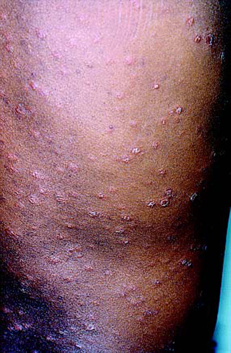
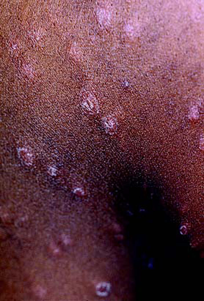
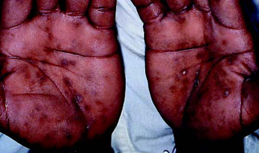
Physical examination revealed a healthy looking man with a diffuse papular eruption and minor associated scaling involving the trunk and limbs (Figures 1 and 2) with discrete erythematous lesions on the palms and soles (Figure 3). The patient had mild scalp scaling, mild photophobia, and a moderate degree of adenopathy, especially in the groin and posterior neck.
Figure 1.

Scattered guttate erythematous psoriasiform plaques on the trunk.
Figure 2.

Closer view of the lesions on the upper trunk.
Figure 3.
Palms with hyperpigmented macules and erythematous psoriasiform plaques.
What is your diagnosis, and what lab evaluations may be helpful in the diagnosis?
DIAGNOSIS: Secondary syphilis.
DISCUSSION
Infection with Treponema pallidum has myriad systemic and cutaneous manifestations (1, 2). Infection is usually due to intimate personal contact. The 2 broad categories of syphilis are acquired and congenital; further subdivisions are noted in Table 1. In the vast majority of patients, a primary sore, or chancre, occurs 3 weeks after inoculation. Without treatment, lymphadenopathy occurs and is followed by a variety of mucocutaneous lesions that develop approximately 3 to 12 weeks after the initial chancre (3). Studies investigating the long-term effects of untreated syphilis have identified a 10% mortality rate (4).
Table 1.
Categories of syphilis
| Acquired syphilis |
| Primary |
| Secondary |
| Latent |
| Late |
| Congenital syphilis |
| Early congenital |
| Late congenital |
Signs and symptoms
Secondary syphilis is characterized by generalized mucocutaneous involvement with a variety of symmetric and usually asymptomatic lesions (Table 2). The coppery red, macular, oval lesions are the earliest and usually occur symmetrically on the trunk with orientation along skin cleavage lines. Papular lesions appear soon after the macular eruption and are considered the most common in secondary syphilis. The appearance of the papules is dictated largely by the body region involved: moist, coalescing papules in the perineum (condyloma lata); split papules at the corners of the lip or angle of the nose; eroded papules on the inner lip or anal rim; and psoriasiform papules on the palms. Late in the second stage of syphilis (a year or more after infection), a widespread micropapular eruption may occur, or alternatively, a large papule may be surrounded by smaller satellite papules. Lesions involving the mucous membranes may be papular but more likely appear as gray oval patches on the palate, inner surface of lips, or buccal mucosa. Finally, the alopecia associated with syphilis may be diffuse or patchy (“moth-eaten”) if on the scalp. Additionally, eyebrow alopecia may be due to syphilis (5, 6).
Table 2.
Mucocutaneous manifestations of secondary syphilis
| • Coppery red, asymptomatic, generalized maculopapular lesions |
| • Split papules (especially at corners of lips or foreskin) |
| • Psoriasiform patches on palms and soles |
| • Moist, hypertrophic, coalescing papules in perineum (condyloma lata) |
| • Depigmented macules on a hyperpigmented background (back or sides of neck) |
| • Alopecia (may be patchy [“moth-eaten”] or diffuse) |
| • Oval mucous patches in mouth (palate, tongue, mucosa) or vaginal introitus |
Nonspecific systemic symptoms such as low-grade fever, headache, malaise, and myalgias and arthralgias are common in secondary syphilis and frequently worse at night. Generalized lymphadenopathy is common, and epitrochlear gland involvement has been described as pathognomonic. Patients may complain of sore throat or hoarseness, which indicates involvement of tonsils and larynx, respectively. Systemic involvement of the central nervous system may be indicated by headache, visual changes (optic neuritis), cranial nerve paralysis or, very rarely, meningomyelitis with paraplegia.
Diagnosis
Given the variety of skin lesions seen in secondary syphilis, there is a large differential diagnosis for the disease (Table 3). Past medical history, medication history, allergies to medications, social and sexual history, and a family history of skin conditions (e.g., guttate psoriasis, pityriasis rosea, drug eruptions, and tinea infections) are the most helpful in establishing the diagnosis.
Table 3.
Differential diagnosis for common lesions of secondary syphilis
| Type of lesion | Differential |
| Coppery red macules following skin cleavage lines | Pityriasis rosea, drug eruption, seborrheic dermatitis, measles, rubella, mycosis fungoides |
| Generalized papules | Sarcoid |
| Moist papules in perineum | Condyloma acuminatum, hemorrhoids |
| Generalized micropapules | Lichen planopilaris, keratosis pilaris, lichen scrofulosorum |
| Psoriasiform patches on palms and soles | Tinea infection, psoriasis |
| Split papules | Sarcoid |
The diagnosis of secondary syphilis usually can be confirmed with serological tests, which are either nonspecific, nontreponemal (reaginic) tests or specific treponemal tests (7). The nontreponemal tests widely used for screening at-risk patients include the VDRL and rapid plasma reagin (RPR) tests. In primary syphilis, the VDRL and RPR test results usually will be positive 6 weeks after the infection or 2 to 3 weeks after the primary chancre is seen. In secondary syphilis, these test results are positive. To confirm the diagnosis of syphilis, a specific treponemal test must be performed. The 2 commonly used specific treponemal tests are Treponema pallidum hemagglutination (TPHA) and fluorescent treponemal antibody absorption (FTA-ABS). Unfortunately, these tests are not always accurate in all patients, so the clinician may have to decide on treatment before the relevance of the test results is completely understood (8). An infectious disease consult can be of great help. Common causes of biologic false-positives for specific and nonspecific tests include autoimmune diseases (lupus, rheumatoid arthritis, and polyarteritis nodosa), malaria leprosy (especially lepromatous), typhus, viral respiratory tract infections, infectious mononucleosis, active pulmonary tuberculosis, hepatitis, subacute bacterial endocarditis, measles, chickenpox, filariasis, trypanosomiasis, leptospirosis, relapsing fever, Lyme borreliosis, pregnancy, old age, and narcotic addiction (9–17). Newer diagnostic tests, which include enzyme-linked immunosorbent assay or polymerase chain reaction technology, are being developed (18).
Treatment
Treatment of early syphilis (primary, secondary, and early latent) of longer than 2 years' duration requires benzathine penicillin G 2.4 million U intramuscularly once or aqueous procaine penicillin G 600,000 U in 1 intramuscular injection daily for 10 consecutive days. For patients allergic to penicillin, tetracycline hydrochloride 500 mg 4 times daily for 15 days or erythromycin (not the estolate) 500 mg 4 times daily for 15 days is indicated (16, 19). Two reactions may occur during the treatment of syphilis: the Jarisch-Herxheimer reaction and reactions to penicillin (anaphylactic and Hoigne reactions) (16, 20). The Jarisch-Herxheimer reaction is encountered approximately 12 hours after the administration of penicillin; typical symptoms include fever and possible worsening of skin lesions. Usually these reactions require no treatment; however, for later-stage patients (i.e., those with neurosyphilis or cardiovascular syphilis), pretreatment with corticosteroids has been helpful. Because anaphylactic reactions may be fatal, patients should be observed at least 15 to 20 minutes after receiving penicillin injections, and emergency kits should be close at hand. Finally, acute psychotic symptoms (Hoigne reactions) due to procaine penicillin have been encountered (20).
We obtained an infectious disease consult after the patient's screening test results were positive, and he was treated successfully with 1 intramuscular injection of benzathine penicillin. Additionally, he was tested for HIV and hepatitis after his social history revealed high-risk sexual activities. These results were negative.
References
- 1.Weir E, Fishman D. Syphilis: have we dropped the ball? CMAJ. 2002;167:1267–1268. [PMC free article] [PubMed] [Google Scholar]
- 2.Birley H, Duerden B, Hart CA, Curless E, Hay PE, Ison CA, Renton AM, Richens J, Wyatt GB. Sexually transmitted diseases: microbiology and management. J Med Mtcrobtol. 2002;51:793–807. doi: 10.1099/0022-1317-51-10-793. [DOI] [PubMed] [Google Scholar]
- 3.Sanchez MR. Infectious syphilis. Semin Dermatol. 1994;13:234–242. [PubMed] [Google Scholar]
- 4.Gjestland T. The Oslo study of untreated syphilis. Acta Dermatol Venereal (Stockh) 1955;35(Suppl):34. doi: 10.2340/00015555343368. [DOI] [PubMed] [Google Scholar]
- 5.Cuozzo DW, Benson PM, Sperling LC, Skelton HG., III Essential syphilitic alopecia revisited. J Am Acad Dermatol. 1995;32:840–843. doi: 10.1016/0190-9622(95)91543-5. [DOI] [PubMed] [Google Scholar]
- 6.Van der Ploeg DE, Stagnone JJ. Eyebrow alopecia in secondary syphilis. Arch Dermatol. 1982;90:172–173. doi: 10.1001/archderm.1964.01600020040008. [DOI] [PubMed] [Google Scholar]
- 7.Larsen SA, Steiner BM, Rudolph AH. Laboratory diagnosis and interpre tation of tests for syphilis. Clin Microbiol Rev. 1995;8:1–21. doi: 10.1128/cmr.8.1.1. [DOI] [PMC free article] [PubMed] [Google Scholar]
- 8.Sequeira PJ. Serological diagnosis of untreated early syphilis. Importance of the differences in THA, TPHA, and VDRL test litres. Br J Vener Dis. 1983;59:145–150. doi: 10.1136/sti.59.3.145. [DOI] [PMC free article] [PubMed] [Google Scholar]
- 9.Shore RN, Faricelli JA. Borderline and reactive FTA-ABS. Results in lu pus erythematosus. Arch Dermatol. 1977;113:37–41. [PubMed] [Google Scholar]
- 10.Garner ME, Backhouse JL, Collins CA, Roeder PJ. Serological tests for treponemal infections in leprosy patients. An evaluation of the fluorescent treponemal antibody absorption (FTA-ABS) test. Br J Vener Dis. 1969;45:19–22. doi: 10.1136/sti.45.1.19. [DOI] [PMC free article] [PubMed] [Google Scholar]
- 11.British Co-operative Clinical Group An examination of the treponemal immobilization test in the investigation of positive Serological tests for syphilis in pregnancy. Br J Vener Dis. 1959;35:162–168. [PMC free article] [PubMed] [Google Scholar]
- 12.Catterall RD. Collagen disease and the chronic biological false positive phenomenon. Q J Med. 1961;30:41–55. [PubMed] [Google Scholar]
- 13.Moore JE, Mohr CE. Biologically false positive serologic tests for syphilis. JAMA. 1951;150:467–473. doi: 10.1001/jama.1952.03680050033010. [DOI] [PubMed] [Google Scholar]
- 14.McKenna CH, Schroeter AL, Kierland RR, Stilwell GO, Pien FD. The fluorescent treponemal antibody absorbed (FTA-ABS) test beading phe nomenon in connective tissue diseases. Mayo Clin Proc. 1973;48:545–548. [PubMed] [Google Scholar]
- 15.Hunter EF, Russell H, Farshy CE, Sampson JS, Larsen RA. Evaluation of sera from patients with Lyme disease in the fluorescent treponemal anti body-absorption test for syphilis. Sex Transm Dis. 1986;13:232–236. doi: 10.1097/00007435-198610000-00005. [DOI] [PubMed] [Google Scholar]
- 16.van Voorst Vader PC. Syphilis management and treatment. Dermatol Clin. 1998;16:699–711. doi: 10.1016/s0733-8635(05)70035-8. [DOI] [PubMed] [Google Scholar]
- 17.Gibowski M, Neumann E. Non-specific positive test results to syphilis in dermatological diseases. Br J Vener Dis. 1980;56:17–19. doi: 10.1136/sti.56.1.17. [DOI] [PMC free article] [PubMed] [Google Scholar]
- 18.Young H. Syphilis: new diagnostic directions. Int J STD AIDS. 1992;3:391–413. doi: 10.1177/095646249200300601. [DOI] [PubMed] [Google Scholar]
- 19.Guthe T, Reynolds FW, Krag P. Mass treatment of treponemal diseases with particular reference to syphilis and yaws. Br Med J. 1953;1:594–598. doi: 10.1136/bmj.1.4810.594. [DOI] [PMC free article] [PubMed] [Google Scholar]
- 20.Schreiber W, Krieg JC. Hoigne syndrome. Case report and current litera ture review. Nervenart. 2001;7:546–548. doi: 10.1007/s001150170080. [DOI] [PubMed] [Google Scholar]


