Abstract
Establishment of intestinal infection with Entamoeba histolytica depends on the mouse strain; C57BL/6 mice are highly resistant, and C3H/HeJ mice are relatively susceptible. We found that resistance to intestinal infection was independent of lymphocyte activity or H-2 haplotype and occurred in the first hours to days postchallenge according to in vivo imaging. At 18 h postchallenge, the ceca of resistant C57BL/6 mice were histologically unremarkable, in contrast to the severe inflammation observed in susceptible C3H/HeJ mice. Comparison of cecal gene expression in C3H/HeJ and C57BL/6 mice demonstrated that there was parasite-induced upregulation of proinflammatory and neutrophil chemotaxis transcripts and there was downregulation of transforming growth factor β signaling molecules. Pretreatment with dexamethasone abrogated the partial resistance of C3H/HeJ or CBA mice through an innate, lymphocyte-independent mechanism, but it had no effect on the high-level resistance of C57BL/6 mice. Similarly, administration of a neutrophil-depleting anti-Gr-1 monoclonal antibody (RB6-8C5) decreased the partial resistance of CBA mice and led to severe pathology compared to control antibody-treated mice, but it had no effect on C57BL/6 resistance. These data indicate that there are discrete mechanisms of innate resistance to E. histolytica depending on the host background and, in contrast to other reports, imply that neutrophils are protective and not damaging in intestinal amebiasis.
Entamoeba histolytica colonizes the intestinal tract of up to 8% of the world's population and leads to invasive disease in approximately 10% of the colonized individuals (3, 6, 13, 16). The reason for this variable resistance to colonization or disease is poorly understood. Human and vaccine data suggest that acquired fecal immunoglobulin A (IgA) plays an important role in resistance in addition to the existence of innate immunity to infection (16, 18), although the latter mechanism is undefined.
We previously showed in a mouse model of amebic colitis that resistance to intestinal E. histolytica infection is dependent on the mouse strain (19). A role for innate immunity in this resistance was suggested by the early clearance of fecal antigen, which was complete by 2 weeks postchallenge in resistant mice. In this work, we confirmed that resistance to E. histolytica is conferred within the first days postchallenge and occurs through innate, lymphocyte-independent mechanisms. We compared the intestine's innate response to the parasite in resistant and susceptible mouse strains by microarray analysis and found that there is an association between susceptibility and proinflammatory cytokine production and neutrophil chemotaxis. The biological significance of proinflammatory cytokines or neutrophils has not been resolved in the amebiasis literature. While neutrophils contribute to hepatocyte or epithelial damage in vitro and to epithelial leakiness in intestinal xenografts (5, 31, 33), neutrophils have been protective early after infection in hepatic or intestinal animal models (34, 36) and can kill trophozoites in vitro if they are stimulated with proinflammatory cytokines (10).
We therefore examined the role of inflammation and neutrophils in the course of infection in this mouse model of amebic colitis. We found that neutrophil depletion with an anti-Gr-1 monoclonal antibody (MAb) diminished the innate resistance in certain mouse strains (e.g., CBA) but had no effect on the high-level resistance of C57BL/6 mice, indicating that mechanisms of innate immunity to intestinal E. histolytica infection vary depending on the host genetic background.
MATERIALS AND METHODS
Mice.
Female CBA, C3H/HeJ, C3H/HeSnJ, C3H SCID (C3Smn.CB17-PrkdcSCID/J), AKR, 129/Sv, C57BL/6, C57BL/6 SCID (C57BL/6J-PrkdcSCID/SzJ), C57BL/6 phagocyte oxidase cytochrome b−/− (B6.129S6-Cybbtm1Din/J), F1 C3H/HeJ × C57BL/6, F1 CBA × C57BL/6, and BALB/c mice were purchased from The Jackson Laboratory (Bar Harbor, ME). C.B-17 SCID-beige and C57BL/6 MyD88−/− mice were obtained from David Camerini (University of California, Irvine) and Ruslan Medzhitov (Yale University, New Haven, CT), respectively. Animals were maintained under specific-pathogen-free conditions at the University of Virginia and were challenged when they were 6 to 10 weeks old. The Institutional Animal Care and Use Committee approved all protocols.
Parasites and intracecal inoculation.
The trophozoites used for intracecal injection were originally laboratory strain HM1:IMSS (American Type Culture Collection, Manassas, VA) that were sequentially passaged in vivo through the mouse cecum. Cecal contents were cultured in trypsin-yeast-iron (TYI-S-33) medium supplemented with 25 U/ml penicillin and 25 mg/ml streptomycin until they were axenic, as confirmed by the absence of bacterial growth on Trypticase soy agar with 5% sheep blood (Becton Dickinson, Sparks, MD). Firefly luciferase-expressing ameba were generated by suspending 2.2 × 10⁵ trophozoites/ml in medium 199 (Invitrogen, Carlsbad, CA) supplemented with 5.7 mM cysteine, 1 mM ascorbic acid, and 25 mM HEPES, pH 6.8. Trophozoites were transfected using a modified version of the Superfect protocol (QIAGEN, Valencia, CA) at a ratio of 3 μg of DNA to 15 μl of Superfect for 3 h. The introduced construct contained the luciferase structural gene under the control of the E. histolytica hgl5 gene with regulatory sequences mutated at the URE3 motif to optimize expression (pHTP.luc [37]). Transfected trophozoites were then washed with TYI-S-33 medium supplemented with 2 U/ml penicillin and 2 mg/ml streptomycin sulfate; a selective antibiotic (6 μg/ml G418) was added at 24 h, and the concentration was increased stepwise until it was 50 μg/ml. For all intracecal inoculations, axenic trophozoites were grown to the log phase and counted with a hemacytometer, and 2 × 10⁶ trophozoites in 150 μl were injected intracecally into each mouse according to the protocol described previously (19).
Imaging of bioluminescent E. histolytica.
Mice were anesthetized with isoflurane and placed inside a charge-coupled device camera (Xenogen IVIS II System). Baseline images were obtained prior to intraperitoneal injection of 1 mg luciferin (Xenogen, California), and sequential images were obtained after injection. Peak luminescence was found to occur 15 to 30 min after luciferin administration. The images were 15-s exposures, and luminescence was quantified for each mouse using the IgorPro 4.09A software (WaveMetrics, Inc., Lake Oswego, OR). No background luminescence was observed in control-transfected trophozoites with or without luciferin or in luciferase-transfected trophozoites without luciferin substrate.
Pathology and scoring of amebic colitis.
Mice were sacrificed, and each cecum was longitudinally bisected. One-half of the cecum was placed in Hollande's fixative, cut into three to five equal cross sections, and paraffin embedded, and 4-μm sections were stained with hematoxylin and eosin. Histopathology was scored blindly for each mouse. Cecal thickness was measured at two or more sites with an ocular micrometer at a magnification of ×40. An ameba score and an inflammation score were determined as described previously (19). The contents of the other half of each cecum were rinsed in phosphate-buffered saline (PBS) and assayed for E. histolytica antigen using an E. histolytica II enzyme-linked immunosorbent assay kit (Techlab, Blacksburg, VA) according to the manufacturer's instructions. Optical density values were normalized to the manufacturer's positive control for each run.
Gene chip analysis.
Affymetrix gene chip analysis was performed according to the manufacturer's instructions using murine U74Av2 arrays. Full public access to the raw data is available at https://genes.med.virginia.edu/public_data/index.cgi under “Eric_Houpt_Acute_Amebic_Colitis.” Minimum information about a microrarray experiment (www.mged.org/miame) was obtained as follows. Ceca were obtained from female 6-week-old C3H/HeJ and C57BL/6 mice 18 h after intracecal parasite challenge (n = 3 and n = 4, respectively) or intracecal sham challenge with 150 μl TYI-S-33 medium (n = 3 and n = 3, respectively) for a total of 13 samples and hybridizations. Cecal tissue was rinsed in sterile PBS to remove the luminal contents and then placed in RNAlater (Ambion, Austin, TX) followed by Trizol (Invitrogen, Carlsbad, CA) and homogenized, and total RNA was extracted using a QIAGEN RNAEasy kit (QIAGEN, Valencia, CA). The ribosomal peaks were intact and showed no evidence of degradation for any samples. The Affymetrix BioB control complementary RNA was added at the detection threshold (1.5 pM) and received a “present” detection call for all samples. The glyceraldehyde-3-phosphate dehydrogenase and β-actin housekeeping genes had 3′-to-5′ detection ratios of <4, indicating that there was no excess 5′ mRNA degradation. cDNA synthesis, in vitro transcription to complementary RNA, and hybridization were performed as described at a website (http://www.healthsystem.virginia.edu/internet/biomolec/genechipprotocols.cfm).No reference samples, additional quality control steps, additional designs, or sample or protocol manipulation was used. Sample gene expression data were analyzed with the D-chip software (23) and averaged among groups, and dysregulated gene expression between groups was defined as a fold change of >1.5, an average signal intensity difference of >100 U, and a P value of <0.05. According to this analysis, 204 of 12,422 Affymetrix gene probe sets were dysregulated between the ceca of C3H mice and the ceca of C57BL/6 mice after E. histolytica challenge (87 genes were upregulated in C3H mice, and 117 genes were upregulated in C57BL/6 mice). Of these 204 candidate genes, 87 were statistically dysregulated in sham-challenged mice, leaving 117 genes that were dysregulated specifically in response to the parasite (52 genes were upregulated in C3H/HeJ mice, and 65 genes were upregulated in C57BL/6 mice). These 117 genes were compared with the nondysregulated 12,305 genes by GenMapp 2.0 (http://www.genmapp.org) for patterns of biological processes by using the methods of Doniger et al., where a Z score of >2 indicated a significantly overrepresented biological process (1, 12).
In vivo manipulations.
For dexamethasone experiments, 0.2 mg dexamethasone (American Regent, Shirley, NY) or PBS was administered intraperitoneally on days −3, −2, −1, and 0 relative to the intracecal challenge. For neutrophil depletion, 300 μg of anti-Gr-1 monoclonal antibody RB6-8C5 (rat IgG2b; a gift from David Askew, University of Cincinnati) or purified rat IgG (Lampire Biologicals, Pipersville, PA) was administered intraperitoneally on days −2, −1, and 2 relative to the intracecal challenge. Neutrophil depletion was confirmed in the peripheral blood on days 0 and 6 by performing blinded manual differential counting with Wright-Giemsa-stained blood smears (CamcoQuik; Cambridge Chemical Products, Ft. Lauderdale, FL). For anti-transforming growth factor β (TGF-β) experiments, 2 mg of clone 1D11 purified from ascites or control mouse IgG was injected on day −2 relative to the challenge by using previously described protocols (21).
Myeloperoxidase assays.
The cecal neutrophil content was quantified by a myeloperoxidase (MPO) assay similar to the methods of Seydel et al. (33). Briefly, ceca were bisected longitudinally, rinsed to remove the luminal contents, weighed, frozen at −70°C, and homogenized for 15 s with a tissue grinder in 50 mM potassium phosphate buffer (pH 6.0) containing 0.5% hexadecyltrimethylammonium bromide. Seven microliters of supernatant was added to 200 μl of 5 mM potassium phosphate buffer containing 33 μg o-dianisidine and 0.005% H2O2, and the results were read sequentially with a microplate reader at 450 nm. The change in optical density per minute was normalized per gram of tissue, where 1 U of MPO equaled a change in the optical density of 1.13 × 10⁻² U per min.
Statistics.
Group averages were compared using a t test or the Mann-Whitney test, and proportions (e.g., infection rates) were compared using Fisher's exact test. The data are expressed below as means ± standard errors unless otherwise indicated. All P values were two tailed.
RESULTS
Resistance to intestinal E. histolytica infection is H-2 haplotype and lymphocyte independent.
We previously showed that susceptibility to intestinal amebiasis depends on the mouse strain; C57BL/6 (H-2d) and BALB/c (H-2b) mice are relatively resistant to the establishment of an amebic infection, while C3H strains (HeJ, HeOuJ, and HeN; all H-2k) are relatively susceptible (19). Therefore, we examined a possible role for H-2k in susceptibility. The H-2k CBA strain exhibited relative susceptibility, while H-2k AKR mice were relatively resistant, indicating that the H-2k haplotype was not sufficient for susceptibility (42 of 69 and 1 of 6 mice, respectively, were infected based on histopathology and culture at 10 days postchallenge; P ≤ 0.01). We also found that the resistance phenotype of C57BL/6 mice (1 of 38 mice was infected at 10 days) was not dependent on lymphocyte activity, as SCID mice with this background remained resistant (one of six mice was infected; difference not significant). Likewise, the SCID-beige mutation, which confers lymphocyte, NK cell, CTL, and macrophage defects (28), did not render C.B-17 mice susceptible (none of 18 mice were infected, compared with none of 12 congenic BALB/c mice; difference not significant). Resistance remained lymphocyte independent in the C3H mice; in fact, wild-type C3H mice had a mildly increased infection rate compared with C3H SCID mice (29 of 64 C3H/HeJ or C3H/HesnJ mice were infected, compared with 8 of 33 C3H SCID mice, P = 0.05), suggesting that naive lymphocytes may even potentiate innate susceptibility in this strain.
Resistance to intestinal E. histolytica infection occurs rapidly after challenge.
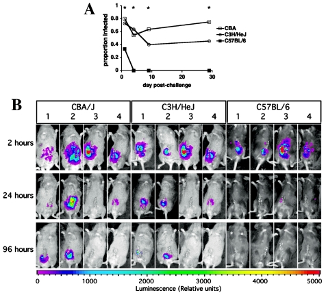
To more directly examine the kinetics of innate resistance, cohorts of mice were challenged at day 0 and sacrificed sequentially thereafter. Clearance, defined as the absence of amebae as determined by cecal histopathology and culture, occurred within 1 day and was complete by 4 days in C57BL/6 mice (Fig. 1A). The clearance that occurred in a minority of CBA and C3H/HeJ mice happened within the first 4 to 9 days, after which clearance did not occur. We confirmed the site and kinetics of clearance by in vivo imaging of E. histolytica trophozoites (Fig. 1B). Mouse-passaged trophozoites transfected with firefly luciferase were intracecally inoculated into C3H, CBA, and C57BL/6 mice. Upon administration of luciferin substrate, all mice exhibited similar luminescence at 2 h postchallenge, after which luminescence persisted only in the ceca of successfully infected C3H and CBA mice (as confirmed by histopathology and culture at 96 h postchallenge). The C3H or CBA mice that cleared infection did so with kinetics similar to those of the highly resistant C57BL/6 strain, raising the question of whether mechanisms of innate resistance were shared.
FIG. 1.
Resistance to E. histolytica occurs within hours in the mouse intestine. (A) CBA, C3H/HeJ, and C57BL/6 mice were challenged on day 0 and sacrificed sequentially on days 1, 4, 9, and 29. The proportions of mice infected as confirmed by culture and histopathology are shown (n = 10 to n = 12 for each mouse strain on each day). An asterisk indicates that the P value is <0.04 for a comparison of the rates of infection of CBA or C3H/HeJ mice and C57BL/6 mice; the infection rates were not significantly different for CBA or C3H/HeJ mice at any time. (B) CBA, C3H/HeJ, and C57BL/6 mice were intracecally inoculated with luciferase-expressing E. histolytica trophozoites and serially imaged with a charge-coupled device camera at 2, 24, and 96 h after intraperitoneal administration of luciferin. Mice were sacrificed at 96 h for confirmation of infection. Four representative mice of each strain are shown. CBA mice 1 and 2 and C3H mice 1 and 2 were infected based on histology and culture, while all other mice were uninfected. Infection was limited to the cecum. Luciferase-expressing trophozoites exhibited no luminescence without administration of luciferin, and trophozoites transfected with the control plasmid did not luminesce (data not shown).
Histology and gene expression profiles indicate that there is acute inflammation in susceptible but not resistant mouse strains after challenge.
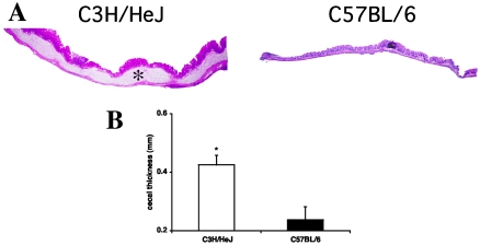
Based on the rapidity with which resistance occurred, we chose to examine the cecal histopathology of C3H/HeJ and C57BL/6 mice 18 h after challenge. At this point greater numbers of amebae were visible in C3H/HeJ mice than in C57BL/6 mice (ameba scores, 2.7 ± 0.5 and 0.9 ± 0.4, respectively; n = 8; P = 0.01), and the pathology differed dramatically, with diffuse inflammation, widespread submucosal edema, and increased cecal thickness in C3H/HeJ mice compared with the relatively normal histology in C57BL/6 mice (Fig. 2).
FIG. 2.
C3H/HeJ mice exhibit rapid intestinal inflammation in response to acute E. histolytica challenge. (A) Histology 18 h after intracecal challenge with E. histolytica trophozoites showed diffuse cecal inflammation with submucosal edema (asterisk) in the susceptible C3H/HeJ strain compared with the normal histology in the C57BL/6 mouse (hematoxylin and eosin staining). Magnification, ×20. (B) Cross-sectional cecal thickness was measured in both strains at 18 h. The data are means and standard errors (n = 8). An asterisk indicates that the P value is 0.007.
We characterized the two strains' cecal responses by gene microarray. Using D-chip analysis, 204 genes were statistically dysregulated between C3H/HeJ and C57BL/6 mice 18 h after intracecal challenge (n = 3 and n = 4, respectively). Eighty-seven of these genes were also dysregulated between the strains after sham challenge, and the remaining 117 parasite-induced dysregulated genes were compared with the 12,305 nondysregulated genes using GenMapp (Table 1). This analysis indicated that the parasite-induced response of the C3H/HeJ intestine was associated with an innate acute-phase inflammatory response and neutrophil chemotaxis, as well as a downregulation of SMAD/TGF-β signaling pathway transcripts relative to C57BL/6 mice.
TABLE 1.
Microarray analysis of the postchallenge cecum of susceptible C3H mice demonstrated that there was a parasite-induced innate inflammatory response with downregulation of TGF-β signaling molecules
| Biological processes (Z score)a | Geneb | Locus identification or accession no. | Fold change (C3H/HeJ/C57BL/6) | C3H/HeJ signal intensity (mean, n = 3)c | C57BL/6 signal intensity (mean, n = 4)c | P |
|---|---|---|---|---|---|---|
| Neutrophil chemotaxis (9.4), chemotaxis (5.9), and cell | Lectin, galactose binding, soluble 1 | 16852 | 2.62 | 4,210 | 1,606 | 0.001 |
| adhesion (3.4) | Small inducible cytokine A6 | 20305 | 1.99 | 902 | 453 | 0.02 |
| S100 calcium binding protein A8 (calgranulin A) | 20201 | 1.83 | 9,093 | 4,960 | 0.03 | |
| Procollagen, type III, alpha 1 | 12825 | 1.74 | 2,818 | 1,623 | 0.03 | |
| Fibronectin 1 | 14268 | 1.52 | 816 | 536 | 0.04 | |
| Colony-stimulating factor 3 receptor (granulocyte) | 12986 | 2.07 | 830 | 402 | 0.05 | |
| Integrin alpha M | 16409 | 1.54 | 783 | 509 | 0.05 | |
| Acute-phase response (8.4), innate immune response (7.0), | Properdin factor, complement | 18636 | 1.81 | 719 | 398 | 0.007 |
| inflammatory response (7.0), immune response (5.0), and | Immunoglobulin joining chain | 16069 | 1.55 | 3,151 | 2,038 | 0.01 |
| defense response (4.1) | Regenerating islet-derived 3 gamma | 19695 | 2.12 | 7,508 | 3,538 | 0.02 |
| Small chemokine (C-C motif) ligand 11 | U77462 | 1.52 | 960 | 632 | 0.03 | |
| P lysozyme structural | 17110 | 2.25 | 2,419 | 1,074 | 0.04 | |
| Pancreatitis-associated protein | 18489 | 1.67 | 16,865 | 10,092 | 0.05 | |
| SMAD protein nuclear translocation | Transducer of ErbB-2.1 | 22057 | 0.58 | 721 | 1,247 | 0.01 |
| (16.8) and TGF-β receptor signaling pathway (5.8) | MAD homolog 4 (Drosophila) | 17128 | 0.64 | 2,445 | 3,835 | 0.02 |
| MAD homolog 1 (Drosophila) | 17125 | 0.51 | 490 | 953 | 0.03 | |
| Fatty acid (8.3), organic acid (5.13), and coenzyme (2.5) metabolism and physiological process (2.5)d |
Gene ontology biological processes were compared for the 117 parasite-induced dysregulated cecal genes and the 12,305 nondysregulated cecal genes with GenMapp 2.0 as described in Materials and Methods. A Z score of >2.0 indicates a significantly overrepresented biological process (12).
Dysregulated genes attributed to the biological processes. Full access to the raw data is available at “Eric_Houpt_Acute_Amebic_Colitis” at https://genes.med.virginia.edu/public_data/index.cgi.
Signal intensities are expressed in relative units of gene expression from ceca after intracecal challenge with E. histolytica.
The genes include genes encoding microsomal glutathione S-transferase 1, lysophospholipase 1, acetyl-coenzyme A dehydrogenase (long chain), 3-phosphoadenosine 5-phosphosulfate synthase 2, stearoyl-coenzyme A desaturase, chloride intracellular channel 4 (mitochondrial), acetyl-coenzyme A acyltransferase, accession no. AI841279, protein C receptor (endothelial), amine N-sulfotransferase, and thrombomodulin.
Dexamethasone decreases innate resistance to infection in C3H and CBA mice.
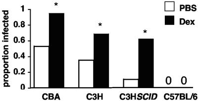
To examine whether the inflammatory response demonstrated by the microarray analysis in susceptible mice was a protective or deleterious response to infection, we chose to pharmacologically inhibit this response with a corticosteroid. Pretreatment of mice on days −2, −1, and 0 with dexamethasone increased susceptibility in C3H/HeJ and CBA mice (Fig. 3), suggesting that the innate inflammatory response in these mice was protective. Dexamethasone also increased susceptibility in C3H SCID mice, indicating that the corticosteroid effect was exerted through inhibition of innate immunity. In contrast, dexamethasone had no effect on C57BL/6 resistance. It was noteworthy that upon sacrifice 10 to 30 days postchallenge, there was no difference in amebic load or inflammation score between infected, dexamethasone-treated mice and infected, PBS-treated mice for the CBA, C3H/HeJ, or C3H SCID strain (data not shown).
FIG. 3.
Dexamethasone decreases innate resistance to intestinal E. histolytica infection in susceptible strains. CBA, C3H/HeJ, C3H SCID, and C57BL/6 mice were treated with 0.2 mg dexamethasone (Dex) or PBS on days −3, −2, −1, and 0 relative to intracecal challenge. The proportions of mice infected upon sacrifice are shown (n = 15, n = 20, n = 34, n = 29, n = 9, n = 8, n = 6, and n = 8 for the groups shown from left to right on the x axis) An asterisk indicates that the P value is ≤0.05 compared with PBS-treated mice. C3H/HeJ mice were sacrificed at 30 days postchallenge, and all other mice were sacrificed at 10 days postchallenge.
Gr-1+ cells mediate innate resistance and control disease in CBA mice.
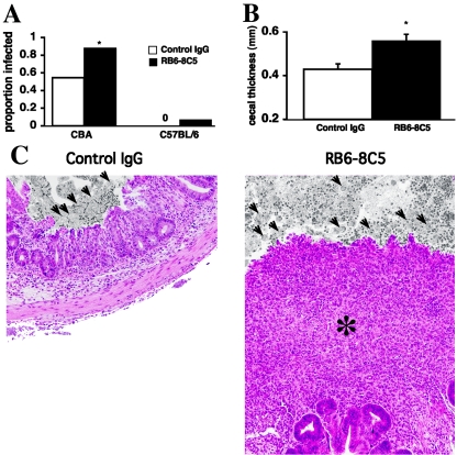
Because neutrophils are key effector cells of innate immunity and can exert amebicidal activity in vitro (10) and the microarray data indicated that there was neutrophil activity in susceptible mice, we chose to examine the role of neutrophils in resistance. Neutrophils were depleted by administering anti-Gr-1 MAb RB6-8C5 on days −2, −1, and 2. Neutrophil depletion was confirmed by testing peripheral blood on days 0 and 6 (3% ± 4% of leukocytes in anti-Gr-1 MAb-treated mice were neutrophils, compared with 32% ± 6% in control rat IgG-treated mice; n = 11; P < 0.0001) and by assaying the myeloperoxidase activity in the intestine at the time of sacrifice (78 ± 10 MPO units in anti-Gr-1 MAb-treated mice, compared with 277 ± 67 MPO units in control rat IgG-treated mice; n = 12; P = 0.01). The mortality after intracecal challenge was high in neutropenic C3H/HeJ mice (17 of 20 mice died by day 1), and therefore, the phenotype of infection in this strain was difficult to evaluate (two of three mice were infected upon sacrifice at day 6). As observed with dexamethasone therapy, anti-Gr-1 MAb administration had no effect on the high-level resistance of C57BL/6 mice (Fig. 4A). Not surprisingly, C57BL/6 mice deficient in respiratory burst oxidase activity in phagocytes (gp91phox−/−) also remained resistant (none of eight mice were infected upon sacrifice at day 7, compared with none of eight wild-type mice). In the CBA strain, however, anti Gr-1 MAb administration increased the susceptibility rate. Furthermore, upon evaluation of histology at sacrifice on day 6, the cecal thickness was significantly greater and the pathology was more severe in infected, anti-Gr-1 MAb-treated mice than in infected, control IgG-treated animals (Fig. 4B and C), and not only the initial susceptibility but also the disease severity were increased. The parasite burden was nonstatistically elevated in infected, anti-Gr-1 MAb-treated mice compared with infected, control IgG-treated mice (cecal E. histolytica antigen optical densities, 1.1 ± 0.2 and 0.8 ± 0.2, respectively; n = 11 and n = 9, respectively; difference not significant).
FIG. 4.
Innate resistance to intestinal E. histolytica infection is diminished by neutrophil-depleting anti-Gr-1 MAb administration in CBA mice. (A) Six- to 10-week-old mice with the CBA and C57BL/6 mice background were given 300 μg of anti-Gr-1 MAb RB6-8C5 or control rat IgG on days −2, −1, and 2 relative to intracecal challenge. Neutrophil depletion was confirmed in the peripheral blood on days 0 and 6 and in the intestine by the MPO assay on day 6. Mice were sacrificed on day 6 for evaluation of infection by histology and culture. An asterisk indicates that the P value is 0.01 for a comparison of anti-Gr-1 MAb-treated and control IgG-treated CBA mice (n = 22, n = 26, n = 13, and n = 13 for the four groups shown from left to right on the x axis). (B) In infected CBA mice, cecal thickness was compared for the anti-Gr-1MAb-treated and control IgG-treated groups. The data are means and standard errors (n = 12 and n = 23 for infected, control IgG-treated mice and infected, anti-Gr-1 MAb-treated mice, respectively) An asterisk indicates that the P value is ≤0.01. (C) Representative photomicrographs showing that there was significant destruction of mucosal architecture with infiltration (asterisk) in a neutrophil-depleted animal. Luminal contents are black and white, and trophozoites are indicated by arrows.
High-level innate resistance in C57BL/6 mice.
To evaluate whether the relative overexpression of SMAD TGF-β signaling gene transcripts in C57BL/6 mice participated in this strain's innate resistance, we administered 2 mg of anti-TGF-β on day −2 prior to challenge (21), but we observed no increase in susceptibility (none of six mice was infected at sacrifice on day 4, compared with none of six control IgG-treated mice). Finally, Toll-like receptor signaling through MyD88 was not required for the C57BL/6 innate defense, as MyD88−/− mice remained resistant (none of eight mice was infected at sacrifice on day 7, compared with none of eight MyD88+/+ mice).
DISCUSSION
The most important finding of this work is that mouse strain-specific immunity to intestinal E. histolytica infection is innate, occurring within hours after challenge. Neutrophils are likely protective effector cells in the innate resistance of CBA mice; however distinct undefined innate immune mechanisms are sufficient for the high-level resistance of C57BL/6 mice.
There are previous conflicting reports on whether neutrophils are protective or deleterious during E. histolytica infection. Neutrophils contribute to hepatocyte and epithelial cell damage upon E. histolytica infection in vitro (5, 7, 31), and in the intestinal xenograft model neutrophils increase epithelial destruction and permeability (33). Indeed, a requirement for neutrophils in amebic ulceration has been hypothesized based on microscopic findings for neutrophils at the leading edge of the intestinal ulcers (24). Other studies have indicated that neutrophils have a protective role in amebiasis. Neutrophils limit the size of amebic liver abscesses in wild-type and SCID mice (34, 36) and appear to hasten parasite clearance from the intestine in BALB/c mice, albeit the effect was observed only at 6 h since all mice cleared the infection thereafter (27). Our data support the latter findings and strongly suggest that neutrophils play a protective role in intestinal amebiasis. Specifically, we found that Gr-1+ cells provided ∼73% of the innate resistance to intestinal E. histolytica infection that exists in CBA mice (i.e., the resistance rate fell from 45% to 12% after anti-Gr-1 MAb administration). Our results are not completely definitive, however, because the anti-Gr-1 antibody used widely to deplete neutrophils can react with subsets of eosinophils, monocytes, and plasmacytoid dendritic cells (14, 17). Nonetheless, our working assumption is that the neutrophil potentially acts in two ways: preventing the infection from becoming established and then limiting or regulating the disease once infection occurs.
The simplest explanation for how neutrophils could prevent an infection from becoming established within hours after challenge is through migration and direct killing of the parasite. In vitro data might not support this scenario since laboratory strain HM1:IMSS trophozoites kill neutrophils readily; however, when there is a numerical excess or proinflammatory cytokine stimulation, neutrophils can be amebicidal (10, 15). One could thus envision that the inflammatory environment described by the microarray data for susceptible C3H/HeJ mice is protective in at least this regard. It is intriguing to speculate that the relative decrease in TGF-β signaling molecules in C3H mice (particularly Smad4, the common intracellular Smad of TGF-β receptor signaling pathways [11]) is permissive to this innate inflammatory response given TGF-β's known effect on suppressing NF-κβ activation in the gut (25). The cell types responsible for the proinflammatory response in this model are not clear, but intestinal epithelial cells are likely important given their known secretion of tumor necrosis factor alpha, interleukin-8 (IL-8), and IL-1β in the E. histolytica xenograft model (32, 38).
Neutrophils could also act indirectly in this model through elaboration of proinflammatory cytokine and chemokines to augment other protective immune responses (30). It is known that CD4+ T cells contribute to immunopathology during chronic infection in this mouse model (19), and we are currently examining whether neutrophil depletion alters the character of the subsequent T-cell response and thereby contributes, or could later contribute, an immune component to the pathology seen with neutrophil depletion. Such things happen in Toxoplasma gondii and Candida infection models (4, 29) and would explain the severe disease seen in this model with neutrophil depletion amidst a relatively unchanged parasite burden. As for the relevance to human infection, genetic or acquired defects in neutrophil function (e.g., migration, effector function, or cytokine secretion) may partially explain the variable innate resistance seen in endemic populations.
We were surprised to find increased innate resistance to infection in C3H SCID mice. The effect was modest, however, so it will be difficult to experimentally identify the mechanisms of increased resistance in the SCID mice. Although highly speculative, these mechanisms might include increased NK cell activity (22) or a potential increase in the protective innate inflammatory response due to loss of inhibition by naturally occurring T regs (e.g., with a further decrease in TGF-β production [26]). It is possible that a similar phenotype may be exhibited by the mouse model of intestinal Citrobacter infection, in which C3H strain mice (both Toll-like receptor 4 mutant and wild type) are highly susceptible to infection, while C3H SCID mice exhibit diminished pathology (35).
We also found that the corticosteroid dexamethasone decreased innate immunity to the parasite. Given the broadly suppressive actions of corticosteroids, we are unlikely to define a specific innate immune defect; however, inhibition of neutrophil chemotaxis, reactive oxygen species production, and NF-κβ activation are reasonable candidates (2, 8, 9). Regardless, corticosteroid use and amebic colitis have been associated in humans (20), and it is validating that this effect is replicated in this new mouse model.
The mechanisms of innate immunity in C57BL/6 mice are unclear, although they remain active despite neutrophil depletion, dexamethasone therapy, anti-TGF-β antibody administration, or a deficiency in NADPH oxidase, MyD88, IL-12, or inducible nitric oxide synthase (19). The rapidity of clearance in these mice is striking, and given the absence of a histological inflammatory response in the mice, we wonder if this reflects primary defects in parasite survival in the C57BL/6 intestinal flora or parasite adherence to C57BL/6 mucin or epithelium, as opposed to an active immune process. Finally, we were not surprised to find similar susceptibilities and innate resistance phenotypes in CBA and C3H mice, given that these strains were derived from a common Bagg albino × DBA cross that occurred in 1920 and share many genes in addition to H-2. We hope that definition of the mechanisms of innate immunity in the mouse, both between and within strains, will translate into a rational understanding of the human spectrum of amebic colonization and disease.
Acknowledgments
This work was supported by grant AI052444-01A1, by an Infectious Diseases Society of America Wyeth-Ayerst Vaccines Young Investigator Award in Vaccine Development, and by the Virginia Commonwealth Technology Research Fund.
We acknowledge the Biomolecular Research Facility for performing the gene chip analysis and the histological support of the Research Histology Core of the Center for Research in Reproduction, both at the University of Virginia. We thank William A. Petri, Jr. for helpful advice and support and David Lyerly, Techlab, for providing the E. histolytica II enzyme-linked immunosorbent assay kits.
Editor: J. F. Urban, Jr.
REFERENCES
- 1.Ashburner, M., C. A. Ball, J. A. Blake, D. Botstein, H. Butler, J. M. Cherry, A. P. Davis, K. Dolinski, S. S. Dwight, J. T. Eppig, M. A. Harris, D. P. Hill, L. Issel-Tarver, A. Kasarskis, S. Lewis, J. C. Matese, J. E. Richardson, M. Ringwald, G. M. Rubin, and G. Sherlock. 2000. Gene ontology: tool for the unification of biology. The Gene Ontology Consortium. Nat. Genet. 25:25-29. [DOI] [PMC free article] [PubMed] [Google Scholar]
- 2.Auphan, N., J. A. DiDonato, C. Rosette, A. Helmberg, and M. Karin. 1995. Immunosuppression by glucocorticoids: inhibition of NF-kappa B activity through induction of I kappa B synthesis. Science 270:286-290. [DOI] [PubMed] [Google Scholar]
- 3.Blessmann, J., I. K. Ali, P. A. Nu, B. T. Dinh, T. Q. Viet, A. L. Van, C. G. Clark, and E. Tannich. 2003. Longitudinal study of intestinal Entamoeba histolytica infections in asymptomatic adult carriers. J. Clin. Microbiol. 41:4745-4750. [DOI] [PMC free article] [PubMed] [Google Scholar]
- 4.Bliss, S. K., L. C. Gavrilescu, A. Alcaraz, and E. Y. Denkers. 2001. Neutrophil depletion during Toxoplasma gondii infection leads to impaired immunity and lethal systemic pathology. Infect. Immun. 69:4898-4905. [DOI] [PMC free article] [PubMed] [Google Scholar]
- 5.Burchard, G. D., G. Prange, and D. Mirelman. 1993. Interaction between trophozoites of Entamoeba histolytica and the human intestinal cell line HT-29 in the presence or absence of leukocytes. Parasitol. Res. 79:140-145. [DOI] [PubMed] [Google Scholar]
- 6.Caballero-Salcedo, A., M. Viveros-Rogel, B. Salvatierra, R. Tapia-Conyer, J. Sepulveda-Amor, G. Gutierrez, and L. Ortiz-Ortiz. 1994. Seroepidemiology of amebiasis in Mexico. Am. J. Trop. Med. Hyg. 50:412-419. [DOI] [PubMed] [Google Scholar]
- 7.Chadee, K., F. Moreau, and E. Meerovitch. 1987. Entamoeba histolytica: chemoattractant activity for gerbil neutrophils in vivo and in vitro. Exp. Parasitol. 64:12-23. [DOI] [PubMed] [Google Scholar]
- 8.Cronstein, B. N., S. C. Kimmel, R. I. Levin, F. Martiniuk, and G. Weissmann. 1992. A mechanism for the antiinflammatory effects of corticosteroids: the glucocorticoid receptor regulates leukocyte adhesion to endothelial cells and expression of endothelial-leukocyte adhesion molecule 1 and intercellular adhesion molecule 1. Proc. Natl. Acad. Sci. USA 89:9991-9995. [DOI] [PMC free article] [PubMed] [Google Scholar]
- 9.Dandona, P., M. Suri, W. Hamouda, A. Aljada, Y. Kumbkarni, and K. Thusu. 1999. Hydrocortisone-induced inhibition of reactive oxygen species by polymorphonuclear neutrophils. Crit. Care Med. 27:2442-2444. [DOI] [PubMed] [Google Scholar]
- 10.Denis, M., and K. Chadee. 1989. Human neutrophils activated by interferon-gamma and tumour necrosis factor-alpha kill Entamoeba histolytica trophozoites in vitro. J. Leukoc. Biol. 46:270-274. [DOI] [PubMed] [Google Scholar]
- 11.Derynck, R., Y. Zhang, and X. H. Feng. 1998. Smads: transcriptional activators of TGF-beta responses. Cell 95:737-740. [DOI] [PubMed] [Google Scholar]
- 12.Doniger, S. W., N. Salomonis, K. D. Dahlquist, K. Vranizan, S. C. Lawlor, and B. R. Conklin. 2003. MAPPFinder: using Gene Ontology and GenMAPP to create a global gene-expression profile from microarray data. Genome Biol. 4:R7. [DOI] [PMC free article] [PubMed] [Google Scholar]
- 13.Gathiram, V., J. T. 1987. A longitudinal study of asymptomatic carriers of pathogenic zymodemes of Entamoeba histolytica. S. Afr. Med. J. 72:669-672. [PubMed] [Google Scholar]
- 14.Gilliet, M., A. Boonstra, C. Paturel, S. Antonenko, X. L. Xu, G. Trinchieri, A. O'Garra, and Y. J. Liu. 2002. The development of murine plasmacytoid dendritic cell precursors is differentially regulated by FLT3-ligand and granulocyte/macrophage colony-stimulating factor. J. Exp. Med. 195:953-958. [DOI] [PMC free article] [PubMed] [Google Scholar]
- 15.Guerrant, R. L., J. Brush, J. I. Ravdin, J. A. Sullivan, and G. L. Mandell. 1981. Interaction between Entamoeba histolytica and human polymorphonuclear neutrophils. J. Infect. Dis. 143:83-93. [DOI] [PubMed] [Google Scholar]
- 16.Haque, R., P. Duggal, I. M. Ali, M. B. Hossain, D. Mondal, R. B. Sack, B. M. Farr, T. H. Beaty, and W. A. Petri, Jr. 2002. Innate and acquired resistance to amebiasis in Bangladeshi children. J. Infect. Dis. 186:547-552. [DOI] [PubMed] [Google Scholar]
- 17.Hestdal, K., F. W. Ruscetti, J. N. Ihle, S. E. Jacobsen, C. M. Dubois, W. C. Kopp, D. L. Longo, and J. R. Keller. 1991. Characterization and regulation of RB6-8C5 antigen expression on murine bone marrow cells. J. Immunol. 147:22-28. [PubMed] [Google Scholar]
- 18.Houpt, E., L. Barroso, L. Lockhart, R. Wright, C. Cramer, D. Lyerly, and W. A. Petri. 2004. Prevention of intestinal amebiasis by vaccination with the Entamoeba histolytica Gal/GalNac lectin. Vaccine 22:611-617. [DOI] [PubMed] [Google Scholar]
- 19.Houpt, E. R., D. J. Glembocki, T. G. Obrig, C. A. Moskaluk, L. A. Lockhart, R. L. Wright, R. M. Seaner, T. R. Keepers, T. D. Wilkins, and W. A. Petri, Jr. 2002. The mouse model of amebic colitis reveals mouse strain susceptibility to infection and exacerbation of disease by CD4+ T cells. J. Immunol. 169:4496-4503. [DOI] [PubMed] [Google Scholar]
- 20.Kanani, S. R., and R. Knight. 1969. Amoebic dysentery precipitated by corticosteroids. Br. Med. J. 3:114. [DOI] [PMC free article] [PubMed] [Google Scholar]
- 21.Kullberg, M. C., D. Jankovic, P. L. Gorelick, P. Caspar, J. J. Letterio, A. W. Cheever, and A. Sher. 2002. Bacteria-triggered CD4+ T regulatory cells suppress Helicobacter hepaticus-induced colitis. J. Exp. Med. 196:505-515. [DOI] [PMC free article] [PubMed] [Google Scholar]
- 22.Lauzon, R. J., K. A. Siminovitch, G. M. Fulop, R. A. Phillips, and J. C. Roder. 1986. An expanded population of natural killer cells in mice with severe combined immunodeficiency (SCID) lack rearrangement and expression of T cell receptor genes. J. Exp. Med. 164:1797-1802. [DOI] [PMC free article] [PubMed] [Google Scholar]
- 23.Li, C., and W. H. Wong. 2001. Model-based analysis of oligonucleotide arrays: expression index computation and outlier detection. Proc. Natl. Acad. Sci. USA 98:31-36. [DOI] [PMC free article] [PubMed] [Google Scholar]
- 24.Martinez-Palomo, A., V. Tsutsumi, F. Anaya-Velazquez, and A. Gonzalez-Robles. 1989. Ultrastructure of experimental intestinal invasive amebiasis. Am. J. Trop. Med. Hyg. 41:273-279. [PubMed] [Google Scholar]
- 25.Monteleone, G., J. Mann, I. Monteleone, P. Vavassori, R. Bremner, M. Fantini, G. Del Vecchio Blanco, R. Tersigni, L. Alessandroni, D. Mann, F. Pallone, and T. T. MacDonald. 2004. A failure of transforming growth factor-beta1 negative regulation maintains sustained NF-kappaB activation in gut inflammation. J. Biol. Chem. 279:3925-3932. [DOI] [PubMed] [Google Scholar]
- 26.Nakamura, K., A. Kitani, I. Fuss, A. Pedersen, N. Harada, H. Nawata, and W. Strober. 2004. TGF-beta 1 plays an important role in the mechanism of CD4+ CD25+ regulatory T cell activity in both humans and mice. J. Immunol. 172:834-842. [DOI] [PubMed] [Google Scholar]
- 27.Rivero-Nava, L., J. Aguirre-Garcia, M. Shibayama-Salas, R. Hernandez-Pando, V. Tsutsumi, and J. Calderon. 2002. Entamoeba histolytica: acute granulomatous intestinal lesions in normal and neutrophil-depleted mice. Exp. Parasitol. 101:183-192. [DOI] [PubMed] [Google Scholar]
- 28.Roder, J. C. 1979. The beige mutation in the mouse. I. A stem cell predetermined impairment in natural killer cell function. J. Immunol. 123:2168-2173. [PubMed] [Google Scholar]
- 29.Romani, L., A. Mencacci, E. Cenci, G. Del Sero, F. Bistoni, and P. Puccetti. 1997. An immunoregulatory role for neutrophils in CD4+ T helper subset selection in mice with candidiasis. J. Immunol. 158:2356-2362. [PubMed] [Google Scholar]
- 30.Salata, R. A., R. D. Pearson, and J. I. Ravdin. 1985. Interaction of human leukocytes and Entamoeba histolytica. Killing of virulent amebae by the activated macrophage. J. Clin. Investig. 76:491-499. [DOI] [PMC free article] [PubMed] [Google Scholar]
- 31.Salata, R. A., and J. I. Ravdin. 1986. The interaction of human neutrophils and Entamoeba histolytica increases cytopathogenicity for liver cell monolayers. J. Infect. Dis. 154:19-26. [DOI] [PubMed] [Google Scholar]
- 32.Seydel, K. B., E. Li, P. E. Swanson, and S. L. Stanley, Jr. 1997. Human intestinal epithelial cells produce proinflammatory cytokines in response to infection in a SCID mouse-human intestinal xenograft model of amebiasis. Infect. Immun. 65:1631-1639. [DOI] [PMC free article] [PubMed] [Google Scholar]
- 33.Seydel, K. B., E. Li, Z. Zhang, and S. L. Stanley, Jr. 1998. Epithelial cell-initiated inflammation plays a crucial role in early tissue damage in amebic infection of human intestine. Gastroenterology 115:1446-1453. [DOI] [PubMed] [Google Scholar]
- 34.Seydel, K. B., T. Zhang, and S. L. Stanley, Jr. 1997. Neutrophils play a critical role in early resistance to amebic liver abscesses in severe combined immunodeficient mice. Infect. Immun. 65:3951-3953. [DOI] [PMC free article] [PubMed] [Google Scholar]
- 35.Vallance, B. A., W. Deng, K. Jacobson, and B. B. Finlay. 2003. Host susceptibility to the attaching and effacing bacterial pathogen Citrobacter rodentium. Infect. Immun. 71:3443-3453. [DOI] [PMC free article] [PubMed] [Google Scholar]
- 36.Velazquez, C., M. Shibayama-Salas, J. Aguirre-Garcia, V. Tsutsumi, and J. Calderon. 1998. Role of neutrophils in innate resistance to Entamoeba histolytica liver infection in mice. Parasite Immunol. 20:255-262. [DOI] [PubMed] [Google Scholar]
- 37.Vines, R. R., G. Ramakrishnan, J. B. Rogers, L. A. Lockhart, B. J. Mann, and W. A. Petri, Jr. 1998. Regulation of adherence and virulence by the Entamoeba histolytica lectin cytoplasmic domain, which contains a beta2 integrin motif. Mol. Biol. Cell 9:2069-2079. [DOI] [PMC free article] [PubMed] [Google Scholar]
- 38.Zhang, Z., S. Mahajan, X. Zhang, and S. L. Stanley, Jr. 2003. Tumor necrosis factor alpha is a key mediator of gut inflammation seen in amebic colitis in human intestine in the SCID mouse-human intestinal xenograft model of disease. Infect. Immun. 71:5355-5359. [DOI] [PMC free article] [PubMed] [Google Scholar]


