Abstract
Preinoculation of susceptible 5-day-old gnotobiotic piglets with Salmonella enterica serovar Infantis strain 1326/28Φr stimulates neutrophil migration into the intestine, which rapidly protects the pigs against a subsequent (normally lethal) challenge with S. enterica serovar Typhimurium strain F98. Here we show that inoculation with either 1326/28Φr or F98 activated reactive oxygen species (ROS) in neutrophils via NADPH pathways in vivo and in vitro and that the survival of both Salmonella strains was increased if neutrophils were cocultured with the ROS inhibitor N-acetylcysteine (captopril). Neither F98 nor 1326/28Φr significantly increased reactive nitrogen species (RNS) levels in neutrophils isolated from uninfected pigs. Our results indicate the following: (i) rapid protection of highly susceptible gnotobiotic piglets against F98-induced gastroenteritis by preinoculation with 1326/28Φr is likely to be due to stimulation of ROS-producing neutrophils in the intestinal epithelium prior to challenge with the lethal strain; (ii) pathological lesions of the intestine during severe gastroenteritis are not necessarily induced by neutrophil migration per se; and (iii) if neutrophil migration into the intestine is responsible for pathology, then neither increased production of ROS or RNS (in pigs inoculated with the lethal strain) nor reduced production (in protected pigs in which pathological lesions are ameliorated by preinoculation with 1326/28Φr) can account for this phenomenon.
Infection of pigs by Salmonella enterica serovar Typhimurium is a significant animal and public health problem. In Europe, Salmonella serovar Typhimurium is the serovar that is most frequently isolated from humans or pigs (25), while in the United States it is estimated that up to 4 million cases of salmonellosis occur annually, of which about 500 are fatal (19). Although vaccination of piglets against Salmonella is a desirable target, gnotobiotic piglets have previously been shown to be a useful model of enteric disease in humans (16, 20, 41). Recently, we showed that oral inoculation of 5-day-old gnotobiotic piglets with a rough lipopolysaccharide (LPS) mutant of S. enterica serovar Infantis 1326/28 (1326/28Φr) protects against a subsequent, normally lethal challenge with Salmonella serovar Typhimurium strain F98 (16). Furthermore, our results indicated that there is an immunological basis for this protection and that the protection is most likely due to the significant infiltration of neutrophils in the intestinal villi prior to challenge with Salmonella serovar Typhimurium strain F98. However, neutrophil infiltration in protected pigs did not cause clinical symptoms or pathological lesions associated with inflammation (e.g, diarrhea, unsteady gait, stunting of plica circularis, or villus exfoliation). The nature of the killing pathways employed by neutrophils during this protection is therefore of considerable immunological importance.
In mice, the innate cell-killing pathways employed by neutrophils following phagocytosis of bacteria involve the induction of both reactive oxygen species (ROS) and reactive nitrogen species (RNS) (9). However, studies have indicated that RNS are not active in porcine myeloid cells (30), and there is no convincing evidence which suggests that this is an important antibacterial pathway in humans (2, 14). Classically, ROS are generated when cell membrane-bound phagocytic oxidases (gp91 phox and p22 phox) combine with cytosolic p40 phox, gp47 phox, and p67 phox (reviewed in reference 4). However, a number of studies using human neutrophils have shown that functional assembly of the NADPH phox system and the subsequent induction of ROS occur in cytoplasmic compartments (23, 33). The combination of these enzymes forms an active enzyme complex which stimulates electron transfer from NADPH to molecular oxygen, thus giving rise to ROS which are toxic to bacteria (4, 9). Porcine, bovine, and murine neutrophils also contain NADPH phagocytic oxidases, and these enzymes have been shown to exhibit a high degree of amino acid sequence homology with each other and human NADPH phox (13).
The aim of the work described here was to determine whether activation of gp91 ROS or RNS pathways in porcine neutrophils by avirulent Salmonella serovar Infantis strain 1326/28Φr was responsible for the previously reported protection of gnotobiotic piglets following Salmonella serovar Typhimurium strain F98 infection or indeed whether induction of neutrophil ROS or RNS may in fact be the cause of the pathology and clinical disease associated with Salmonella serovar Typhimurium strain F98 challenge.
MATERIALS AND METHODS
Bacterial strains.
Salmonella serovar Typhimurium strain F98 (phage type 14) has been extensively studied (5). Salmonella serovar Infantis strain 1326/28Φr is a nonvirulent poultry strain (6) in which virulence is further reduced by induction of roughness by lytic bacteriophage activity (7, 8). In all cases, mutants of these strains were made resistant to either nalidixic acid (Salmonella serovar Typhimurium) or spectinomycin (Salmonella serovar Infantis) to facilitate enumeration (37). These mutations did not affect the virulence of the strains (7, 37). Both strains were cultured for 24 h in 10 ml LB broth (Difco laboratories, Detroit, MI) in a shaking incubator (150 rpm). They reached densities of 3 × 10⁹ to 5 × 10⁹ CFU/ml. Bacteria used to infect isolated neutrophils were obtained from subcultures (1/10) grown to the late log phase for 4 h in the conditions described above. The bacteria were then washed in phosphate-buffered saline (PBS) and used at a multiplicity of infection (MOI) of 10.
Experimental animals.
Gnotobiotic large white pigs were obtained by hysterectomy and were reared in metal floor cages in positive-pressure isolators (39). They were reared at an ambient temperature of 25 to 28°C and were given increasing amounts of a mixture of equal volumes of sterilized condensed milk and water with a mineral supplement. They were checked for the absence of bacterial contamination and were infected when they were 5 days old.
Infection protocol and quantitative bacteriological analyses.
All animal experiments were performed under Home Office guidelines and surgical licenses. Animals were infected orally with a syringe by introducing 1-ml portions of dilutions of a bacterial culture into the back of the mouth, allowing the animal to swallow the inoculum, and then immediately giving the animal access to milk. In all cases the first strain was inoculated at a dose of 106 CFU/ml, and the second (challenge) strain was inoculated at a dose of 103 CFU/ml. Generally, the second strain was administered 24 h after the first strain.
The clinical conditions and temperature were monitored closely, and animals were killed for postmortem examination 48 h after administration of the first inoculum. After the animals were anesthetized with a standard oxygen-halothane anesthetic machine fitted with a face mask, 20 ml of blood was withdrawn via cardiac puncture into a syringe containing 200 μl heparin (Sigma, Poole, United Kingdom). The abdomen of each pig was then washed with alcohol prior to laparotomy. A 5-cm section of the terminal ileum (not including the ileocecal Peyer's patches) was removed, and the lumen was flushed with a 5% paraformaldehyde solution before the tissue was fully immersed in paraformaldehyde prior to sectioning. The animal was then killed by injection of sodium pentobarbital (5 ml directly into heart) prior to removal of a spleen sample and ileum scraping for microbiological examination. The latter samples were mixed with PBS and homogenized by vigorous mixing (gut contents) or with a pestle and mortar (spleen). The densities of the viable bacteria in the homogenates were estimated by plating serial dilutions onto LB agar containing spectinomycin (50 μg/ml) or sodium nalidixate (20 μg/ml; Sigma, St. Louis, MO). Altogether, three or four piglets were used for each experimental group and uninfected controls. The experiment was repeated on six separate occasions, resulting in a minimum of 18 piglets per test group.
Isolation and culture of neutrophils.
Neutrophils were isolated from the blood of members of all experimental groups (blood samples from uninfected pigs, pigs inoculated with only F98 or 1326/28Φr, or pigs preinoculated with 1326/28Φr prior to F98 challenge) by a method described previously (3, 28). Briefly, neutrophils were isolated from whole blood diluted 1/10 following removal of monocytes by density centrifugation in Ficoll-Hypaque 1.077 (Sigma) at 2,900 × g. Red blood cells in the neutrophil-containing fraction were lysed with cold ammonium chloride. The layer containing neutrophils was assessed for neutrophil purity and viability by propidium iodide and trypan blue exclusion each time the experiment was replicated, and both the purity and the viability were found to be greater than 90% in every assessment. A total of 1.8 × 10⁵ cells per well were plated into 96-well plates (Nunclon, Roskilde, Denmark) containing Dulbecco's minimal essential medium (Sigma) and 5% heat-killed fetal calf serum (total volume, 100 ml). The cells were then incubated at 37°C in 5% CO2 for 60 min prior to use. All neutrophils were analyzed between 2 and 7 h after the beginning of culture.
Assessment of bacterial numbers in neutrophils.
Neutrophils isolated from each piglet were split into two groups (three replicates per group). All neutrophils were washed three times in PBS prior to lysis in PBS-Tween (0.5%) for 10 min. The first group was lysed after 2 or 7 h of culture and was grown on both spectinomycin agar and nalidixic acid agar plates. The number of bacteria was assessed by counting the CFU/ml at each time, and the values obtained indicated the number of Salmonella serovar Infantis strain 1326/28Φr Specr and Salmonella serovar Typhimurium strain F98 Nalr cells within each neutrophil (in vivo) at the times examined. The second group of neutrophils was also infected in vitro with Salmonella serovar Typhimurium strain F98 Nalr or Salmonella serovar Infantis strain 1326/28Φr Specr (MOI, 10) for 60 min prior to three washes in PBS and incubation in Dulbecco's minimal essential medium containing 100 μg/ml gentamicin sulfate (Sigma) for 60 min to kill the remaining external bacteria and prevent further invasion. Therefore, the effect of a secondary strain on the primary neutrophil infection could be determined. The numbers of Salmonella serovar Infantis strain 1326/28Φr and Salmonella serovar Typhimurium strain F98 cells were assessed as described above.
Measurement of RNS activity in neutrophils.
The nitrite ion accumulation in the culture media above infected or control neutrophils was measured with a Greiss reaction kit (Promega, Madison, WI) as previously described (22), using the manufacturer's instructions. Briefly, equal volumes (100 μl) of neutrophil culture medium and Greiss reagent (1:1 mixture of 1% sulfanilamide in 5% phosphoric acid and a 0.1% napthylethylenediamine dihydrochloride solution) were mixed and incubated at room temperature for 15 min. The absorbance at 532 nm was determined with a plate reader (Anthos Labtech Instruments, Hamburg, Germany). All values were compared to values obtained from a nitrite ion standard curve constructed with reagents supplied with the kit.
Measurement of ROS activity in neutrophils.
A standard nitroblue tetrazolium (NBT) reduction assay (31) was used to measure ROS activity in porcine neutrophils. Neutrophils from each experimental group, treated as described above (in vivo and in vitro infection), and from two or three uninfected (control) animals were washed in PBS. The cells were then incubated for 45 min with 50 μl NBT (Sigma, Poole, United Kingdom) (10 mg/ml NBT in PBS) at 37°C in 5% (vol/vol) CO2. The cells were then washed in PBS and incubated in 100 μl hydrochloric acid (1 M solution) for 10 min to stop the reaction. The cells were then washed in PBS, and 150 μl dimethyl sulfoxide (Sigma, Poole, United Kingdom) was added and thoroughly mixed prior to addition of 10 μl sodium hydroxide (5 M solution) to develop the color. Upon removal from pigs or culture media, neutrophils were not reacted further with bacteria for the test. Any ROS measured should therefore have been present. The optical density at 620 nm of the reaction mixture was determined with a plate reader (Anthos Labtech Instruments, Hamburg, Germany). As a positive control, neutrophils removed from uninfected animals were incubated with 1 μg/ml zymosan (Sigma, Poole, United Kingdom) for the times indicated below.
Microscopic examination of ROS and gp91 phox activity in isolated neutrophils.
Isolated neutrophils were grown in chamber slides (Nunc, Naperville, IL) under the conditions described above and placed into the same experimental groups described above. To examine ROS activity at each time, the cells were washed in PBS and incubated in 50 μl NBT as described above. The cells were washed in PBS and incubated in a 5% paraformaldehyde solution at 4°C for 60 min. The cells were then lysed with Triton X-100 (0.05%, vol/vol; Sigma, Poole, United Kingdom) for 10 min and washed in PBS prior to temporary mounting on microscope slides. The cells were then covered with Vectashield (Vectashield Laboratories, Burlingame CA) and coverslips. Each sample was examined for the presence of dark (formazan) staining, which is indicative of NBT reduction, with an Eclipse 400 microscope (Nikon, Japan). To assess gp91 phox activity, cells were washed, fixed, and lysed as described above (excluding incubation with NBT) prior to incubation for 60 min at the ambient room temperature in PBS-0.05% Tween 20 (Sigma, Poole, United Kingdom) (PBS-T) containing polyclonal goat anti-mouse gp91 phox antibody (1/100 dilution; Autogenbioclear, Calne, Wilts, United Kingdom). This antibody reacts with mouse, rat, and human gp91 phox, all of which exhibit greater than 93% amino acid homology. The amino acid sequences of porcine gp91 phox have been shown to exhibit 95% homology with the sequences of human gp91 phox (13), and therefore, we considered the antibody to be appropriate. After 60 min, the cells were washed three times in PBS-Tween and then incubated for 45 min at room temperature in the dark in PBS-Tween containing mouse anti-goat immunoglobulin G conjugated to fluorescein isothiocyanate (Sigma, Poole, United Kingdom) at a 1/200 dilution. The cells were then washed and counterstained with PBS containing 100 μg/ml propidium iodide (Sigma, Poole, United Kingdom) for 5 min at room temperature prior to washing and mounting as described above. Microscopic assessment of gp91 phox activity was performed using dual-channel confocal laser scanning microscopy with a TCSNT confocal microscope fitted with an argon laser (Leica, Hamburg, Germany).
Microscopic examination of intestinal sections.
Paraformaldehyde-fixed ileal tissue was mounted in paraffin wax and sectioned with a Leica cm 1900 cryostat (without freezing). Ten sections (thickness, 6 to 8 μm) per sample were placed onto microscope slides and stored at 4°C prior to use. Dewaxed sections were assessed for gp91 phox activity as described above. Other sections were stained with hematoxylin and eosin by standard methods and assessed for pathologic lesions (see below) using a Nikon Eclipse E400 microscope.
Expression of gp91 phox mRNA in porcine polymorphonuclear leukocytes.
Expression of gp91 phox mRNA in porcine neutrophils was measured by reverse transcription-PCR (RT-PCR) using standard methods. Briefly, 6 × 10⁶ neutrophils were suspended in 3 ml TRI reagent (Sigma, Poole, United Kingdom) and stored at −70oC until they were required (they were used within 14 days). Samples were centrifuged at 12,000 × g for 10 min in a bench top centrifuge at 4°C. The supernatants were transferred to separate tubes, and 200 μl chloroform per ml TRI reagent was added prior to incubation for 10 min at the ambient room temperature. Each sample was then centrifuged at 1,200 × g and 4°C for 15 min, the aqueous phase was removed, and an equal volume of propan-2-ol was added. The sample was centrifuged at 12,000 × g for 10 min, and the RNA pellet was washed in 75% ethanol (1:1 [vol/vol] ethanol-TRI reagent). The mixture was then centrifuged for 10 min at 7,500 × g, and after removal of the supernatant, the pellet was allowed to air dry for 10 min. The pellet was then resuspended in diethyl pyrocarbonate-treated water. RNA purity was determined using an Ultraspec III spectrophotometer (Pharmacia, Herts, United Kingdom), and the RNA was found to have a typical A260/A280 ratio of 1.9, resulting in yields of around 100 μg/ml. The RNA concentration was adjusted to 1 μg/μl prior to the RT reaction. RT-PCR was performed by standard methods using the following primers: gp91 Forward (GAGCAGGGATTGGGGTCACG), gp91 gp91 Reverse (CCTGGCCTTCGGAACCGRC) (GenBank accession number U02476), GADPH Forward (CCTGTACCACCAACTGCTT), and GADPH Reverse (CTTCGTCCGCAGGCTCC) (GenBank accession number AF017079).
Inhibition of ROS by ACC.
The effect of N-acetyl-l-cysteine (ACC) (Sigma, Poole, United Kingdom), an ROS inhibitor, on Salmonella growth in neutrophils was tested. Neutrophils isolated from uninfected piglets were infected in vitro with F98 or 1326/28Φr (MOI, 10) and cultured as described above or in the presence of ACC (10 mM). In a parallel study the same experimental groups and conditions were used to test the effect of ACC on ROS production with an NBT assay (as described above).
Pathological index.
To determine the extent of pathology due to Salmonella serovar Typhimurium strain F98 infection or the reductions in pathological changes due to protection by Salmonella serovar Infantis strain 1326/28Φr, a scoring system was used as described previously (11, 16). Briefly, scores of 0 to 3 were used to describe the extent of flattening of the plica circularis and whether epithelial cell exfoliation had occurred at the tips or sides of the villi, as follows: 0, no flattening of plica and long slender villi with no exfoliation at the tips or sides; 1, flattened plica with stunted villi and no exfoliation; 2, flattened plica with stunted villi and exfoliation of the villus tips; and 3, flattened plica with stunted villi and exfoliation of both villus sides and tips.
Statistical analysis.
Student t tests were performed (Minitab program) to analyze the significance of differences between optical densities obtained for control and test samples during NBT reduction analysis of ROS activity and following Greiss determination of nitrite ion concentrations. Differences were considered significant when the difference between the control and test values exceeded tabulated values at P = 0.05. In further analyses correlation coefficients for survival and ROS production during the postinfection period were calculated. Significance was also tested at P = 0.05.
RESULTS
Survival of Salmonella in neutrophils and induction of ROS.
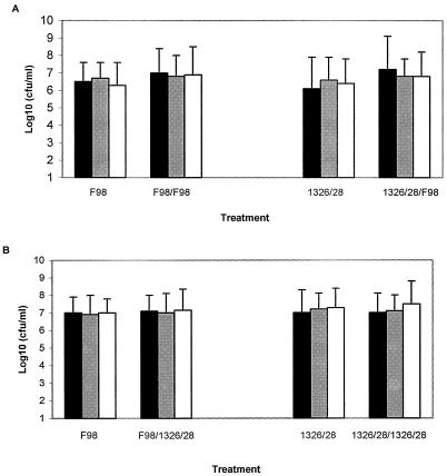
We examined the number of Salmonella cells in isolated neutrophils (48 h postinfection) and the effect of rechallenge with a second strain (ex vivo) on survival of the first strain (the strain with which pigs were infected). Rechallenge with a second strain (MOI, 10) had no effect on the survival of the first strain phagocytosed by neutrophils (Fig. 1). Also, the number of Salmonella cells present in isolated neutrophils did not change significantly (P < 0.05) over the culture period (Fig. 1). Therefore, our results indicate that infected neutrophils isolated from infected porcine blood carried the maximal number of Salmonella cells and that this level could not be altered (increased or decreased) by challenge with a second strain (ex vivo) regardless of the order in which the test strains were used.
FIG. 1.
Survival of Salmonella in neutrophils removed from F98- or 1326/28Φr-inoculated pigs following in vitro culture for 24 h or following in vitro reinfection with F98 (A) or 1326/28Φr (B) (MOI, 10). Solid bars, 2 h postinfection; shaded bars, 7 h postinfection; hollow bars, 24 h postinfection. Each experiment was replicated three times on six separate occasions (18 pigs per group).
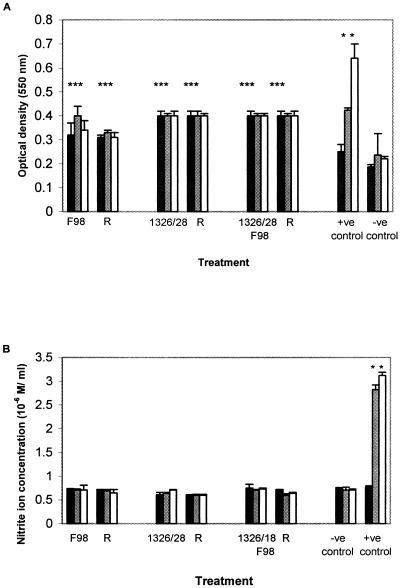
Neutrophils isolated from blood taken from all infected groups following rechallenge with F98 or 1326/28Φr were assessed for production of ROS and nitrite ions. Significant increases (P < 0.05) in the ROS level were observed for neutrophils isolated from all test strains when they were compared with neutrophils isolated from control (uninfected) pigs. However, the elevation of the neutrophil ROS level by test strains did not significantly change (P > 0.05) with time in culture (2 to 7h). The ROS level was also not increased by ex vivo reinfection of neutrophils (which had been isolated from infected pigs) with virulent F98, nor was there a significant difference (P > 0.05) between the three test groups (Fig. 2A). However, when neutrophils removed from uninfected control pigs were cocultured with zymosan, there was a time-dependent increase in the level of ROS, and 7 h postinfection the ROS production was significantly greater than the ROS production in neutrophils isolated from any of the test strains (Fig. 2A).
FIG. 2.
Induction of ROS (A) and nitrite ions (B) in cultured neutrophils removed from F98- or 1326/28Φr-inoculated pigs or following in vitro rechallenge (R) with F98 (MOI, 10). Neutrophils taken from pigs were also protected by 1326/28Φr inoculation prior to challenge with F98 (bars labeled with both 1326/28 and F98); these neutrophils were also reinfected (R) or not reinfected. +ve, neutrophils isolated from uninfected pigs cocultured with 1 μg/ml zymosan (A) or 1 μg/ml phorbol myristate acetate (B); −ve, neutrophils removed from uninfected pigs. Solid bars, 2 h postinfection; shaded bars, 7 h postinfection; hollow bars, 24 h postinfection. Each experiment was replicated three times on six separate occasions (18 pigs per group). An asterisk indicates a significant increase (P < 0.05) compared with uninfected control levels.
The nitrite ion concentrations in supernatants obtained from cultured neutrophils were not significantly different (P > 0.05) for test strains and uninfected controls (Fig. 2B), and although phorbol myristate acetate significantly increased the supernatant nitrite ion concentration (Fig. 2A), the concentration was considered negligible (less than 3.5 μM/ml). No further studies to assess inducible nitric oxide synthase expression or nitrite ion production were performed.
ROS and gp91 protein were detected ex vivo in neutrophils isolated from protected and unprotected pigs.
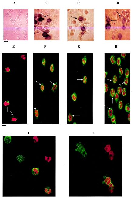
Microscopic examination was used to further analyze ROS induction in infected neutrophils. Neutrophils isolated from uninfected (control) pigs did not reduce NBT to formazan (a dark blue pigment) (Fig. 3A) when they were tested for ROS, and immunofluorescence showed that gp91 phox expression was detected only in the cell membrane of control neutrophils (Fig. 3D). However, neutrophils isolated from the control pigs were able to generate ROS, since removal and stimulation with zymosan or Salmonella caused a positive NBT reaction (data not shown). In contrast, neutrophils isolated from F98-challenged pigs (unprotected) or from pigs protected by 1326/28Φr inoculation prior to F98 challenge showed positive NBT reduction without further stimulation, gp91 phox immunofluorescence was localized to the cell membrane, and some immunofluorescence was observed in the cytoplasm (Fig. 3E and F). gp91 immunoreactivity was also detected in neutrophils migrating into the villi in both protected (Fig. 3H) and unprotected (Fig. 3I) pigs but was not observed in uninfected (control) pigs (Fig. 3G).
FIG.3.
Expression of ROS and gp91 phox immunoreactivity in porcine neutrophils. (A to D) NBT reduction due to ROS activity. The arrows indicate formazan-positive neutrophils removed from 1326/28Φr-inoculated pigs (B), F98-inoculated pigs (C), and 1326/28Φr-protected pigs after F98 challenge (D). Formazan was not observed in NBT reactions used to examine blood neutrophils taken from uninfected pigs (A) unless they were challenged with bacteria in vitro (not shown). (E to H) Confocal microscope images showing gp91 (green) immunofluorescence associated with the cell membrane (arrows) in neutrophils isolated from uninfected protected pigs (E). In neutrophils isolated from 1326/28Φr-inoculated pigs (F), F98-inoculated pigs (G), and 1326/28Φr-protected pigs following F98 challenge (H), gp91 immunofluorescence appears to be increased throughout the cytoplasm (arrows). (I to J) Overlaid images of gp91 (green) and propidium iodide nuclear stain (red) activities in neutrophils removed from 1326/28Φr-protected pigs following F98 challenge (I) or neutrophils isolated from unprotected F98-infected pigs (J). Scale bars = 10 μm.
Comparable levels of expression of gp91 phox mRNA were observed in porcine neutrophils isolated from protected and unprotected pigs.
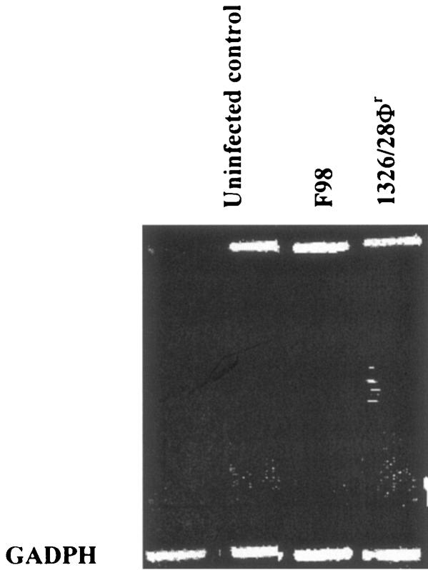
The expression of gp91 mRNA in porcine polymorphonuclear leukocytes removed from the blood of test groups was also measured by RT-PCR. Although we did observe some individual variation within experimental groups, we consistently measured elevated levels of gp91 mRNA in neutrophils isolated from pigs infected with F98 or 1326/28Φr or protected pigs (Fig. 4).
FIG. 4.
RT-PCR showing gp91 mRNA expression in porcine neutrophils. For each experiment gp91 phox mRNA expression is shown for neutrophils from one pig, although comparable results were obtained for two different pigs per group on three separate occasions. GADPH, glyceraldehyde-3-phosphate dehydrogenase.
Induction of ROS pathways in porcine neutrophils bacteriostatically affect Salmonella growth within the first 7 h postinfection.
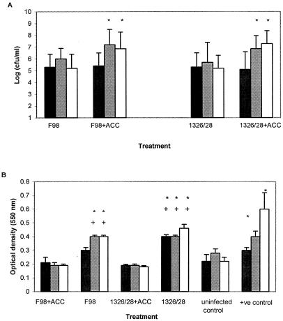
Induction of ROS and the Salmonella load in neutrophils isolated from all test groups were equivalent and did not change following a further 7-h ex vivo culture period. Therefore, we needed to ascertain the effect of ROS on the survival dynamics of Salmonella in porcine neutrophils over the first 7 h postinfection. Neutrophils isolated from uninfected control pigs were infected with either F98 or 1326/28Φr (MOI, 10), and parallel infections were also cocultured with ACC (an ROS inhibitor). The number of Salmonella cells (F98 or 1326/28Φr) isolated 2 to 7 h postinfection did not change significantly (P > 0.05), and there was no significant difference between the number of cells of F98 (virulent Salmonella serovar Typhimurium) and the number of cells of 1326/28Φr (avirulent Salmonella serovar Infantis) isolated from neutrophils (Fig. 5A). However, the numbers of either F98 or 1326/28Φr cells recovered from neutrophils increased significantly (P < 0.05) over the postinfection period following coculture of infected cells with ACC (Fig. 5A). This increase in Salmonella recovery due to coculture with ACC was associated with a significant reduction in ROS production by infected cells (Fig. 5B).
FIG. 5.
Effect of ACC on Salmonella survival (A) and ROS production (B) in porcine neutrophils. Neutrophils were isolated from uninfected pigs and cultured in vitro with F98 or 1326/28Φr for 24 h with or without ACC. The positive control was 1 μg/ml zymosan. Each bar represents the mean for at least three piglets measured on six separate occasions. Solid bars, 2 h postinfection; shaded bars, 7 h postinfection (production of ROS); hollow bars, 24 h postinfection. In panel A, an asterisk indicates a significant increase (P < 0.05) in the number of Salmonella cells following coculture of neutrophils with ACC. In panel B, an asterisk indicates a significant increase (P < 0.05) in the ROS level compared with the levels in uninfected (control) neutrophils, and a plus sign indicates a significant decrease (P < 0.05) in ROS production in Salmonella-infected neutrophils cocultured with ACC compared with the production in Salmonella-infected neutrophils without ACC at the same time.
Histopathological examination of porcine intestine.
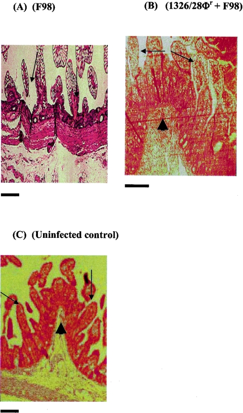
As previously observed (16), hematoxylin and eosin staining of fixed porcine ileum showed that F98 challenge caused stunting and flattening of intestinal plicae. In some cases the lesions were so severe that they caused an apparent loss of the plicae and exfoliation of the villi (Fig. 6A). In contrast, intestines removed from pigs inoculated with 1326/28Φr prior to challenge with F98 (protected animals) showed no pathological lesions (Fig. 6B), and upon examination the villi were similar to villi from uninfected (control) pigs (Fig. 6C).
FIG. 6.
Hematoxylin- and eosin-stained ileal sections isolated from F98-inoculated piglets (A), piglets inoculated with 1326/28Φr and then with F98 24 h later (B), and uninfected (control) piglets (C). The arrows indicate long slender villi in panels B and C and stunted villi in panel A. The arrowheads indicate well-formed pilcae in panels B and C. Scale bars = 10 μm.
To score the pathological lesions observed following histopathological examination of porcine intestines (Fig. 6), a pathological index was used. Scores of 3 (stunting and flattening of intestinal plicae with exfoliation of villi) were consistent with unprotected F98 challenge, while scores of 0 were consistent with 1326/28Φr inoculation with or without subsequent F98 challenge.
DISCUSSION
Previously, we showed that inoculation of 5-day-old gnotobiotic piglets with Salmonella serovar Typhimurium F98 results in severe diarrhea and death and is associated with significant intestinal lesions. However, amelioration of intestinal lesions and clinical symptoms could be achieved by preinoculation of the piglets with an avirulent rough mutant of Salmonella serovar Infantis 1326/28 (1326/28Φr) (16). In the previous study we also showed that neutrophil infiltration was comparable in the intestines of piglets inoculated with F98 and in the intestines of piglets preinoculated with 1326/28Φ and subsequently challenged with F98, thus suggesting that neutrophils may be responsible for protection but that infiltration by neutrophils is not associated with pathology. In this study we examined the role of neutrophils during F98 infection and during protection by 1326/28Φr to ascertain whether neutrophils killed Salmonella and/or whether differential nitrosative or oxidative responses in neutrophils caused intestinal pathology or were responsible for disease progression.
In accordance with our previous studies, piglets which were protected by preinoculation with 1326/28Φr did not show the severe clinical symptoms associated with F98 infection following subsequent F98 inoculation. The pathological index scores, determined following immunohistochemical examination of intestines, were 0 for groups of piglets inoculated with 1326/28Φr alone and for groups of piglets inoculated with 1326/28Φr followed by F98. In contrast, unprotected piglets infected with F98 had severe clinical symptoms and required euthanasia, and the pathological index scores determined for this group were consistently 3 or 4. These results are similar to previously observed results which showed that a rough mutant of Salmonella serovar Typhimurium strain SF1591 can protect pigs against lethal challenge with Salmonella serovar Typhimurium strain LT2 (15). This phenomenon has been explained by a shift in the type of immune response induced by inoculation with a rough mutant from Th1 (which causes inflammatory pathology) to Th2 (40). However, these studies differ from ours in a number of respects since piglets were challenged with virulent strain LT2 8 days after the protective inoculation, whereas the effect that we report here occurred within 48 h after challenge. Although numerous studies have shown that enterocyte pathology is associated with Th1 cytokines, such as gamma interferon and tumor necrosis factor alpha (TNF-α) (1, 17, 21, 36), insignificant numbers of lymphocytes are found in the intestines of gnotobiotic piglets (29, 42). This may indicate that within the first week after birth (the duration of our study) the production of inflammatory cytokines is not sufficient to induce the severe pathology that we observed in F98-infected piglets. Furthermore, neutrophil infiltration in the intestine was not measured in the previous studies and is unlikely to be prominent 8 days after challenge with the first strain.
The numbers of Salmonella cells contained in neutrophils isolated from blood were comparable whether the piglets were infected with virulent strain F98 or with avirulent strain 1326/28Φr and were not altered following rechallenge with a second strain in vitro. This indicates that preinoculation of piglets with 1326/28Φr induces maximal phagocytic activity. We then examined the output of reactive species by infected neutrophils and found that the nitrite ion production was not greater than control levels in piglets infected with a virulent (F98) or avirulent (1326/28Φr) strain of Salmonella, which is consistent with a previous study which indicated that nitrite ion pathways may not be an important factor in the biology of porcine myeloid cells (30). However, the levels of ROS activity were shown to be significantly greater than the control levels in neutrophils isolated from piglets infected with 1326/28Φr and/or F98, but the level of ROS induction could not be further increased by in vitro rechallenge. However, Salmonella did not induce maximal levels of ROS since zymosan (used as a positive control) increased ROS levels in neutrophils above the levels recorded for any of the infection groups studied. The level of gp91 phox mRNA was elevated in all test groups, and this result was in accordance with our confocal microscopy studies which showed that cytoplasmic gp91 phox activity was induced by F98 infection and also following 1326/28Φr protection. However, further confocal analyses showed that there was cytoplasmic gp91 activity in neutrophils which were isolated from inoculated animals but were not themselves associated with Salmonella. This indicates that ROS was probably induced (in at least some neutrophil populations) by LPS or cytokines in the circulation as a result of preinoculation with 1326/28Φr.
Stimulation of human neutrophil ROS has previously been shown to occur in response to LPS (27, 43), particularly rough forms (33), and the stimulatory effect of gamma interferon on neutrophil gp91-induced ROS has been widely studied with respect to chronic granulomatous disease (12, 32, 34). TNF-α production in response to Staphylococcus aureus also stimulates neutrophil ROS, although the ROS pathway in the latter case appears to involve myeloperoxidase rather than NADPH phox (24). TNF-α has also been shown to prime ROS in human and porcine (but not rabbit, rat, or mouse) neutrophils for subsequent stimulation by N-formylmethionyl-leucyl-phenylalanine (45), further demonstrating the relevance of the porcine model for human neutrophil studies. gp91-active neutrophils were present in the villus tips following F98 infection, but the neutrophils were associated with very large numbers of Salmonella cells in comparison with gp91-active neutrophils in the villus tips of 1326/28Φr-protected piglets. We therefore hypothesize that inoculation of piglets with 1326/28Φr stimulates neutrophil migration into the intestine, probably followed by stimulation of the NADPH-ROS pathway initially; however, subsequent blood neutrophils are probably stimulated by other factors (cytokines or LPS) prior to migration into the intestine. Inoculation of gnotobiotic piglets with 1326/28Φr, therefore, causes neutrophils to migrate into the intestine (possibly in an active form), which protects against a subsequent intestinal challenge with a normally lethal F98 inoculum.
We then examined the effect of inhibiting ROS during the first 7 h after infection of neutrophils with Salmonella in vitro. Our results show that when the cells were cultured with ACC, the levels of ROS were significantly reduced in F98- or 1326/28Φr-infected neutrophils, and this coincided with a significant increase in survival of both Salmonella strains in porcine neutrophils. However, in the absence of ACC, the numbers of Salmonella cells did not significantly change from 2 to 7 h postinfection in vitro or, as discussed above, in culture for 7 h after removal from experimentally infected animals. Taken together, these results indicate that ROS act bacteriostatically (rather than bacteriocidally) on virulent strain F98 or avirulent strain 1326/28Φr in porcine neutrophils and that neither neutrophil-derived ROS nor inducible nitric oxide synthase induces the pathology associated with F98 infection.
However, neutrophils produce other substances that may cause host pathology. In the mouse model of colitis (CD4+/SCID adoptive transfer) neutrophil elastase is associated with disease pathology (38), while in vitro studies have shown that transmigration of neutrophils across intestinal epithelial monolayers causes epithelial cell destruction which can been reversed by the addition of elastase inhibitors (18). It has also been proposed that measurement of fecal neutrophil elastase may be used to monitor the activity of Crohn's disease or ulcerative colitis (35). Therefore, differential production of elastase (or other neutrophil-derived proteases) may occur following inoculation of pigs with F98 or 1326/28Φr, and this requires further investigation. Our observation that neutrophil influx into the intestine does not necessarily cause pathology is supported by the results of previous studies. Wallis et al. (44) showed that this was the case in rabbit intestinal loops infected with Salmonella serovar Typhimurium, while neutrophil influx into the lung is the basis for protection of the respiratory tract following treatment with RU41740 (Biostim) (10, 26).
Our results indicate that activation of neutrophil ROS (via NADPH phox) by avirulent Salmonella serovar Infantis strain 1326/28Φr may protect gnotobiotic piglets against lethal challenge with Salmonella serovar Typhimurium strain F98. Furthermore, they indicate that neutrophil nitrite ion production is not important in protection by 1326/28Φr and that neither nitrite ion production, ROS production, nor intestinal influx by neutrophils per se induces the pathological lesions associated with nonprotected F98 challenge. We hope to elucidate the cause of pathology and pathological amelioration in future studies.
Acknowledgments
We thank Elaine Bennett for assistance with cell culture and Helen Mathews for histological preparations.
This work was funded by European Union grant Fair 98-4006 and by the Department of Food, the Environment and Rural Affairs, United Kingdom.
Editor: F. C. Fang
REFERENCES
- 1.Adams, R. B., S. M. Planchon, and J. K. Roche. 1993. IFN-gamma modulation of epithelial barrier function. Time course, reversibility, and site of cytokine binding. J. Immunol. 150:2356-2363. [PubMed] [Google Scholar]
- 2.Albina, J. E. 1995. On the expression of nitric oxide synthase by human macrophages. Why no NO? J. Leukoc Biol. 58:643-649. [DOI] [PubMed] [Google Scholar]
- 3.Anderson, G. J., W. T. Roswit, M. J. Holtzman, J. C. Hogg, and S. F. van Eeden. 2001. Effect of mechanical deformation of neutrophils on their CD18/ICAM-1 dependant adhesion. J. Appl. Physiol. 91:1084-1090. [DOI] [PubMed] [Google Scholar]
- 4.Babior, B. M. 1999. NADPH oxidase: an update. Blood 93:1464-1476. [PubMed] [Google Scholar]
- 5.Barrow, P. A., J. F. Tucker, and J. M. Simpson. 1987. Inhibition of colonisation of the chicken alimentary tract with Salmonella typhimurium by Gram-negative facultatively anaerobic bacteria. Epidemiol. Infect. 98:311-322. [DOI] [PMC free article] [PubMed] [Google Scholar]
- 6.Barrow, P. A., M. B. Huggins, and M. A. Lovell. 1994. Host specificity of Salmonella infection in chickens and mice is expressed in vivo primarily at the level of the reticuloendothelial system. Infect. Immun. 62:4602-4610. [DOI] [PMC free article] [PubMed] [Google Scholar]
- 7.Barrow, P. A., J. O. Hassan, and A. Berchieri. 1990. Reduction in faecal excretion of Salmonella typhimurium F98 in chickens vaccinated with live and killed S. typhimurium organisms. Epidemiol. Infect. 104:413-426. [DOI] [PMC free article] [PubMed] [Google Scholar]
- 8.Barrow, P. A., J. M. Simpson, and M. A. Lovell. 1988. Intestinal colonisation in the chicken by food-poisoning Salmonella serotypes; microbiological characteristics associated with faecal excretion. Avian Pathol. 17:571-588. [DOI] [PubMed] [Google Scholar]
- 9.Bogdan, C., M. Rollinghoff, and A. Diefenbach. 2000. Reactive oxygen and reactive nitrogen intermediates in innate and specific immunity. Curr. Opinion. Immunol. 12:64-76. [DOI] [PubMed] [Google Scholar]
- 10.Capsoni, F., F. Minonzio, E. Venegoni, A. M. Ongari, P. M. Meroni, G. Guidi, and C. Zanussi. 1988. In vitro and ex vivo effect of RU41740 on human polymorphonuclear leukocyte function. Int. J. Immunopharmacol. 10:121-133. [DOI] [PubMed] [Google Scholar]
- 11.Clark, R. C., and C. L. Gyles. 1987. Virulence of wild and mutant strains of Salmonella typhimurium in ligated intestinal segments of calves, pigs and rabbits. Am. J. Vet. Res. 48:504-510. [PubMed] [Google Scholar]
- 12.Condino-Neto, A., and P. E. Newburger. 2000. Interferon-gamma improves splicing efficiency of CYBB gene transcripts in an interferon responsive variant of chronic granulomatous disease due to splice site consensus region mutation. Blood 95:3548-3554. [PubMed] [Google Scholar]
- 13.Davis, A. R., P. L. Mascolo., P. L. Bunger, K. M. Sipes, and M. Quinn. 1998. Cloning and sequencing of the bovine flavocytochrome b subunit proteins, gp91-phox and p22-phox: comparison with other known flavocytochrome b sequences. J. Leukoc. Biol. 64:114-123. [DOI] [PubMed] [Google Scholar]
- 14.Denis, M. 1994. Human monocytes/macrophages: NO or no NO? J. Leukoc. Biol. 55:682-684. [DOI] [PubMed] [Google Scholar]
- 15.Dlabac, V., I. Trebichavsky, Z. Rehakova, B. Hofman, I. Splinchal, and B. Cukrowska. 1997. Pathogenicity and protective effect of rough mutants of Salmonella species in germ-free pigs. Infect. Immun. 65:5238-5243. [DOI] [PMC free article] [PubMed] [Google Scholar]
- 16.Foster, N., M. A. Lovell, K. L. Marston, S. D. Hulme, A. J. Frost, P. Bland, and P. A. Barrow. 2003. Rapid protection of gnotobiotic pigs against experimental salmonellosis following induction of polymorphonuclear leucocytes by avirulent Salmonella. Infect. Immun. 71:2182-2191. [DOI] [PMC free article] [PubMed] [Google Scholar]
- 17.Garside, P., C. Bunce, R. C. Tomlinson, B. L. Nichols, and A. M. Mowat. 1993. Analysis of enteropathy induced by tumor necrosis factor alpha. Cytokine 5:24-30. [DOI] [PubMed] [Google Scholar]
- 18.Ginzberg, H. H., V. Cherapanov, Q. Dong, A. Cantin, C. A. McCulloch, P. T. Shannon, and G. P. Downey. 2001. Neutrophil-mediated epithelial injury during transmigration role of elastase. Am. J. Physiol. Gastrointest. Liver Physiol. 281:G705-G717. [DOI] [PubMed] [Google Scholar]
- 19.Glynn, M. K., C. Bopp, W. Dewitt, P. Dabney, M. Mokhtar, and F. J. Angulo. 1992. Emergence of multidrug resistance Salmonella enterica serotype Typhimurium DT104 infections in the United states. N. Engl. J. Med. 338:1331-1338. [DOI] [PubMed] [Google Scholar]
- 20.Gomez, G. C. 1997. The colostrum-deprived, artificially reared, neonatal pig as a model animal for studying rotavirus gastroenteritis. Front. Biosci. 2:471-481. [DOI] [PubMed] [Google Scholar]
- 21.Guy-Grand, D., J. P. DiSanto, P. Hencoz, M. Malassis-seris, and P. Vassalli. 1998. Small bowel enteropathy: role of intraepithelial lymphocytes and of cytokines (IL-12, IFN-γ, TNF-α) in the induction of epithelial cell death and renewal. Eur. J. Immunol. 28:730-744. [DOI] [PubMed] [Google Scholar]
- 22.Huang, A., C. Li, R. L. Kao, and W. L. Stone. 1999. Lipid hydroperoxide inhibits nitric oxide production in RAW 264.7 macrophages. Free Radic. Biol. Med. 26:526-537. [DOI] [PubMed] [Google Scholar]
- 23.Kobayashi, T., and H. Seguchi. 1999. Novel insight into current models of NADPH oxidase regulation, assembly and localization in human polymorphonuclear leukocytes. Histol. Histopathol. 14:1295-1308. [DOI] [PubMed] [Google Scholar]
- 24.Kowanko, I. C., A. Ferrante, G. Clemente, and L. K. Kumaratilake. 1996. Tumor necrosis factor primes neutrophils to kill Staphylococcus aureus by an oxygen-dependent mechanism and Plasmodium falciparum by an oxygen-independent mechanism. Infect. Immun. 64:3435-3437. [DOI] [PMC free article] [PubMed] [Google Scholar]
- 25.Laval, A., H. Morvan, G. Desprez, and B. Corbion. 1992. Salmonellosis in swine, p. 164-175. In Proceedings of the International Symposium on Salmonella and Salmonellosis, Ploufragan, France. World Veterinary Poultry Association, Zoopole, St. Brieuc, France.
- 26.Minonzio, F., A. M. Ongar, R. Palmieri, D. Bochicchio, G. Guidi, and F. Capsoni. 1991. Immunostimulation of neutrophil phagocytic function by RU41740 (Biostim) in elderly subjects. Allergol. Immunopathol. 19:58-62. [PubMed] [Google Scholar]
- 27.Nielsen, H., S. Birkholz, L. P. Andersen, and A. P. Moran. 1994. Neutrophil activation by Helicobacter pylori lipopolysaccharide. J. Infect. Dis. 170:135-139. [DOI] [PubMed] [Google Scholar]
- 28.Otto, M., B. Wahn, and C. J. Kirkpatrick. 2003. Modification of human polymorphonuclear neutrophilic cell (PMN)—adhesion on biomaterial surfaces by protein preadsorption under static and flow conditions. J. Mater. Sci. Mater. Med. 14:263-270. [DOI] [PubMed] [Google Scholar]
- 29.Pabst, R., and H. J. Rothkotter. 1999. Postnatal development of lymphiocyte subsets in different compartments of the small intestine of piglets. Vet. Immunol. Immunopathol. 72:167-173. [DOI] [PubMed] [Google Scholar]
- 30.Pampusch, M. S., A. M. Bennaars, S. Harsch, and M. P. Murtaugh. 1998. Inducible nitric oxide synthase expression in porcine immune cells. Vet. Immunol. Immunopathol. 61:279-289. [DOI] [PubMed] [Google Scholar]
- 31.Park, B. H., S. M. Fikrig, and E. M. Smithwick. 1968. Infection and nitroblue-tetrazolium reduction by neutrophils. A diagnostic acid. Lancet ii:532-534. [DOI] [PubMed] [Google Scholar]
- 32.Rae, J., P. E. Newburger, M. C. Dinauern, D. Noack, P. J. Hopkins, R. Kuruto, and J. T. Curnutte. 1998. X-linked chronic granulomatous disease: mutations in the CYBB gene encoding the gp91-phox component of the respiratory burst oxidase. Am. J. Hum. Genet. 62:1320-1331. [DOI] [PMC free article] [PubMed] [Google Scholar]
- 33.Robinson, J. M., T. Kobayashi, H. Seguchi, and T. Takizawa. 1999. Evaluation of neutrophil structure and function by electron microscopy: cytochemical studies. J. Immunol. Methods 232:169-178. [DOI] [PubMed] [Google Scholar]
- 34.Roos, D., M. de Boer, F. Kuribayashi, C. Meischel, R. Weening, A. Segal, A. Ahlin, K. Nemet, J. Hossle, E. Bernatowska-Matuszkiewicz, and H. Middleton-Price. 1996. Mutations in the X-linked and autosomal recessive forms of chronic granulomatous disease. Blood 87:1663-16681. [PubMed] [Google Scholar]
- 35.Saitoh, O., K. Sugi, R. Matsuse, K. Uchida, H. Matsumoto, K. Nakagawa, K. Takada, M. Yoshizumi, I. Hirata, and K. Katsu. 1995. The forms and levels of feacal PMN-elastase in patients with colorectal diseases. Am. J. Gastroenterol. 90:388-393. [PubMed] [Google Scholar]
- 36.Siegmund, B., G. Fantuzzi, F. Rieder, F. Gambioni-Robertson, H. A. Lehr, G. Hartmann, C. A. Dinarello, S. Endres, and A. Eigler. 2001. Neutralization of interleukin-18 reduces severity in murine colitis and intestinal IFN-γ and TNF-α production. Am. J. Pathol. 281:1264-1273. [DOI] [PubMed] [Google Scholar]
- 37.Smith, H. W., and J. F. Tucker. 1980. The virulence of Salmonella strains for chickens; their excretion by infected chickens. J. Hyg. 84:479-488. [DOI] [PMC free article] [PubMed] [Google Scholar]
- 38.Tarlton, J. F., C. V. Whiting, D. Tunmore, S. Bregenholt, J. Reimann, M. H. Claesson, and P. W. Bland. 2000. The role of up-regulated serine proteases and matrix metalloproteinases in the pathogenesis of a murine model of colitis. Am. J. Pathol. 157:1927-1935. [DOI] [PMC free article] [PubMed] [Google Scholar]
- 39.Tavernor, W. D., P. C. Trexler, L. C. Vaughan, J. E. Cox, and D. G. C. Jones. 1971. The production of gnotobiotic piglets and calves by hysterectomy under general anaesthesia. Vet. Rec. 88:10-14. [DOI] [PubMed] [Google Scholar]
- 40.Trebichavsky, I., V. Dlabac, Z. Rehakova, M. Zahradnickova, and I. Spinchal. 1997. Cellular changes and cytokine expression in the ilea of gnotobiotic piglets resulting from peroral Salmonella typhimurium challenge. Infect. Immun. 65:5244-5249. [DOI] [PMC free article] [PubMed] [Google Scholar]
- 41.Tzipori, S., J. Montanaro, R. M. Robins-Browne, P. Vial, R. Gibson, and M. M. Levine. 1992. Studies with enteroaggregative Escherichia coli in the gnotobiotic piglet gastroenteritis model. Infect. Immun. 60:5302-5306. [DOI] [PMC free article] [PubMed] [Google Scholar]
- 42.Vega-Lopez, M. A., G. Arenas-Contreras, M. Bailey, S. Gonzalez-Pozos, C. R. Stokes, M. G. Ortega, and R. Mondragob-Flores. 2001. Development of intraepithelial cells in the porcine small intestine. Dev. Immunol. 8:147-158. [DOI] [PMC free article] [PubMed] [Google Scholar]
- 43.Vosbeck, K., P. Tobias, H. Mueller, R. A. Allen, K. E. Arfors, R. J. Ulevitch, and L. A. Sklar. 1990. Priming of polymorphonuclear granulocytes by lipopolysaccharides and its complexes with lipopolysaccharide binding protein and high density lipoprotein. J. Leukoc. Biol. 47:97-104. [DOI] [PubMed] [Google Scholar]
- 44.Wallis, T. S., R. J. H. Hawker, D. C. A. Candy, G. M. Qi, G. J. Clarke, K. J. Worton, M. P. Osborne, and J. Stephen. 1989. Quantification of the leucocyte influx into rabbit ileal loops induced by strains of Salmonella typhimurium of different virulence. J. Med. Microbiol. 30:149-156. [DOI] [PubMed] [Google Scholar]
- 45.Yaffe, M. B., J. Xu, P. A. Burke, and G. E. Brown. 1999. Priming of the neutrophil respiratory burst is species-dependent and involves MAP kinase activation. Surgery 126:248-254. [PubMed] [Google Scholar]


