

ABSTRACT
Background
In small animals such as cats, fundus imaging plays a critical role in early diagnosis of eye diseases and monitoring of treatment processes. Traditional methods usually require dilation of the pupil, which in some cases may require anaesthesia or cause discomfort for the animals. In recent years, non‐mydriatic fundus cameras, especially with the development of handheld models, offer a new alternative that simplifies this process. These cameras allow high‐quality fundus images to be obtained without the need for pupil dilation. This helps veterinarians save time and resources during examinations, while also providing a less stressful experience for the animals.
Objectives
Evaluation of the use and image quality of the new non‐mydriatic handheld fundus camera (Aurora, Optomed, Oulu, Finland) for fundus imaging in cats.
Methods
Fundus photographs of 208 cats were obtained using a handheld, non‐mydriatic fundus camera. Undilated colour images of each eye centred on the optic disc were taken in a dimly lit room using a non‐mydriatic fundus camera. The image acquisition protocol consisted in a single 50° colour photo centred on the cats optic disc. Images were evaluated for normal and pathological fundus. In addition, the obtained fundus images were evaluated in terms of centration, clarity and visible range parameters, with the quality score of each parameter varying between 2 (excellent) and 0 (ungradable).
Results
Although 204 of 208 cats had a normal fundus, four cats had retinal pathology. The optic disc was defined as well‐centred in 412 of 416 eyes, partially centred in 3 eyes and not centred in 1 eye. Fundus vessels were clearly visible in 409 eyes, recognisable in 6 eyes and not recognisable in 1 eye. The entire image was visible in 413 eyes; more than 80% of the image was visible in 2 eyes, and less than 80% of the image was visible in 1 eye. The mean image quality scores for image centration, clarity and visible range were 1.99, 1.98 and 1.99, respectively, in 416 eyes.
Conclusions
The evaluated fundus camera appears to be suitable for use in fundus imaging in non‐dilated eyes of cats, and the quality of the images obtained is significantly satisfactory.
Keywords: cat, fundus imaging, handheld fundus camera, non‐mydriatic, Optomed Aurora
A non‐mydriatic handheld fundus camera (Aurora, Optomed, Oulu, Finland) was evaluated for feline fundus imaging, providing high‐quality images without pupil dilation. The device demonstrated excellent centration, clarity and visibility in 416 eyes, making it a practical and less stressful alternative for veterinary ophthalmic examinations
.
1. Introduction
Visualization of the posterior segment of the eye and the information it provides is fundamental to both medical and veterinary medicine (Pirie and Pizzirani 2011). On this basis, the development of digital fundus cameras has facilitated the rapid acquisition and interpretation of fundus images (Ruan et al. 2022). The collection and recording of images is commonly performed in ophthalmology to document lesions, monitor their progression and share information with other colleagues. Recent advances in technology have enabled the development of new cameras integrated into a pocket‐sized computer with numerous other functionalities, which also works as a mobile phone (Espinheira and Ledbetter 2019). Smartphone technologies used in veterinary medicine require mydriatic agents to perform fundus scans (Espinheira and Ledbetter 2019; Balland et al. 2017; Kanemaki et al. 2017; Guyomard et al. 2008; Ocelli and Petersen 2014), which can prolong the examination time and increase the stress factor in animals (Sebbag et al. 2024).
In this study, we describe a new handheld non‐mydriatic Optomed Aurora fundus camera (Aurora, Optomed, Oulu, Finland) for imaging the fundus in cats and evaluate its image quality.
2. Materials and Methods
2.1. Animals
Medical records from 2023 to 2024 were reviewed. Informed consent was obtained from the owners of each animal. A total of 208 cats who underwent routine ophthalmological examinations had no ocular problems that could interfere with fundus imaging, and whose fundus photographs were obtained with the handheld, non‐mydriatic Optomed Aurora and brought to Burdur Mehmet Akif Ersoy University Veterinary Faculty Animal Hospital by their owners between 2023 and 2024 were included in the study. Opthalmic examinations include the dazzle reflex, pupillary light responses, direct ophthalmoscopy (Heine mini 3000F.O, Heine Optotechnik, Gilching, Germany), rebound tonometry (Icare TONOVET Plus, Icare, Finland) and fluorescein staining (Fluorescite 10%, Alcon Laboratories Inc., Texas, USA). Cats that might interfere with adequate fundus imaging (e.g., cataracts and cornea or conditions that interfere with the visual axis) or cats with a history of any ophthalmological disease and poor cooperation were excluded.
2.2. Photographic Equipment
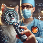
Optomed Aurora is a handheld, non‐mydriatic (50°) digital fundus camera that provides general, ophthalmoscopic, otoscopic and dermatoscopic imaging with a five megapixel image sensor. It weighs 400 g (0.88 pounds), giving the freedom to move around and take the device anywhere (Figure 1). The Optomed Aurora has autofocus, an internal LED light source, is Wi‐Fi enabled and produces an image resolution of 2560 × 1920 pixels, which is compressed to 1280 × 960 pixels after transmission (Balland et al. 2017). Non‐mydriatic handheld Optomed Aurora includes a camera, retinal module, anterior module, charging station, USB cable, power booster, eye cups, batteries and USB flash drive. The anterior module is used to visualize the surface of the eye. The anterior module can be used by attaching directly to the camera. Retinal module is used to visualize the fundus. The retinal module part is attached to the camera, and after attaching to the camera, it can be used by attaching an eye cup to the retinal module. After all these modules are attached, it is ready to be used after the battery is attached. The device is switched on by pressing the power button. You can switch between the menus with the button on it. Menu section includes fixation target, brightness, focus mode, patient and setting sections. Patient information can be saved in the patient section. In the fixation target section, the targeted peripheral or central area can be selected, and the image can be taken. Brightness can be adjusted manually and automatically. Focus mode can be adjusted manually and automatically. After the photos are obtained, the images can be transferred by connecting to the computer via Wi‐Fi connection or USB cable.
FIGURE 1.

Non‐mydriatic handheld fundus camera (Aurora, Optomed, Oulu, Finland).
2.3. Photographic Application
Undilated colour images of each eye centred on the optic disc were taken in a dimly lit room using a non‐mydriatic fundus camera. The image acquisition protocol consisted of a single 50° colour photograph centred on the cat's optic disc. No mydriatic drug was used, whereas images were obtained. The same person (general practitioner) acquired all of the images. After routine eye examinations were completed, information about the cat and its owner was recorded in the patient section of the Optomed Aurora menu. A photograph of the eye surface was obtained by attaching a front module to the camera for permanent storage and later review. To obtain fundus images, the retinal module was attached to the camera, and the eye cup was attached to the retinal module. Then the fixation target was set by selecting the central area target. The brightness was set automatically. Focus mode was set automatically. An assistant held the cat's head still while at the same time opening the eyelids. All cats were kept in the same position, whereas the images were acquired. The image taker stood in front of the cat and supported the optical disc with one left hand to minimize the movement of the camera while using the right hand to bring the camera closer to the cat's eye. The correct distance of the camera was adjusted with the frame on the screen. At the correct distance, the frames are coloured green. After the frames turn green, the device automatically focuses the eye. After focusing, a green square appears on the screen. An image can be obtained with a single shot (Figure 2).
FIGURE 2.

Fundus images of two different cats were obtained using a non‐mydriatic handheld fundus camera (Aurora, Optomed, Oulu, Finland).
2.4. Fundus Photographic Quality Assessment
All images from Optomed Aurora were transmitted and stored in their original state at Joint Photographic Expert Groups (JPEG) 100 quality (1280 × 960 pixels). All images were then analysed by a single blind operator (an experienced ophthalmologist) using a 17.4‐in. high‐resolution computer screen. At the same time, in Fundus Photographic Quality Assessment, three parameters were evaluated to investigate various aspects of image quality. The quality score for each parameter ranged from 2 (excellent) to 0 (ungradable). Images were considered to be of excellent quality if the optic disc was well centred, showed clear fundus vessels, there was no retinopathy, and the entire image was visible. Images were defined as acceptable if the optic disc was partially centred and fundus vessels and any retinopathy could be recognized and more than 80% of the image was visible. If images were not centred, blurred without recognition of retinal vessels or retinopathy features, or if less than 80% of the image was visible, they were defined as ungradable (Figure 3) (Upadhyaya et al. 2022).
FIGURE 3.
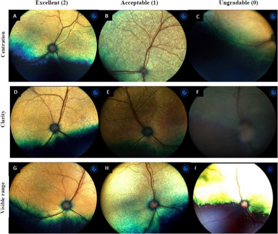
Fundus Photographic Quality Assesment (A–I). (A) Optic disc was well‐centred. (B) Optic disc was partially centred. (C). Optic disc was not centred. (D) Image clear focused. (E) Image recognizable. (F) Image unrecognizable. (G) Visible range is the whole image. (H) Visible range >80%. (I) Visible range <80%.
2.5. Statistical Analysis
Statistical analysis was performed using statistical software (IBM SPSS Statistics 27). Data are presented as median for continuous variables and frequency (%) for categorical variables.
3. Results
3.1. Demographic
Non‐mydriatic fundus photographs centred on the optic disc were obtained from both eyes of 208 cats, for a total of 416 eyes, using a handheld non‐mydriatic fundus camera. The average age of the cats was 2.63 years (0.6 months–3 years), and their average weight was 3.45 kg (1.5–5 kg). Of the cats, 46.2% (n = 96) were female and 53.8% (n = 112) were male.
3.2. Fundus Imaging Findings
The non‐mydriatic handheld Optomed Aurora is easy to use and provides quality images that allow photographic documentation of both the ocular surface and the ocular fundus. In the screening performed by an experienced ophthalmologist, 204 of 208 cats had a normal fundus (Figure 4), whereas pathology was detected in the fundus of 4 cats. These pathologies consisted of retinal degeneration, retinal degeneration due to taurine deficiency, feline central retinal degeneration and retinopathy (Figure 5). Eight of 204 cats with normal fundus had subalbinotic fundus.
FIGURE 4.
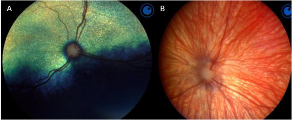
The normal fundus of the cat. (A) Normal fundus of a cat with a green‐yellow tapetum. (B) Subalbinotic fundus.
FIGURE 5.
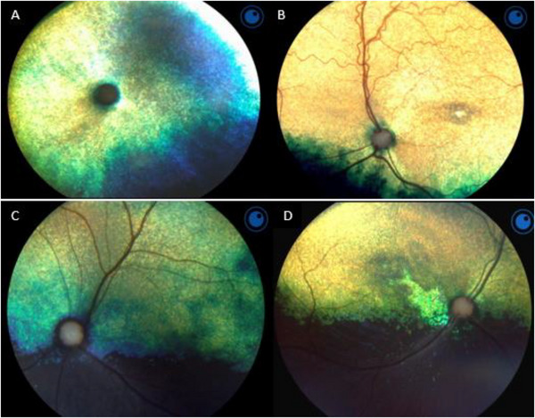
Feline retinal pathologies (A–D). (A) Retinal degeneration. (B) Retinal degeneration due to taurine deficiency. (C) Retinopathy. (D) Feline central retinal degeneration.
Of a total of 416 non‐mydriatic images, 416 (100%) were graded. The optic disc was defined as well centred in 411 of the 416 eyes evaluated, the optic disc was defined as partially centred in 3 eyes, and the optic disc was not centred in 2 eye. Fundus vessels could be clearly seen in 408 eyes, fundus vessels could be recognized in 6 eyes, and fundus vessels were not recognized in 2 eye. The entire image was visible in 412 eyes, more than 80% of the image was visible in 2 eyes, and less than 80% of the image was visible in 2 eye. The mean image quality scores for image centration, clarity and visible range were 1.99, 1.98 and 1.99, respectively, using a non‐mydriatic handheld Optomed Aurora fundus camera in 416 eyes. In 10 of the 208 cats, images of the tapetal region were sometimes overexposed, with the fundus appearing artificially over‐reflective. They varied in size but did not involve the entire tapetal region.
4. Discussion
This study reported that a new non‐mydriatic handheld Optomed Aurora fundus camera was able to visualize the fundus in cats without the use of a mydriatic agent, and the fundus images obtained were of good quality.
Fundus imaging devices such as direct ophthalmoscopy (5°), d‐EYE (20°; Si14), PanOptic i‐Examiner (25°; Welch Allyn), Kowa Genesis‐D (25°–30°; Kowa Company) and Smartscope VET2 (40°; Optimum) provide a specific field of view (Sebbag et al. 2024). The non‐mydriatic handheld Optomed Aurora has a viewing angle of 50° and offers a wider field of view than other fundus imaging cameras.
The fact that the d‐EYE system is compatible with only two smartphone brands may represent a limitation for potential users with different smartphones. The design of the d‐EYE also matches to a specific, fixed distance between the lens and the LED of the smartphone. If this distance changes in the future on newer generation of smartphones, the d‐EYE might be unsuitable (Şengöz Şirin 2020). The fact that some systems produced for fundus imaging are not compatible with all smartphones and have major limitations for the future makes handheld Optomed Aurora fundus imaging device, which do not require any phone for fundus imaging, even more important.
Although the 3D‐printed lens adapter for smartphone provided faster and wider fundus images in cats and dogs, there was a reflection of polymer colour in the collected image. This was mostly attributed to slightly incorrect placement of the 3D‐printed lens adapter on the smartphone flash (Espinheira and Ledbetter 2019). Placing extra equipment on smartphones for fundus imaging can affect image quality. No such problem was encountered with Optomed Aurora as no extra apparatus was needed for fundus imaging.
In indirect ophthalmoscopy with a smartphone, images need to be rotated 180° to show the natural anatomical orientation (Kanemaki et al. 2017; Das et al. 2023). Optomed Aurora provides direct vertical images of the fundus, making it easier to evaluate the findings.
The exposure issue manifests as the presence of light artefacts on the images. Those artefacts might be reduced by dimming the intensity of the light source provided by the smartphone, consequently leading to an underexposure of the nontapetal region (Balland et al. 2017). In the study, overexposure occurred in 10 cats, and it was thought that this may be due to the automatic adjustment of the light source and overexposure of the light. In such cases, it was thought that these problems could be overcome by manually adjusting the light.
A recent study reported that administration of 0.5% tropicamide in cats causes a significant elevation in IOP in both the treated and the untreated eyes of normal cats. This conclusion may reflect systemic absorption of the topically administered drug, or it may be related to other systemic factors, such as increased stress level resulting in elevations in IOP, or systemic blood pressure, or both (Schmidt et al. 2006). The non‐mydriatic handheld Optomed Aurora can visualize the fundus without the use of any mydriatic agents, thus eliminating the problems that may arise due to the use of topical mydriatic agents.
When the fundus is imaged, a mydriatic agent should be used to facilitate visualization of the peripheral retina. In a busy clinical practice, veterinarians are likely to spend extra time dilating the pupils before using the fundus camera for scanning (Sebbag et al. 2024). In cats, maximum pupillary dilation is achieved 30–60 min after topical atropine use. In cats, horizontal pupil diameter begins to decrease 24 h after topical atropine application and the pupil returns to its original diameter 96 h after application (Kovalcuka et al. 2015). The use of mydriatic agents in cats can prolong a veterinarian's examination time, and it is anticipated that this can be reduced to a very short time with the non‐mydriatic handheld Optomed Aurora, which does not require mydriatic agents. Fundus examination with the non‐mydriatic handheld Optomed Aurora can be performed in about 30 s with the help of an assistant. This increases both cat and physician comfort. At the same time, as the effect of the mydriatic agent will last for a long time, the discomfort of cats may continue for long periods of time. The use of the non‐mydriatic handheld Optomed Aurora can prevent such situations.
Phone‐based imaging systems used in veterinary medicine to visualize the fundus require a mydriatic agent prior to the examination (Espinheira and Ledbetter 2019; Balland et al. 2017; Kanemaki et al. 2017). Compared to conventional cameras, the Optomed Aurora has a smaller minimum pupil size requirement. The minimum pupil size required for image acquisition in humans was determined as Optomed Aurora (≥3.1 mm), Zeiss Visucam 200 (≥4.0 mm), Topcon TRC‐50DX (≥4.0 mm), Canon CR‐2 (≥3.3 mm) and Newvision Reticam 3100 (≥3.3 mm) (Upadhyaya et al. 2022). The minimum pupil diameter required to obtain images of non‐mydriatic handheld Optomed Aurora in cats is unknown. However, considering the pupil sizes of cats and humans, it is thought that images can be obtained with smaller pupil diameters than humans.
In a study using a non‐mydriatic fundus camera to evaluate the optic disc for glaucoma in humans, the mean scores for non‐mydriatic image quality in terms of image centration, sharpness and visible range for the handheld fundus camera were 1.47, 1.48 and 1.40 (Upadhyaya et al. 2022). Image quality averages in cats were better than those obtained in humans. Mean scores of 1.99, 1.98 and 1.99 were obtained for centring, clarity and visible range, respectively. While obtaining these averages, images were evaluated by a single person, and these data may change according to the experience of the person. Fundus images were obtained by holding the cats still by an assistant and waiting for the most still moment, and the aim here was to eliminate image quality deterioration due to movement. In addition, in order to minimize overexposure in the image caused by the tapetal reflex, care was taken to ensure that the frame and frame on the camera were green, that is, at sufficient distance and fundus focus.
Limitations of this study include that fundus photographs were evaluated by a single person, and this may not be valid for other evaluators due to differences in experience.
5. Conclusion
The study showed that the non‐mydriatic handheld Optomed Aurora fundus camera was able to visualize the fundus in cats without the use of mydriatic agents and although there were problems such as overexposure and unrecognizability of the image in some images, a high‐quality image could be obtained. The use of Optomed Aurora in non‐dilated eyes was important to prevent cats from being kept in the clinic for a long time and causing more stress and to enable veterinarians to use their time more efficiently.
Author Contributions
Özlem ŞENGÖZ ŞİRİN: study design, data interpretation, data management, preparation of manuscript. Mehmet Nur ÇETİN: study design, data interpretation, preparation of manuscript. Muhammed Yusuf ŞİRİN: data management, statistical analysis, preparation of manuscript.
Ethics Statement
The authors have nothing to report.
Conflicts of Interest
The authors declare no conflicts of interest.
Peer Review
The peer review history for this article is available at https://www.webofscience.com/api/gateway/wos/peer‐review/10.1002/vms3.70348.
Acknowledgements
The authors have nothing to report.
Funding: This study was supported by Türkiye Bilimsel ve Teknolojik Araştırma Kurumu.
Data Availability Statement
The datasets used and/or analysed in the current study are available from the corresponding author upon reasonable request.
References
- Balland, O. , Russo A., Isard P. F., Mathieson I., Semeraro F., and Dulaurent T.. 2017. “Assessment of a Smartphone‐Based Camera for Fundus Imaging in Animals.” Veterinary Ophthalmology 20, no. 1: 89–94. 10.1111/vop.12350. [DOI] [PubMed] [Google Scholar]
- Das, S. , Kuht H. J., De Silva I., et al. 2023. “Feasibility and Clinical Utility of Handheld Fundus Cameras for Retinal Imaging.” Eye (London, England) 37, no. 2: 274–279. 10.1038/s41433-021-01926-y. [DOI] [PMC free article] [PubMed] [Google Scholar]
- Espinheira, G. F. , and Ledbetter E.. 2019. “Canine and Feline Fundus Photography and Videography Using a Nonpatented 3D Printed Lens Adapter for a Smartphone.” Veterinary Ophthalmology 22, no. 1: 88–92. 10.1111/vop.12577. [DOI] [PubMed] [Google Scholar]
- Guyomard, J. L. , Rosolen S. G., Paques M., et al. 2008. “A Low‐Cost and Simple Imaging Technique of the Anterior and Posterior Segments: Eye Fundus, Ciliary Bodies, Iridocorneal Angle.” Investigative Ophthalmology & Visual Science 49, no. 11: 5168–5174. 10.1167/iovs.07-1340. [DOI] [PubMed] [Google Scholar]
- Kanemaki, N. , Inaniwa M., Terakado K., Kawarai S., and Ichikawa Y.. 2017. “Fundus Photography With a Smartphone in Indirect Ophthalmoscopy in Dogs and Cats.” Veterinary Ophthalmology 20, no. 3: 280–284. 10.1111/vop.12399. [DOI] [PubMed] [Google Scholar]
- Kovalcuka, L. , Birgele E., Bandere D., and Williams D. L.. 2015. “Comparison of the Effects of Topical and Systemic Atropine Sulfate on Intraocular Pressure and Pupil Diameter in the Normal Canine Eye.” Veterinary Ophthalmology 18, no. 1: 43–49. 10.1111/vop.12138. [DOI] [PubMed] [Google Scholar]
- Occelli, L. M. , and Petersen‐Jones S. M.. 2014. “Altered Fundus Appearance Resulting From Autofluorescence Imaging With the Confocal Scanning Laser Ophthalmoscope (cSLO) in Cats.” Veterinary Ophthalmology 17, no. 5: 385–388. 10.1111/vop.12192. [DOI] [PubMed] [Google Scholar]
- Pirie, C. G. , and Pizzirani S.. 2011. “Reflex‐Free Digital Fundus Photography Using a Simple and Portable Camera Adaptor System. A Viable Alternative.” Journal of Visual Communication in Medicine 34, no. 4: 146–155. 10.3109/17453054.2011.635293. [DOI] [PubMed] [Google Scholar]
- Ruan, S. , Liu Y., Hu W. T., et al. 2022. “A New Handheld Fundus Camera Combined With Visual Artificial Intelligence Facilitates Diabetic Retinopathy Screening.” International Journal of Ophthalmology 15, no. 4: 620–627. 10.18240/ijo.2022.04.16. [DOI] [PMC free article] [PubMed] [Google Scholar]
- Schmidt, K. S. , Hacker D. V., Kass P. H., and Barkhoodarian A. L.. 2006. “Effects of Systemic Administration of 0.5% Tropicamide on Intraocular Pressure, Pupillary Diameter, Blood Pressure, and Heart Rate in Normal Cats.” Veterinary Ophthalmology 9, no. 2: 137–139. 10.1111/j.1463-5224.2006.00447.x. [DOI] [PubMed] [Google Scholar]
- Sebbag, L. , Ofri R., Arad D., Handel K. W., and Pe'er O.. 2024. “Using a Smartphone‐Based Digital Fundus Camera for Screening of Retinal and Optic Nerve Diseases in Veterinary Medicine: A Preliminary Investigation.” Veterinary Record 194, no. 9: e4088. 10.1002/vetr.4088. [DOI] [PubMed] [Google Scholar]
- Şengöz Şirin, Ö. 2020. “Evaluation of Fundus Examination of Hunting Dogs' Eyes Using a Smartphone‐Based Camera.” Harran Üniversitesi Veteriner Fakültesi Dergisi 9, no. 2: 183–188. 10.31196/huvfd.807349. [DOI] [Google Scholar]
- Upadhyaya, S. , Agarwal A., Rengaraj V., Srinivasan K., Newman Casey P. A., and Schehlein E.. 2022. “Validation of a Portable, Non‐Mydriatic Fundus Camera Compared to Gold Standard Dilated Fundus Examination Using Slit Lamp Biomicroscopy for Assessing the Optic Disc for Glaucoma.” Eye (London, England) 36, no. 2: 441–447. 10.1038/s41433-021-01485-2. [DOI] [PMC free article] [PubMed] [Google Scholar]
Associated Data
This section collects any data citations, data availability statements, or supplementary materials included in this article.
Data Availability Statement
The datasets used and/or analysed in the current study are available from the corresponding author upon reasonable request.