

ABSTRACT
Lingual treatment is becoming more and more common, but it is a field in which not all orthodontists excel. Moreover, patients wearing lingual appliances experience more pain, speech difficulties, and problems maintaining adequate oral hygiene, which may cause them to lose their patience with the technique. As a result, iatrogenic treatment outcomes are sometimes observed. In this case report, the patient's initial chief complaint was spacing. She had been wearing lingual brackets for 1.5 years with minimal improvement. The previous doctor had informed her that the total treatment time would be about 1–1.5 years. However, the patient lost her patience with the treatment and came to us seeking an alternative solution that maintains a similar focus on aesthetics. After discussing potential alternative treatments with the patient, we decided to proceed with in‐house aligners. Initially, the patient opted to distribute the spaces for veneers on the anterior teeth to save time. However, after the first series of aligners, she changed her mind and decided to close all the spaces because she found the aligners effective. After 7 months with three series of aligners, the achieved outcomes included closing all spaces, improving the midlines, achieving a Class I canine relationship, and better intercuspation. The treatment outcomes remained stable at the 1‐year follow‐up. Using in‐house aligners is an effective alternative to lingual brackets in certain cases.
Keywords: alternative, case report, iatrogenic, in‐house aligners, lingual treatment
Summary.
In‐house clear aligners present a highly effective and aesthetically pleasing alternative for patients who are dissatisfied with lingual orthodontic treatment.
While lingual braces offer the advantage of being hidden from view, some patients may find them uncomfortable, difficult to clean, or challenging to adapt to due to their placement on the tongue side of the teeth.
In contrast, in‐house clear aligners can be custom‐designed, 3D‐printed, and adjusted according to each patient's specific needs—allowing for improved comfort, better oral hygiene, and greater flexibility during treatment.
Additionally, because these aligners are produced in‐house, clinicians can exercise greater control over the treatment process, make faster modifications when needed, and reduce turnaround time, leading to a more efficient and patient‐centered orthodontic experience.
1. Introduction
As the number of patients who prefer to avoid the unaesthetic appearance of conventional orthodontic appliances continues to grow, so does the popularity of lingual fixed appliances [1].
With more examples of successful treatment being seen, dental practitioners will be more apt to refer patients to orthodontists proficient in this technique. However, patients wearing lingual appliances experience more pain, speech difficulties, and problems maintaining adequate oral hygiene [2], which may cause them to lose their patience with the technique. A recent cohort study demonstrated that while bracket type (conventional vs. self‐ligating) did not significantly affect oral health‐related quality of life (OHRQoL) during early treatment stages, self‐ligating brackets were associated with worse OHRQoL at 6 months (IRR = 1.23; 95% CI: 1.12–1.36) [3]. This aligns with reports of patient discomfort with fixed appliances, reinforcing the need for alternatives like clear aligners in aesthetics‐focused cases. Moreover, lingual orthodontic treatment is a field that not all orthodontists can excel in. As a result, iatrogenic treatment outcomes are sometimes observed.
Iatrogenic is described as a situation that leads to reversible or irreversible damage to patients who undergo any type of treatment. Iatrogenic usually occurs due to inaccurate growth prediction, incorrect choice of orthodontic appliances, technical failure by the dentist, poor patient cooperation, or lack of control of space and anchorage [4]. This is recognized in orthodontic publications mainly in terms of failures in patient compliance that result in poor treatment, no improvement, or damage. For this case, the patient has been wearing lingual brackets for 1,5 years with minimal improvement. We need to find an alternative solution that maintains a similar focus on aesthetics.
Clear aligner in‐house is an alternative approach to lingual orthodontics in cases where patients have high aesthetic demands. Indeed, the most obvious advantage of in‐house aligner (IHA) is that it provides a more comfortable feeling than lingual orthodontics and does not cause tongue damage and tooth decay due to difficulty inoral hygiene. Moreover, there was no difference in the treatment duration with in‐house aligners and fixed appliances [5].
This case report presents an approach using IHA to close space, correct midlines, and canine relationships after treatment with lingual bracket therapy with minimal improvement in 1,5 years. The reasons for discontinuing lingual bracket treatment in this case include the poor quality of the previous lingual fixed appliance therapy, difficulty maintaining oral hygiene, and discomfort, which led to the patient's exhaustion with the treatment.
2. Case Presentation
A 25‐year‐old female patient presented with a chief complaint of spacing issues and midline deviation that had not improved after 1.5 years of fixed lingual treatment (Figure 1). The previous doctor informed her that the total treatment time would be about 1 to 1.5 years. However, despite following the doctor's instructions, she had seen only minimal improvement so far. Therefore, she wanted to find an alternative treatment.
FIGURE 1.

(a–h) Records from the initial consultation.
After the initial consultation, the patient decided to return to her previous doctor for further discussion. Six months later, she returned to our clinic with the brackets removed and chose to proceed with clear aligners.
On extraoral evaluation, the chin had shifted to the left. In the lateral view, the lower lip was prominent relative to the E line. No signs of temporomandibular joint disorder were noted.
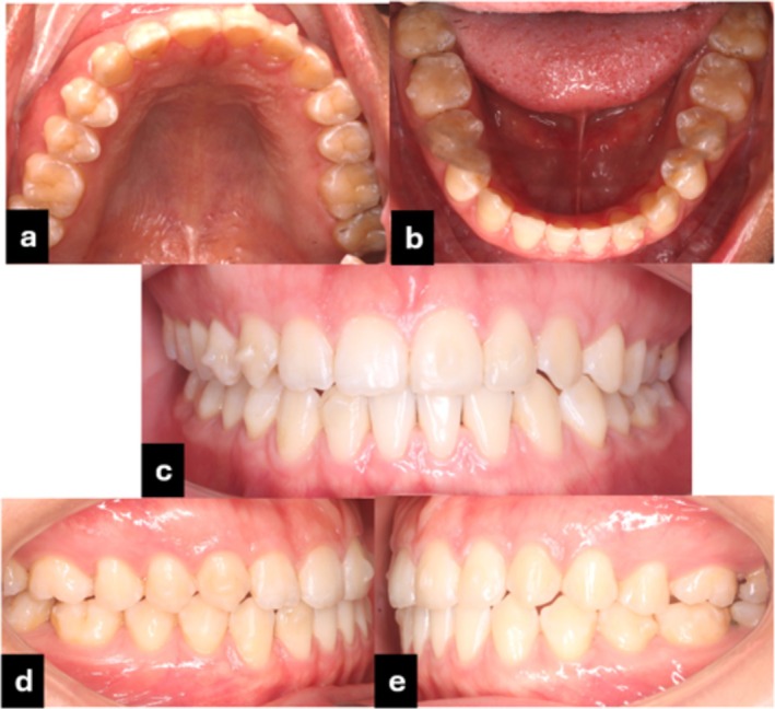
On intraoral evaluation, the patient had a mild class III molar relationship on both sides, a mild Class III canine relationship on the right side, and a mild Class II canine relationship on the left side (Figure 2). Overjet and overbite were 0.5 mm. The upper and lower arch forms were ovoid. There were spaces in both arches of 2,1 mm and 4,2 mm in the upper and lower arch, respectively. The upper dental midline was deviated 1 mm to the right, while the lower midline deviated 1 mm to the left of the facial midline. The pretreatment records of the patient are shown in Figure 2a–k.
FIGURE 2.

Pretreatment facial and intraoral photographs. (a–d) initial extraoral, (f–j) intraoral photographs, (e and k) panoramic and lateral cephalometric X‐rays.
On pretreatment model evaluation, we observed an increased posterior overjet on the right side (Figure 3). The 3–3 Bolton discrepancy was 75.7%.
FIGURE 3.

Pre‐treatment 3D models.
On a lateral cephalometric evaluation, the patient had a skeletal class III relationship (point A‐nasion‐point B angle, 0.8°), normal position of the maxilla and mandible (SNA 83.6°, SNB 82.8°) normal lower facial height (Frankfort mandibular angle (FMA), 26.9°), proclination of the upper and lower incisors (U1‐SN 114.3°, IMPA 95.11°) (Table 1). The panoramic radiograph showed the presence of all teeth including the third molars (Figure 2k). The patient is diagnosed with anterior diastemas in both arches and bilateral occlusal discrepancies due to transverse and vertical mandibular skeletal deviations.
TABLE 1.
Cephalometric analysis pre‐treatment.
| Measurement | Norm | Pre‐treatment | |
|---|---|---|---|
| SNA (°) | 81.1 ± 3.7 | 83.6 | Skeletal |
| SNB (°) | 79.2 ± 3.8 | 82.8 | |
| ANB (°) | 2.5 ± 1.8 | 0.8 | |
| FMA (°) | 25 ± 4 | 26.9 | |
| U1‐SN (°) | 105.3 ± 6.6 | 114.3 | Dental |
| U1‐APo (mm) | 5 ± 2 | 9 | |
| U1‐A vertical(mm) | 4 ± 2 | 9.1 | |
| U1‐ L1 (°) | 128 ± 5.3 | 114.4 | |
| L1‐NB (mm) | 4 ± 2 | 7.7 | |
| L1‐ APo(mm) | 2 ± 2 | 7.4 | |
| IMPA (°) | 90 ± 3.5 | 95.11 | |
| Nasiolabial Angle (°) | 100 ± 10 | 83.2 | Soft tissue |
| UL‐E line (mm) | 0 ± 2 | 0 | |
| LL – E line (mm) | 0 ± 2 | 5.2 |
Abbreviations: ANB, A point nasion B point; FMA, Frankfort mandibular plane angle; IMPA, Incisor mandibular plane angle; L1, Lower central incisor; LL, Lower lip; MP, Mandibular plane; NA, nasion point A; NB, nasion point B; SNA, Sella nasion point A; SNB, Sella nasion point B; U1, Upper central incisor; UL, Upper lip E‐line Ricketts.
2.1. Treatment Objectives
The objectives of the treatment were (1) to close spaces; (2) to increase overjet to 1.5 mm; (3) to improve dental midlines deviation; (4) to correct canine relationship to Class I; (5) to achieve satisfactory smile aesthetics; and (6) to achieve stable occlusion and treatment outcome in the long term.
Limitations of non‐surgical orthodontic treatment option: Inability to correct skeletal discrepancies that cause facial asymmetry.
2.2. Potential Alternative Treatments
We presented two treatment options, including (1) closing all the spaces combined with interproximal reduction (IPR) and (2) redistributing spaces to do veneers on the anterior teeth, as per the treatment plan of the previous doctor.
We suggest two types of appliances: (1) lingual brackets (keeping the current brackets) and (2) in‐house clear aligners to provide aesthetic orthodontic treatment as per the patient's requirements.
The patient was exhausted with lingual brackets, so she chose clear aligners.
Initially, the patient opted to distribute spaces for veneers on the anterior teeth to save time. However, after the first series of aligners, she changed her mind and wanted to close all the spaces because she felt the aligners were effective.
2.3. Treatment Progress
The initial step involved intraoral scanning, followed by model preparation and the setup of a 3D simulation using Autolign software (Autolign, Diorco, Gyeonggi‐do, Korea) before treatment to assess the biomechanics and movement of the teeth.
After discussing the detailed treatment plan with the patient, we began producing the aligners in our office. First, we created a virtual setup of the dental arches. Typically, we design around 8–12 steps per setup, allowing the patient to wear the aligners for 2–3 months. During the setup process, the necessary attachments for tooth movement were placed. In the final stage, sequential labeling was added to each virtual model before printing.
Next, we exported the file for 3D model printing. During this process, we used a 3D printer (RXDent‐L230, Guangzhou Riton Biotech Co. Ltd., China). It is a UV‐based 3D printer designed for dental applications, utilizing DLP or LCD‐based masked stereolithography (mSLA) technology with a light wavelength of 385–405 nm.
After printing the working model, the next step was aligner thermoforming. We used plastic foils (Duran, Scheu‐Dental, Iserlohn, Germany) with a thickness of 0.625 mm. A separate template aligner was made from a thinner foil (0.5 mm) to facilitate easier removal. Subsequently, we manually trimmed and polished the aligners. Finally, we packaged the aligners and delivered them to the patient (Figure 4).
FIGURE 4.

Procedure of in‐house aligners.
It took us about 5 to 7 days to complete the procedure for one set of aligners.
2.3.1. The First Setup
In the first setup (Figure 5), we aimed to:
Distalize tooth #43 to correct Class III canine on the right side
Move the upper midline 1 mm to the left.
Distribute spaces in the lower anterior area.
Contract quadrant I slightly to correct the increased posterior overbite on the right side.
FIGURE 5.

The first setup in Autolign software.
With this setup, we divided the treatment into 8 aligners, with attachments. The patient was instructed to change aligners every 7 days, with the duration of the first set of aligners being 2 months.
There are three types of attachments in this setup:
Optimized attachments on teeth #21, #22, and #23 to support movement to the left.
An optimized attachment on tooth #42 to support extrusion.
Retention attachments on the remaining teeth to gain retention.
After 2 months, the intraoral examination revealed significant improvement. The results were shown in Figure 6 (an attachment on tooth #16 fell off, but it was not necessary to reattach it).
FIGURE 6.

(a–e) Intraoral photographs after the first series of aligners.
The patient returned to our clinic with a happy smile and said, “I like these aligners, and I want to close all the spaces despite the longer treatment time”.
We agreed with the patient's request and adjusted our treatment plan with the following key points:
Performing interproximal reduction (IPR) on the upper arch. Prior to treatment, there was a 3–3 Bolton discrepancy of 75.7%. Therefore, IPR on the upper arch was appropriate for the mechanics and ensured good occlusion after treatment.
Closing all spaces on the lower arch.
2.3.2. The Second Setup
In the second setup, we performed IPR of 0.2 mm between teeth #21, #22, and #23 to correct the midline. There were 12 steps in this stage, and the treatment time was 3 months. The result after 3 months for the patient was shown in Figure 7.
FIGURE 7.
(a–e) Intraoral photographers after the second setup.
2.3.3. The Third Setup—The Final Setup
In this setup, we want to continue contracting in quadrant I (even though the patient hasn't acquired it yet, but we want to try), close all remaining spaces in the lower arch by moving lower incisors lingually, and correct the midlines by shifting the upper midline 0.5 mm to the left. We performed IPR distally, reducing 0.15 mm from tooth #21 to tooth #23 (Figure 8).
FIGURE 8.

The final setup.
There were 8 aligners in this setup, and the treatment time was 2 months.
3. Result & Follow up
The achieved outcomes included closing all spaces, improving the midlines, achieving a class I canine relationship, and decreasing the posterior overjet on the right to achieve better intercuspation. The patient also had a better smile (Figure 9).
FIGURE 9.

(a–d) post‐treatment extraoral photographs; (f–j) Intraoral phophotographs; (e, k) panoramic and lateral cephalometric X‐rays.
The limitation of the treatment result is that the midline has not coincided. After about 7 months of treatment, we recommended that the patient correct the lower midline a little more, but she did not want to proceed. The patient was happy with the results and chose to finish the treatment.
Cephalometric analysis and superimposition showed that lower incisors tipped backward, as confirmed by the difference between the initial (95.11) and final (86.51) IMPA angles (Figure 10 and Table 2).
FIGURE 10.

Overall and regional cephalometric superimpositions of the initial (black) and final (red) lateral cephalometric tracings.
TABLE 2.
Cephalometric analysis pre‐treatment and post‐treatment.
| Measurement | Norm | Pre‐treatment | Post‐treatment | |
|---|---|---|---|---|
| SNA (°) | 81.1 ± 3.7 | 83.6 | 83.7 | Skeletal |
| SNB (°) | 79.2 ± 3.8 | 82.8 | 82.4 | |
| ANB (°) | 2.5 ± 1.8 | 0.8 | 0.9 | |
| FMA (°) | 25 ± 4 | 26.9 | 26.8 | |
| U1‐SN (°) | 105.3 ± 6.6 | 114.3 | 113.7 | Dental |
| U1‐APo (mm) | 5 ± 2 | 9 | 8.6 | |
| U1‐A vertical(mm) | 4 ± 2 | 9.1 | 8.3 | |
| U1‐ L1 (°) | 128 ± 5.3 | 114.4 | 127.8 | |
| L1‐NB (mm) | 4 ± 2 | 7.7 | 4.8 | |
| L1‐ APo(mm) | 2 ± 2 | 7.4 | 4.5 | |
| IMPA (°) | 90 ± 3.5 | 95.11 | 86.51 | |
| Nasiolabial Angle (°) | 100 ± 10 | 83.2 | 83.4 | Soft tissue |
| UL‐E line (mm) | 0 ± 2 | 0 | −0.3 | |
| LL – E line (mm) | 0 ± 2 | 5.2 | 4.2 |
Abbreviations: ANB, A point nasion B point; FMA, Frankfort mandibular plane angle; IMPA, Incisor mandibular plane angle; L1, Lower central incisor; LL, Lower lip; MP, Mandibular plane; NA, nasion point A; NB, nasion point B; SNA, Sella nasion point A; SNB, Sella nasion point B; U1, Upper central incisor; UL. Upper lip E‐line: Ricketts.
Fixed lingual retainers (multistranded stainless‐steel wire) and a clear retainer were used after orthodontic treatment. Retainer protocol was as follows: full‐time wear of the clear retainer for 3 months followed by gradual tapering of the retainer. The patient was encouraged to wear the bonded retainer for the long term.
After 1 year follow‐up, the results remain stable (Figure 11).
FIGURE 11.

One‐year post‐retention facial (a–d) and intraoral (f–j) photographs, (e, k) panoramic and lateral cephalometric X‐rays.
4. Discussion
This case demonstrates the successful retreatment of an iatrogenic case involving lingual brackets. The patient had worn lingual brackets for 1.5 years with minimal improvement and wanted to change the treatment method.
As digital dentistry is evolving, contemporary orthodontics is embracing clear aligners as a tool more than ever. Digital technologies are extensively utilized in dentistry and orthodontics, improving treatment results, enhancing control, and minimizing clinical time. Among these innovations, in‐house clear aligners have emerged as the most widely used digital solution in orthodontics, offering increased efficiency, better customization, and greater cost‐effectiveness. Previously, most orthodontic manufacturers offered an aligner product [2, 4]. In essence, all third‐party aligner manufacturers functioned as orthodontic labs by providing aligners to orthodontists. However, delays can occur between the clinician and technician during discussions about the setup. Moreover, the time required for manufacturing, packaging, and shipping must also be considered. An alternative to paying an outside lab to manufacture aligners is for the orthodontist to handle all aligner fabrication in their own lab. The term commonly used to refer to this process is in‐house aligners [6].
With the development of technology, in‐house fabrication will bring advantages in the price, delivery time, and doctor's time if it is done correctly [7, 8]. For patients, in‐house aligners are invisible, comfortable to wear, and removable for eating [8, 9].
While aligners reduce physical discomfort by 50%–60% [10], their success depends on active patient participation (22 h/day wear). Notably, splint surface properties play an overlooked role. Polishing can improve patient compliance by enhancing comfort, but care must be taken not to compromise the aligner's ability to effectively deliver controlled forces to the teeth.
Spacing and midline deviation are common orthodontic issues that can be effectively treated using clear aligners. Studies have shown that mesiodistal movement, which was used in this case to correct midlines and close spaces, is the most accurate tooth movement with in‐house aligners [11]. In comparison with braces, in‐house aligners have the prominent advantage of allowing precise staging of tooth movements. Therefore, we can close spaces by moving only the anterior teeth without altering the position of the posterior teeth. Since we do not intend to move the posterior teeth in this case, the treatment time can be shortened.
The use of clear aligners from the beginning might have prevented the compliance issues encountered during the initial treatment. The physical discomfort and hygiene challenges of lingual brackets directly contributed to this patient's non‐compliance. If clear aligners had been used from the beginning, these obstacles might have been avoided. Studies indicate that clear aligners reduce pain by 43%–58% [12] and appear more favorable in protecting periodontal tissues, improving plaque control, and reducing gingival inflammation [13] compared to fixed appliances. However, patient motivation is paradoxical: while aligners eliminate fixed‐appliance frustrations, their efficacy depends on strict patient compliance with wear‐time requirements (22 h/day). In this case, the patient's prior negative experience with lingual brackets may have enhanced patient commitment to aligners—a motivation that might not exist if aligners were the first‐choice treatment. Thus, aligners could theoretically prevent compliance issues but require careful patient selection and education.
Root resorption is a multifactorial risk, often aggravated by excessive orthodontic forces, prolonged treatment, and patient‐specific vulnerabilities, such as genetic syndromes or short roots. Several studies have suggested that clear aligners may be associated with a lower risk of apical root resorption compared to fixed appliances. For instance, Krieger et al. (2013) reported that “patients treated with clear aligners exhibited significantly less root resorption compared to those treated with fixed appliances [14].” Moreover, digital treatment planning helps control force staging [15], minimizing the risk of simultaneous heavy forces on multiple teeth—a known trigger for resorption. In this case, the patient's prolonged lingual treatment (1.5 years) and minimal progress raised concerns about potential root damage. IHA provided a safer alternative, with aligners showing 30%–50% less apical root shortening compared to fixed appliances.
For patients with syndromes or previous root damage, aligners may be preferred, though they are not entirely risk‐free. Pretreatment CBCT and mid‐treatment radiographic monitoring are essential for assessing root morphology and preventing further damage. Therefore, careful patient selection, risk assessment, and regular monitoring are crucial in managing root resorption during orthodontic retreatment.
In‐house aligners provide a high level of patient comfort during treatment. Clear aligners focus on aesthetics, similar to lingual brackets. Moreover, clear aligners have been shown to cause less pain during the initial stages of treatment than fixed orthodontic appliances, among other advantages [16, 17, 18]. This is a good alternative to lingual braces in many cases.
Unfortunately, even in this clinical case, the follow‐up period was not long (1 year). However, the patient's age, cooperation, and lack of a tongue‐thrusting habit suggest good stability.
5. Conclusion
In this clinical case, successful space closure, midline correction, and Class I canine relationship were achieved using in‐house clear aligners as an alternative to lingual brackets.
Author Contributions
Pham Thi Thuy Dung: conceptualization, data curation, resources, software, writing – original draft, writing – review and editing. Cao Thi Linh: conceptualization, resources, software, supervision, writing – original draft, writing – review and editing. Nguyen Hanh Hoa: conceptualization, resources, software, visualization, writing – original draft, writing – review and editing. Le Thi Bich Phuong: conceptualization, resources, software, visualization, writing – original draft, writing – review and editing. Hoang Viet: conceptualization, formal analysis, supervision, writing – original draft, writing – review and editing. Nicolas Salesse: resources, software, visualization, writing – original draft, writing – review and editing.
Disclosure
Use of Artificial Intelligence (AI)‐assisted Technology for Manuscript Preparation: The authors confirm that there was no use of artificial intelligence (AI)‐assisted technology for assisting in the writing or editing of the manuscript, and no images were manipulated using AI.
Ethics Statement
The Institutional Review Board has waived ethical approval for this study.
Consent
Written informed consent was obtained from the patient to publish this report in accordance with the journal's patient consent policy.
Conflicts of Interest
The authors declare no conflicts of interest.
Acknowledgments
The authors would like to thank the patient for giving his or her consent.
Funding: The authors received no specific funding for this work.
Data Availability Statement
Data related to the study can be provided at reasonable request.
References
- 1. Gorman J. C., “Treatment of Adults With Lingual Orthodontic Appliances,” Dental Clinics of North America 32 (1988): 589–620. [PubMed] [Google Scholar]
- 2. Ata‐Ali F., Ata‐Ali J., Ferrer‐Molina M., Cobo T., De Carlos F., and Cobo J., “Adverse Effects of Lingual and Buccal Orthodontic Techniques: A Systematic Review and Meta‐Analysis,” American Journal of Orthodontics and Dentofacial Orthopedics 149 (2016): 820–829, 10.1016/j.ajodo.2015.11.031. [DOI] [PubMed] [Google Scholar]
- 3. Barrera‐Chaparro J. P., Plaza‐Ruíz S. P., Parra K. L., et al., “Orthodontic Treatment Need, the Types of Brackets and the Oral Health‐Related Quality of Life,” Dental and Medical Problems 60, no. 2 (2023): 287–294, 10.17219/dmp/151577. [DOI] [PubMed] [Google Scholar]
- 4. Behrents R. G., “Iatrogenics in Orthodontics,” American Journal of Orthodontics and Dentofacial Orthopedics 110 (1996): 235–238, 10.1016/S0889-5406(96)80005-9. [DOI] [PubMed] [Google Scholar]
- 5. Jaber S. T., Hajeer M. Y., and Burhan A. S., “The Effectiveness of In‐House Clear Aligners and Traditional Fixed Appliances in Achieving Good Occlusion in Complex Orthodontic Cases: A Randomized Control Clinical Trial,” Cureus 14 (2022): e30147, 10.7759/cureus.30147. [DOI] [PMC free article] [PubMed] [Google Scholar]
- 6. Cope J. B. and Groth C., “Weighing the Options of an In‐Office Versus an Outsourced Aligner Manufacturing Approach,” Seminars in Orthodontics 27 (2021): 179–183, 10.1053/j.sodo.2021.09.003. [DOI] [Google Scholar]
- 7. Tozlu M. and Ozdemir F., “In‐House Aligners: Why we Should Fabricate Aligners in Our Clinics?,” Turkish Journal of Orthodontics 34 (2021): 199–201, 10.5152/TurkJOrthod.2021.21157. [DOI] [PMC free article] [PubMed] [Google Scholar]
- 8. Viet H., Lam T. H., Phuc N. N., Ngoc Lenh N., and Thao D. T. N., “Class II Correction and Crowding Treatment Using In‐House Direct Printed Clear Aligners: A Literature Review and Case Report,” Cureus 16 (2024): e65024, 10.7759/cureus.65024. [DOI] [PMC free article] [PubMed] [Google Scholar]
- 9. AlMogbel A. M., “Clear Aligner Therapy: Up to Date Review Article,” Journal of Orthodontic Science 12 (2023): 37, 10.4103/jos.jos_30_23. [DOI] [PMC free article] [PubMed] [Google Scholar]
- 10. Fujiyama K., Honjo T., Suzuki M., Matsuoka S., and Deguchi T., “Analysis of Pain Level in Cases Treated With Invisalign Aligner: Comparison With Fixed Edgewise Appliance Therapy,” Progress in Orthodontics 15, no. 1 (2014): 64, 10.1186/s40510-014-0064-7. [DOI] [PMC free article] [PubMed] [Google Scholar]
- 11. Sachdev S., Tantidhnazet S., and Saengfai N. N., “Accuracy of Tooth Movement With In‐House Clear Aligners,” Journal of the World Federation of Orthodontists 10 (2021): 177–182, 10.1016/j.ejwf.2021.08.003. [DOI] [PubMed] [Google Scholar]
- 12. Chan V., Shroff B., Kravitz N. D., et al., “Orthodontic Pain With Fixed Appliances and Clear Aligners: A 6‐Month Comparison,” American Journal of Orthodontics and Dentofacial Orthopedics 166, no. 5 (2024): 469–479, 10.1016/j.ajodo.2024.07.002. [DOI] [PubMed] [Google Scholar]
- 13. Giannini L., Galbiati G., Tartaglia F. C., Grecolini M. E., Maspero C., and Biagi R., “Orthodontic Treatment With Fixed Appliances Versus Aligners: An Experimental Study of Periodontal Aspects,” Dentistry Journal 13, no. 2 (2025): 70, 10.3390/dj13020070. [DOI] [PMC free article] [PubMed] [Google Scholar]
- 14. Laskowska J., Paradowska‐Stolarz A., Miralles‐Jordá L., Schutty D., and Mikulewicz M., “Complication of Orthodontic Treatment: A Case Report on Severe Apical Root Resorption (ARR) in a Patient With Turner Syndrome,” Children 11, no. 3 (2024): 358, 10.3390/children11030358. [DOI] [PMC free article] [PubMed] [Google Scholar]
- 15. Tran V. H. B., Lam T. H., Khue T. N., Phi T. N. Q., and Viet H., “Accuracy and Reliability of Digital Dental Models Obtained by Intraoral Scans Compared With Plaster Models,” Applied Sciences 15, no. 6 (2025): 2927, 10.3390/app15062927. [DOI] [Google Scholar]
- 16. Papageorgiou S. N., Koletsi D., Iliadi A., Peltomaki T., and Eliades T., “Treatment Outcome With Orthodontic Aligners and Fixed Appliances: A Systematic Review With Meta‐Analyses,” European Journal of Orthodontics 42 (2020): 331–343, 10.1093/ejo/cjz094. [DOI] [PubMed] [Google Scholar]
- 17. Nguyen V. A., “3D‐Printed Indirect Bonding Trays and Transfer Jigs for Lingual Brackets: Digital Workflows and Two Case Reports,” Heliyon 10, no. 11 (2024): e32035, 10.1016/j.heliyon.2024.e32035. [DOI] [PMC free article] [PubMed] [Google Scholar]
- 18. Viet H., Marya A., Apuzzo F. d.’, and Nucci L., “The Clinical Applications and Outcomes of Digital MARPE in Orthodontics: A Scoping Review,” Seminars in Orthodontics 31, no. 2 (2025): 299–309, 10.1053/J.SODO.2024.10.002. [DOI] [Google Scholar]
Associated Data
This section collects any data citations, data availability statements, or supplementary materials included in this article.
Data Availability Statement
Data related to the study can be provided at reasonable request.