



Abstract
Vibrio cholerae (V. cholerae), the etiological agent of cholera, uses cholera toxin (CT) to cause severe diarrheal disease. Cholera is still a significant cause of mortality worldwide with about half of all cholera cases and deaths occurring in children under five. Owing to the lack of cost-effective vaccination and poor vaccine efficacy in children, there is a need for alternative preventative and therapeutic strategies. Recent advances in our knowledge of the interplay between CT-induced disease and host–pathogen metabolism have opened the door for investigating how modulation of intestinal metabolism by V. cholerae during disease impacts host intestinal immunity, the gut microbiota, and pathogen–phage interactions. In this review article, we examine recent progress in our understanding of host–pathogen interactions during V. cholerae infection and discuss future work deciphering how modulation of gut metabolism during cholera intersects these processes to enable successful fecal–oral transmission of the pathogen.
Introduction
Vibrio cholerae is the causative agent of cholera, an intestinal bacterial disease characterized by severe diarrhea, vomiting, and hypovolemic shock [1]. Cholera continues to be a significant global health problem causing seasonal and sporadic outbreaks in countries with poor sanitary conditions and limited access to safe drinking water [1]. Despite majority of cases being mild, severe cholera can cause extreme dehydration that results in high incidences of cholera-associated deaths [1]. During infection, V. cholerae uses toxin co-regulated pilus to adhere to the small intestinal epithelium and initiate colonization. There, V. cholerae secretes an AB5 multimeric toxin, cholera toxin (CT), which can bind directly to intestinal epithelial cells via ganglioside GM1 [1,2]. The catalytic activity of the A subunit (CtxA) activates adenylate cyclase that results in accumulation of 3′,5′-cyclic AMP (cAMP) and downstream cAMP-dependent activation of protein kinase A (PKA). Activation of PKA leads to phosphorylation of cystic fibrosis transmembrane conductance regulator (CFTR), which causes chloride secretion and massive fluid loss out of the intestinal lumen [2]. Secretory diarrhea results from massive fluid loss that is the hallmark symptom of cholera. Our group has recently made the surprising discovery that during infection, CT-induced disease promotes the growth of V. cholerae in the gut and induces a distinct transcriptome signature in the pathogen that includes the upregulation of a suite of genes involved in virulence and metabolism [3]. Here, we will review the most recent advances in host–pathogen interactions during V. cholerae infection and discuss how modulation of intestinal metabolism by CT may intercept with processes to drive communicability of disease (Figure 1).
Figure 1.
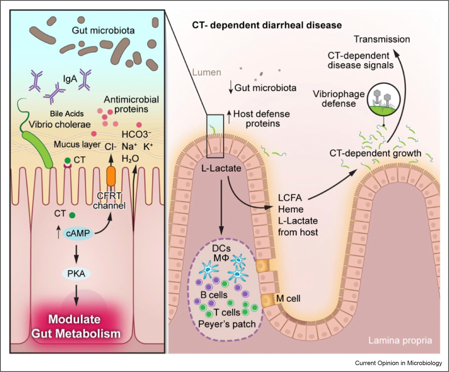
Host–pathogen interactions and metabolism during Vibrio cholerae infection. During infection, Vibrio cholerae colonizes the ileum of the SI and produces high levels of CT, which activates adenylate cyclase, increasing cellular levels of cAMP. Increased concentrations of cAMP lead to activation of PKA, which phosphorylates and activates the CFTR to secrete chloride ions out of epithelial cells, resulting in an electrolyte imbalance and massive water loss. Increased cAMP and activation of PKA leads to metabolic changes in target cells, which are predicted to lead to the secretion of host-derived LCFAs, L-lactate, and glycerol. During early stages of colonization (left side), V. cholerae encounters host factors, including bile acids, which act as signaling molecules to activate ToxT-dependent virulence factor expression. V. cholerae has to overcome components of the innate immune system, including antimicrobial proteins and secretory antibodies, such as IgA, after penetration of the mucus layer. The microbiota in the SI likely imposes ‘colonization resistance’ that V. cholerae overcomes by unknown mechanisms. The gut microbiota also may compete with V. cholerae for host-derived nutrients during CT-induced disease that the pathogen overcomes by evolutionary adaptation of the diseased gut as well as its T6SS. CT-induced disease leads to increased secretion of host defense proteins, which the pathogen overcomes by unknown mechanisms. CT-induced modulation of host metabolism promotes the luminal growth of V. cholerae during infection by acquisition of host-derived nutrients, including LCFAs, L-lactate, and heme. Host-derived L-lactate that is produced during CT-induced disease may play a role in ‘lactylation’ of immune cells in the gut. Furthermore, we propose that CT-induced growth of V. cholerae plays a role in pathogen susceptibility to phage during infection that the pathogen overcomes by deploying phage defense systems. Altogether, these complex chain events that lead to CT-mediated disease and successful replication of V. cholerae have been selected for during the evolution of the pathogen in order to ensure fecal–oral transmission of a new host.
Modulation of host–pathogen metabolism by cholera toxin
Modulation of host–microbe metabolism by CT has been recently reviewed [2]ece. The severe diarrhea produced during V. cholerae infection is thought to be important for the fecal–oral transmission of the pathogen, as up to 109 viable bacteria are excreted per milliliter of stool and remain infectious up to 24 hours after excretion [4]. Recently, it has been found that CT-induced disease promotes the growth of V. cholerae in the gut by the acquisition of host-derived nutrients, including long-chain fatty acids (LCFAs) and L-lactate [3]. Furthermore, during infection, CT induces a distinct transcriptome signature in the pathogen in both the small intestine (SI) and large intestine. In that study, a suite of V. cholerae metabolism genes were found to be upregulated in a CT-dependent manner during colonization of the ileum. Interestingly, a unique set of genes were also found to be upregulated in V. cholerae after it transitions to the large intestine, including genes involved in biofilm formation and chemotaxis [3]. Previous studies have shown that V. cholerae that is shed during infection is in a ‘hyperinfectious’ state that elevates the fecal–oral transmissibility potential of the pathogen [5]. A more recent study found that growth of V. cholerae in a biofilm increases hyperinfectivity of the pathogen by the upregulation of virulence genes [6]. Future studies will determine whether CT-induced modulation of host–pathogen metabolism drives fecal–oral pathogen transmission. These studies may point at both host and bacterial metabolism as critical new targets for the development of potential therapies or preventive measures against cholera and other diarrheal diseases.
Interactions of Vibrio cholerae with host immune system in the gut
Cholera is not an inflammatory disease and thus is not associated with a robust recruitment of immune cells during acute infection in humans [7]. This is also observed in animal models of disease where infection with V. cholerae and CT-induced diarrheal disease does not elicit a notable immune response in the intestine [8]. However, the mechanisms by which V. cholerae is able to dampen the host immune response during replication in the gut remain poorly understood. The A and B subunits of CT are known to have anti-inflammatory properties by independent mechanisms [9,10]. Consistent with this, infection with a V. cholerae mutant that is unable to produce CT leads to reactogenic diarrhea in both humans and animals [11,12]. Furthermore, V. cholerae encodes a multifunctional autoprocessing repeat (MARTX) toxin that has been shown to silence intestinal inflammation and immune cell recruitment [13]. Although infection with V. cholerae does not elicit a robust immune response, the innate immune system, including neutrophils, is believed to be important for controlling the pathogen during disease. The recruitment of neutrophils has been observed in cholera patients and shown to be important for controlling V. cholerae in animal models of disease [14–17].
In the SI, the intestinal mucus layer serves as a physical barrier against V. cholerae. Additionally, secretion of antimicrobial peptides (AMPs) by epithelial cells and production of antibodies in the gut could contribute to defense against V. cholerae colonization. It is likely that V. cholerae encounters AMPs, including human defensin-5 (HD-5), one of the most abundant in the SI, and plays a crucial role in the defense against pathogens [18]. It has been observed that V. cholerae can sense HD-5 and other cationic AMPs in vitro [19]. In the presence of AMPs, V. cholerae virulence gene expression, including expression of CT, is upregulated, suggesting that sensing of AMPs may contribute to CT-induced host metabolism changes and CT-induced diarrheal disease [19]. A recent study found that quorum sensing signals in V. cholerae also play a role in the activation of host innate immune responses by repressing pathogen tryptophan uptake and fueling host intestinal serotonin synthesis [20]. As a result of serotonin synthesis, there is increased expression of AMPs, which contributes to increased host cell survival [20]. It is unclear to what extent AMPs impact bacterial colonization and whether CT-induced changes in host and bacterial metabolism genes contribute to AMP secretion. Furthermore, the mechanisms by which V. cholerae is able to evade AMP-mediated killing remain unknown. Thus, further assessment of V. cholerae infection in mice deficient in AMPs may provide further insight into how AMP-mediated V. cholerae gene regulation contributes to V. cholerae metabolism and pathogenesis.
During infection, V. cholerae likely encounters Peyer’s patches that are aggregates of lymphoid cells, including B cells, T cells, macrophages, and antigen-sampling dendritic cells. Immunoglobulin A (IgA) is one of the most secreted antibodies by differentiated plasma cells (PCs). PCs can respond to luminal cues and intestinal metabolism to modulate IgA secretion. Notably, dietary cholesterol absorption and production of oxysterols contribute to IgA production [21,22]. 25-hydoxycholesterol (25-HC), an oxysterol metabolite generated by cholesterol 25-hydoxylase (CH25H), is necessary for antigen-specific IgA responses [21]. In a CT-treated mouse model, mice lacking CH25H showed increased CT-specific IgA, suggesting that CH25H and production of 25-HC controls antigen-specific IgA responses [21]. CT modulates intestinal metabolism that can alter downstream metabolic cues necessary for antigen-specific IgA responses. However, whether alterations in gut metabolism by CT play a role in IgA secretion remains unexplored. In addition to luminal metabolism-mediated modulation of IgA, nongenetic transfer of IgA through the entero-mammary axis can influence IgA responses in the gut [23]. Therefore, future studies should examine how metabolite-induced IgA secretion and entero-mammary transfer of antigen-specific IgA impacts CT-induced disease.
High-fat diets (HFD) are associated with downregulation of defensins, and downregulation of peroxisome proliferator-activated receptor (PPAR-y), a nuclear receptor that is involved in lipid metabolism and mucosal defense regulation [24]. HFD can also induce fatty acid-binding proteins (FABP), which have been shown to be involved in downregulation of defensins through PPAR-y degradation [25]. A recent study found that activation of PPAR-y in the SI by a Lactobacillus-derived metabolite, phenyllactic acid, alters lipid metabolism in the ileum of the SI [24]. CT is predicted to alter lipid metabolism in the ileum [3]. However, whether PPAR-y signaling, and downstream targets, are altered during CT-induced disease remains unknown. Additionally, studies should investigate whether modulation of PPAR-y-mediated lipid metabolism and mucosal defense regulation impacts V. cholerae colonization.
V. cholerae-secreted proteases have been identified in infected animal and human stool and may be involved in V. cholerae evasion of host innate immunity [26]. Recently, intelectin, which is involved in the clearance of pathogens, was found to bind V. cholerae in vitro [26,27]. V. cholerae proteases target intelectin for degradation, suggesting that host V. cholerae interactions may be modulated by pathogen-secreted enzymes [27]. Analysis of cecal fluid from WT V. cholerae and CT-mutant-infected rabbits demonstrated that CT induces secretion of host defense proteins [28]. Surfactant protein D (SP-D), a host lectin involved in innate immunity, was also found to bind V. cholerae in a CT-dependent manner and alter V. cholerae virulence gene expression [28]. However, the mechanisms by which CT-induced disease leads to increased host defense factors including SP-D and whether this depends on modulation of intestinal metabolism remains unknown. In the absence of proteases, SP-D remained bound to V. cholerae for longer, suggesting that proteases may prevent host defense factors from targeting V. cholerae [28]. Future studies should characterize secreted proteases and their targets to identify unexplored V. cholerae interactions. Additionally, several identified V. cholerae-secreted proteases are secreted by the type-II secretion system (T2SS), whose role in V. cholerae host–microbe interactions in the gut remains poorly understood [29,30]. Thus, future studies should explore how T2SS-secreted factors, including CT, act in concert during V. cholerae infection to modulate host responses, gut metabolism, and bacterial pathogenesis.
Infection with V. cholerae leads to a CT-dependent switch in anaerobic glycolysis and increased concentrations of L-lactate in the SI of infected animals [3,31]. V. cholerae genetically encodes the ability to take up and utilize L-lactate, and these genes are upregulated in a CT-dependent manner during infection [3]. Recent studies have found that increased lactate concentrations can lead to a reprogramming of immune cells to mediate immunosuppression through lactate-induced histone modification, termed ‘lactylation’ [32,33]. It is possible that immunosuppression by CT is in part due to CT-induced modulation of epithelial cell metabolism leading to lactate-mediated lactylation of immune cells in the gut.
How the gut microbiota in the small intestine impacts V. cholerae infection
V. cholerae survival in the intestinal tract is impacted by the gastrointestinal environment. V. cholerae encounters bile acids, changes in pH, and limited oxygen availability, all which the bacterium must overcome to promote its colonization. V. cholerae also encounters commensal microbes that can impact disease susceptibility. Commensal microbes encoding bile salt hydrolases (BSH) can convert taurine-conjugated taurocholate (TC) and glycine-conjugated (glycocholate) forms of bile into cholate/cholic acid (CA). Previous studies have shown that bile salts, specifically TC, the predominant bile molecule in humans and mice, can induce the expression of V. cholerae virulence genes [34,35]. Recently, it was reported that a gut commensal, Blautia obeum, can suppress V. cholerae virulence by BSH-mediated degradation of TC to CA [36]. Interestingly, humans with cholera have decreased concentrations of Blautia obeum and other obligate anaerobes compared with healthy individuals or individuals recovering from the disease [37]. V. cholerae can also employ its antibacterial type-VI secretion system (T6SS) to directly kill members of the microbiota [38,39]. Recently, it was discovered that CT-induced disease can lead to increased concentrations of host-derived nutrients [3], that likely also play a role in altering the small intestinal microbiota during V. cholerae infection. However, because V. cholerae is a human-specific pathogen, microbiota studies have been limited to the use of neonatal animals that have an underdeveloped microbiota (and immune system). Remarkably, a recent study found that targeted depletion of obligate anaerobes, specifically members of the phylum Bacteroidetes, leads to susceptibility of adult mice to colonization upon oral challenge by V. cholerae through the modulation of microbiota-derived short-chain fatty acids [40]. Thus, this study highlights the role of microbiota-derived metabolites in the susceptibility to V. cholerae and opens the door to mechanistic characterization of the role of the gut microbiota and the host immune system during cholera.
Antiviral immunity implications in the context of cholera toxin disease
V. cholerae inhabits warm estuaries and is in constant conflict with bacteriophages in both the aquatic and intestinal environments [41]. Recently, a plethora of studies have revealed a fascinating look at the arms race between Vibrio species and their phages. Bacteriophage therapy is a promising strategy for combating V. cholerae infection. A study found that a cocktail of bacteriophages prevented V. cholerae infection in an animal model of disease [42]. However, some V. cholerae strains can employ a CRISPR/Cas system and secrete outer membrane vesicles as a defense mechanism against phage predation [43,44]. V. cholerae also uses a bacterial cyclic oligonucleotide-based antiphage signaling system (CBASS), which provides the pathogen with antiviral immune protection [45]. The CBASS operon encodes a cGAS/DncV-like nucleotidyltransferase (CD-NTase) enzyme that synthesizes a nucleotide second messenger in response to viral infection in which an associated Cap effector protein then binds the nucleotide signal and executes cell death to destroy the host cell and block phage propagation [46]. During infection, CT-induced disease promotes the explosive growth of V. cholerae in the gut [3]. Thus, V. cholerae is likely highly susceptible to phage attack during CT-dependent replication in the intestine. It is tempting to speculate that some phage defense mechanisms may have evolved regulatory mechanisms that allow them to be employed during CT-dependent diarrheal disease. V. cholerae encodes several toxin–antitoxin (TA) systems that are encoded in the ‘superintegron’ of the small chromosome [47]. Recently, it has been proposed that TA systems may also serve as phage defense elements [48]. Interestingly, expression of the small chromosome is amplified in a CT-dependent manner, which may be important for phage defense during CT-dependent growth in the gut [3]. A better understanding of the evolutionary arms race between phages and V. cholerae antiphage defense mechanisms would be important for the future design of phage therapy to combat cholera.
Concluding remarks and remaining questions
Although the causative agent of cholera, V. cholerae, was identified over 140 years ago, we are still learning how this human pathogen causes disease. Recent advances in our understanding of how the pathogen’s central virulence factor, CT, acts as a multifunctional toxin that couples severe diarrheal disease with modulation of gut metabolism represent a paradigm shift in our understanding of V. cholerae. These recent findings open the door into investigating the interplay between modulation of gut metabolism by CT, host intestinal immunity, the gut microbiota, as well as phage–pathogen interactions during disease. It is important to note that strain-level differences within pathogenic V. cholerae exist and not all strains encode identical virulence factors and antiphage systems [49]. However, all pathogenic V. cholerae strains cause disease by utilizing CT which likely represents an important evolutionary determinant in all pathogenic strains. Further understanding of the molecular mechanisms by which CT-induced modulation of gut metabolism drives the growth and fecal–oral transmission of the pathogen will be key for the future design of therapeutic strategies that target both host and bacterial metabolic pathways.
Acknowledgements
F.R-C is funded by UC San Diego, the Edward Mallinckrodt, Jr. Foundation, and the Hellman Family Foundation. S.R.D is funded by the Institutional Research and Academic Career Development Award, NIH/National Institute of General Medical Sciences (NIGMS) (K12 GM068524).
Footnotes
Declaration of Competing Interest
The authors declare no competing interests.
CRediT authorship contribution statement
Fabian Rivera-Chávez: Conceptualization, Writing – original draft, Writing – review & editing. Sedelia R Dominguez: Conceptualization, Writing – original draft, Writing – review & editing. Phillip N Doan: Writing – review & editing.
Data Availability
No data were used for the research described in the article.
References and recommended reading
Papers of particular interest, published within the period of review, have been highlighted as:
• of special interest
•• of outstanding interest
- 1.Montero DA, Vidal RM, Velasco J, George S, Lucero Y, Gómez LA, et al. : Vibrio cholerae, classification, pathogenesis, immune response, and trends in vaccine development. Front Med 2023, 10:1155751, 10.3389/fmed.2023.1155751 [DOI] [PMC free article] [PubMed] [Google Scholar]
- 2.Chapman CML, Kapinos A, Rivera-Chávez F: Modulation of host-microbe metabolism by cholera toxin. Infect Immun (5) 2023, 91:e00435–22, 10.1128/iai.00435-22 [DOI] [PMC free article] [PubMed] [Google Scholar]
- 3.Rivera-Chávez F, Mekalanos JJ: Cholera toxin promotes pathogen acquisition of host-derived nutrients. Nature 2019, 572:244–248, 10.1038/s41586-019-1453-3 [DOI] [PMC free article] [PubMed] [Google Scholar]
- 4.Harris JB, LaRocque RC, Qadri F, Ryan ET, Calderwood SB: Cholera. Lancet 2012, 379:2466–2476, 10.1016/S0140-6736(12)60436-X [DOI] [PMC free article] [PubMed] [Google Scholar]
- 5.Merrell DS, Butler SM, Qadri F, Dolganov NA, Alam A, Cohen MB, et al. : Host-induced epidemic spread of the cholera bacterium. Nature 2002, 417:642–645, 10.1038/nature00778 [DOI] [PMC free article] [PubMed] [Google Scholar]
- 6.Gallego-Hernandez AL, DePas WH, Park JH, Teschler JK, Hartmann R, Jeckel H, et al. : Upregulation of virulence genes promotes Vibrio cholerae biofilm hyperinfectivity. Proc Natl Acad Sci 2020, 117:11010–11017, 10.1073/pnas.1916571117 [DOI] [PMC free article] [PubMed] [Google Scholar]
- 7.Weil AA, Becker RL, Harris JB: Vibrio cholerae at the Intersection of Immunity and the microbiome. mSphere 2019, 4:e00597–19, 10.1128/mSphere.00597-19 [DOI] [PMC free article] [PubMed] [Google Scholar]
- 8.Ritchie JM, Rui H, Bronson RT, Waldor MK: Back to the future: studying cholera pathogenesis using infant rabbits. mBio 2010, 1:e00047–10, 10.1128/mBio.00047-10 [DOI] [PMC free article] [PubMed] [Google Scholar]
- 9.Tavares LP, Negreiros-Lima GL, Lima KM, E Silva PMR, Pinho V, Teixeira MM, et al. : Blame the signaling: role of cAMP for the resolution of inflammation. Pharmacol Res 2020, 159:105030, 10.1016/j.phrs.2020.105030 [DOI] [PubMed] [Google Scholar]
- 10.Baldauf K, Royal J, Hamorsky K, Matoba N: Cholera toxin B: one subunit with many pharmaceutical applications. Toxins 2015, 7:974–996, 10.3390/toxins7030974 [DOI] [PMC free article] [PubMed] [Google Scholar]
- 11.Fakoya B, Hullahalli K, Rubin DHF, Leitner DR, Chilengi R, Sack DA, et al. : Nontoxigenic Vibrio cholerae challenge strains for evaluating vaccine efficacy and inferring mechanisms of protection. mBio 2022, 13:e00539–22, 10.1128/mbio.00539-22 [DOI] [PMC free article] [PubMed] [Google Scholar]
- 12.Rui H, Ritchie JM, Bronson RT, Mekalanos JJ, Zhang Y, Waldor MK: Reactogenicity of live-attenuated Vibrio cholerae vaccines is dependent on flagellins. Proc Natl Acad Sci 2010, 107:4359–4364, 10.1073/pnas.0915164107 [DOI] [PMC free article] [PubMed] [Google Scholar]
- 13.Woida PJ, Satchell KJF: The Vibrio cholerae MARTX toxin silences the inflammatory response to cytoskeletal damage before inducing actin cytoskeleton collapse. Sci Signal 2020, 13:eaaw9447, 10.1126/scisignal.aaw9447 [DOI] [PMC free article] [PubMed] [Google Scholar]
- 14.Bishop AL, Patimalla B, Camilli A: Vibrio cholerae-induced inflammation in the neonatal mouse cholera model. Infect Immun 2014, 82:2434–2447, 10.1128/IAI.00054-14 [DOI] [PMC free article] [PubMed] [Google Scholar]
- 15.Bourque DL, Bhuiyan TR, Genereux DP, Rashu R, Ellis CN, Chowdhury F, et al. : Analysis of the human mucosal response to cholera reveals sustained activation of innate immune signaling pathways. Infect Immun 2018, 86:e00594–17, 10.1128/IAI.00594-17 [DOI] [PMC free article] [PubMed] [Google Scholar]
- 16.Ellis CN, LaRocque RC, Uddin T, Krastins B, Mayo-Smith LM, Sarracino D, et al. : Comparative proteomic analysis reveals activation of mucosal innate immune signaling pathways during cholera. Infect Immun 2015, 83:1089–1103, 10.1128/IAI.02765-14 [DOI] [PMC free article] [PubMed] [Google Scholar]
- 17.Seper A, Hosseinzadeh A, Gorkiewicz G, Lichtenegger S, Roier S, Leitner DR, et al. : Vibrio cholerae evades neutrophil extracellular traps by the activity of two extracellular nucleases. PLoS Pathog 2013, 9:e1003614, 10.1371/journal.ppat.1003614 [DOI] [PMC free article] [PubMed] [Google Scholar]
- 18.Cui C, Wang F, Zheng Y, Wei H, Peng J: From birth to death: the hardworking life of Paneth cell in the small intestine. Front Immunol 2023, 14:1122258, 10.3389/fimmu.2023.1122258 [DOI] [PMC free article] [PubMed] [Google Scholar]
- 19.•. Liu Y, Xu T, Wang Q, Huang J, Zhu Y, Liu X, et al. : Vibrio cholerae senses human enteric α-defensin 5 through a CarSR two-component system to promote bacterial pathogenicity. Commun Biol 2022, 5:559, 10.1038/s42003-022-03525-3. The authors demonstrated that Vibrio cholerae gene expression is upregulated in the presence of HD-5. Furthermore, the authors demonstrate that a two-component system, CarSR, in V. cholerae is essential for this upregulation. This paper demonstrates a virulence regulating pathway which may depend on presence and sensing of antimicrobial peptides such as HD-5.
- 20.•. Jugder B-E, Batista JH, Gibson JA, Cunningham PM, Asara JM, Watnick PI: Vibrio cholerae high cell density quorum sensing activates the host intestinal innate immune response. Cell Rep 2022, 40:111368, 10.1016/j.celrep.2022.111368. Using an oral infection of Drosophila, the authors demonstrate that V. cholerae quorum sensing regulator, HapR, mediates bacterial anabolic metabolism. The authors found that HapR represses pathogen tryptophan consumption which stimulates host intestinal serotonin production to activate intestinal immunity.
- 21.•. Trindade BC, Ceglia S, Berthelette A, Raso F, Howley K, Muppidi JR, et al. : The cholesterol metabolite 25-hydroxycholesterol restrains the transcriptional regulator SREBP2 and limits intestinal IgA plasma cell differentiation. Immunity 2021, 54:2273–2287.e6, 10.1016/j.immuni.2021.09.004. Using a knockout mouse model, the authors find that a cholesterol metabolite can shape humoral immunity during immunization and intestinal infection.
- 22.Ceglia S, Berthelette A, Howley K, Li Y, Mortzfeld B, Bhattarai SK, et al. : An epithelial cell-derived metabolite tunes immunoglobulin A secretion by gut-resident plasma cells. Nat Immunol 2023, 24:531–544, 10.1038/s41590-022-01413-w [DOI] [PMC free article] [PubMed] [Google Scholar]
- 23.••. Ramanan D, Sefik E, Galván-Peña S, Wu M, Yang L, Yang Z, et al. : An immunologic mode of multigenerational transmission governs a gut Treg setpoint. Cell 2020, 181:1276–1290.e13, 10.1016/j.cell.2020.04.030. Using a comprehensive multigenerational mouse model, the authors demonstrate an entero-mammary axis which contributes to the transmission of protective components of immunity. Specifically, the authors find that regulatory T cells can be transmitted by the mother for multiple generations to control gut inflammation against microbial and/or cellular perturbations.
- 24.••. Shelton CD, Sing E, Mo J, Shealy NG, Yoo W, Thomas J, et al. : An early-life microbiota metabolite protects against obesity by regulating intestinal lipid metabolism. Cell Host Microbe 2023, 31:1604–1619.e10, 10.1016/j.chom.2023.09.002. The authors demonstrate that Lactobacillus impacts susceptibility against antibiotic induced obesity. The authors find that antibiotics and HFD exacerbate obesity through depletion of intestinal PPAR-y. The authors further find that Lactobacillus-derived metabolite protects against antibiotic induced obesity.
- 25.•. Su X, Jin M, Xu C, Gao Y, Yang Y, Qi H, et al. : FABP4 in Paneth cells regulates antimicrobial protein expression to reprogram gut microbiota. Gut Microbes 2022, 14:2139978, 10.1080/19490976.2022.2139978. The authors find that FABP4 in gut epithelial cells can mediate the expression of defensins. Specifically, the authors find that FABP4-mediated downregulation of defensins is through degradation of PPAR-y. Furthermore, the authors demonstrate that during HFD, defensins are downregulated by inducing FABPR in gut epithelial cells. This paper provides a comprehensive understanding of the regulation of defensins during HFD diets and impact of defensins on enteric infections.
- 26.Hatzios SK, Abel S, Martell J, Hubbard T, Sasabe J, Munera D, et al. : Chemoproteomic profiling of host and pathogen enzymes active in cholera. Nat Chem Biol 2016, 12:268–274, 10.1038/nchembio.2025 [DOI] [PMC free article] [PubMed] [Google Scholar]
- 27.Howell M, Dumitrescu DG, Blankenship LR, Herkert D, Hatzios SK: Functional characterization of a subtilisin-like serine protease from Vibrio cholerae. J Biol Chem 2019, 294:9888–9900, 10.1074/jbc.RA119.007745 [DOI] [PMC free article] [PubMed] [Google Scholar]
- 28.••. Zoued A, Zhang H, Zhang T, Giorgio RT, Kuehl CJ, Fakoya B, et al. : Proteomic analysis of the host–pathogen interface in experimental cholera. Nat Chem Biol 2021, 17:1199–1208, 10.1038/s41589-021-00894-4. Using a surface-biotinylation approach coupled with mass spectrometry, the authors identify host proteins present on the surface of V. cholerae. Through this analysis, the authors find that cholera toxin induces the presence of host defense proteins. Additionally, the authors characterize a C-type lectin to be important for host intestinal defense against V. cholerae.
- 29.Sikora AE: Proteins secreted via the type II secretion system: smart strategies of Vibrio cholerae to maintain fitness in different ecological niches. PLoS Pathog 2013, 9:e1003126, 10.1371/journal.ppat.1003126 [DOI] [PMC free article] [PubMed] [Google Scholar]
- 30.Sikora AE, Zielke RA, Lawrence DA, Andrews PC, Sandkvist M: Proteomic analysis of the Vibrio cholerae type II secretome reveals new proteins, including three related serine proteases. J Biol Chem 2011, 286:16555–16566, 10.1074/jbc.M110.211078 [DOI] [PMC free article] [PubMed] [Google Scholar]
- 31.Snider RM, McKenzie JR, Kraft L, Kozlov E, Wikswo JP, Cliffel DE: The effects of cholera toxin on cellular energy metabolism. Toxins 2010, 2:632–648, 10.3390/toxins2040632 [DOI] [PMC free article] [PubMed] [Google Scholar]
- 32.Chen A-N, Luo Y, Yang Y-H, Fu J-T, Geng X-M, Shi J-P, et al. : Lactylation, a novel metabolic reprogramming code: current status and prospects. Front Immunol 2021, 12:688910, 10.3389/fimmu.2021.688910 [DOI] [PMC free article] [PubMed] [Google Scholar]
- 33.Zhang D, Tang Z, Huang H, Zhou G, Cui C, Weng Y, et al. : Metabolic regulation of gene expression by histone lactylation. Nature 2019, 574:575–580, 10.1038/s41586-019-1678-1 [DOI] [PMC free article] [PubMed] [Google Scholar]
- 34.Hung DT, Zhu J, Sturtevant D, Mekalanos JJ: Bile acids stimulate biofilm formation in Vibrio cholerae. Mol Microbiol 2006, 59:193–201, 10.1111/j.1365-2958.2005.04846.x [DOI] [PubMed] [Google Scholar]
- 35.Yang M, Liu Z, Hughes C, Stern AM, Wang H, Zhong Z, et al. : Bile salt–induced intermolecular disulfide bond formation activates Vibrio cholerae virulence. Proc Natl Acad Sci 2013, 110:2348–2353, 10.1073/pnas.1218039110 [DOI] [PMC free article] [PubMed] [Google Scholar]
- 36.•. Alavi S, Mitchell JD, Cho JY, Liu R, Macbeth JC, Hsiao A: Interpersonal gut microbiome variation drives susceptibility and resistance to cholera infection. Cell 2020, 181:1533–1546.e13, 10.1016/j.cell.2020.05.036. The authors show that human microbiome species can shape the chemical environment of the gut. The authors specifically identified that bile salt hydrolase activity and the availability of taurocholate can be shaped by specific microbiome species to impact colonization of V. cholerae. These findings highlight the interactions between V. cholerae and the host microbiome.
- 37.Hsiao A, Ahmed AMS, Subramanian S, Griffin NW, Drewry LL, Petri WA, et al. : Members of the human gut microbiota involved in recovery from Vibrio cholerae infection. Nature 2014, 515:423–426, 10.1038/nature13738 [DOI] [PMC free article] [PubMed] [Google Scholar]
- 38.Zhao W, Caro F, Robins W, Mekalanos JJ: Antagonism toward the intestinal microbiota and its effect on Vibrio cholerae virulence. Science 2018, 359:210–213, 10.1126/science.aap8775 [DOI] [PMC free article] [PubMed] [Google Scholar]
- 39.Toska J, Ho BT, Mekalanos JJ: Exopolysaccharide protects Vibrio cholerae from exogenous attacks by the type 6 secretion system. Proc Natl Acad Sci 2018, 115:7997–8002, 10.1073/pnas.1808469115 [DOI] [PMC free article] [PubMed] [Google Scholar]
- 40.You JS, Yong JH, Kim GH, Moon S, Nam KT, Ryu JH, et al. : Commensal-derived metabolites govern Vibrio cholerae pathogenesis in host intestine. Microbiome 2019, 7:132, 10.1186/s40168-019-0746-y [DOI] [PMC free article] [PubMed] [Google Scholar]
- 41.Boyd CM, Angermeyer A, Hays SG, Barth ZK, Patel KM, Seed KD: Bacteriophage ICP1: a persistent predator of Vibrio cholerae. Annu Rev Virol 2021, 8:285–304, 10.1146/annurev-virology-091919-072020 [DOI] [PMC free article] [PubMed] [Google Scholar]
- 42.Yen M, Cairns LS, Camilli A: A cocktail of three virulent bacteriophages prevents Vibrio cholerae infection in animal models. Nat Commun 2017, 8:14187, 10.1038/ncomms14187 [DOI] [PMC free article] [PubMed] [Google Scholar]
- 43.Bourgeois J, Lazinski DW, Camilli A: Identification of spacer and protospacer sequence requirements in the Vibrio cholerae Type I-E CRISPR/Cas system. mSphere 2020, 5:e00813–20. [DOI] [PMC free article] [PubMed] [Google Scholar]
- 44.Reyes-Robles T, Dillard RS, Cairns LS, Silva-Valenzuela CA, Housman M, Ali A, et al. : Vibrio cholerae outer membrane vesicles inhibit bacteriophage infection. J Bacteriol 2018, 200, 10.1128/JB.00792-17 [DOI] [PMC free article] [PubMed] [Google Scholar]
- 45.Whiteley AT, Eaglesham JB, De Oliveira Mann CC, Morehouse BR, Lowey B, Nieminen EA, et al. : Bacterial cGAS-like enzymes synthesize diverse nucleotide signals. Nature 2019, 567:194–199, 10.1038/s41586-019-0953-5 [DOI] [PMC free article] [PubMed] [Google Scholar]
- 46.••. Ledvina HE, Ye Q, Gu Y, Sullivan AE, Quan Y, Lau RK, et al. : An E1–E2 fusion protein primes antiviral immune signalling in bacteria. Nature 2023, 616:319–325, 10.1038/s41586-022-05647-4. The authors demonstrate that bacteria CD-NTase can sense CD-NTase associated protein 2 (Cap2). The authors characterize this interaction and reveal that Cap2 has distinct domains that resemble E1 and E2 proteins. Further, the authors find endopeptidase, Cap3, balances Cap2 activity. Overall, the authors find that diverse bacterial CBASS use positive and negative regulators to control the activation of CD-NTase and mediate antiphage immunity.
- 47.Iqbal N, Guérout A-M, Krin E, Le Roux F, Mazel D: Comprehensive functional analysis of the 18 Vibrio cholerae N16961 toxin-antitoxin systems substantiates their role in stabilizing the superintegron. J Bacteriol 2015, 197:2150–2159, 10.1128/JB.00108-15 [DOI] [PMC free article] [PubMed] [Google Scholar]
- 48.LeRoux M, Laub MT: Toxin-antitoxin systems as phage defense elements. Annu Rev Microbiol 2022, 76:21–43, 10.1146/annurev-micro-020722-013730 [DOI] [PubMed] [Google Scholar]
- 49.In: Vibrio spp. Infections. Edited by Almagro-Moreno S, Pukatzki S. vol. 1404, Springer International Publishing; 2023, , 10.1007/978-3-031-22997-8 [DOI] [PubMed] [Google Scholar]
Associated Data
This section collects any data citations, data availability statements, or supplementary materials included in this article.
Data Availability Statement
No data were used for the research described in the article.