



Abstract
The early diagnosis of opportunistic infections is a critical concern for patient care worldwide, particularly in the context of the COVID-19 pandemic. This review examines the challenges and advancements in the management and early diagnosis of opportunistic fungal infections, which have become increasingly prominent during the pandemic. Using multiple sources, including curated databases such as PubMed and Scopus, as well as Google Scholar for broader literature searches, we systematically reviewed studies on COVID-19-associated fungal infections, with a focus on candidiasis, mucormycosis, and aspergillosis. The inclusion criteria encompassed peer-reviewed articles, clinical case reports, and cohort studies that discussed diagnostic methods, clinical outcomes, and treatment responses. Data were systematically extracted and analyzed to identify key trends and gaps in current diagnostic practices. Given the significance of opportunistic fungal infections—particularly the selected species—this review provides a comprehensive analysis of diagnostic challenges and advancements in the context of COVID-19 and beyond. Currently, there is no definitive strategy for effectively addressing these opportunistic pathogens, highlighting the need for continued research and innovation. Despite advancements in medical technology, opportunistic fungal infections continue to pose significant challenges to early and accurate diagnosis. The COVID-19 pandemic has exacerbated these challenges, with secondary fungal infections contributing to increased morbidity and mortality rates. This review highlights the complexities of diagnosing fungal coinfections and emphasizes the urgent need for improved diagnostic strategies. Enhancing the early and accurate detection of these infections is critical for effective patient management, particularly during viral pandemics. Addressing the challenges outlined in this review requires innovative diagnostic approaches to improve patient outcomes and reduce the burden of opportunistic infections on global healthcare systems.
Keywords: COVID-19, fungal coinfections, intensive care units (ICUs), invasive fungal infections (IFIs), opportunistic infections, SARS-CoV-2
Summary
• Diagnosing opportunistic fungal infections during the COVID-19 pandemic poses significant challenges.
• Accurate and timely fungal coinfection diagnosis is crucial to prevent increased mortality.
• The review the analyzed literature to understand challenges in fungal disease diagnosis.
• Innovative diagnostics are needed to improve patient care and reduce healthcare impact.
1. Introduction
Throughout history, the prevalence and recurring outbreaks of infectious diseases have had substantial and long-lasting consequences on society [1]. Coronaviruses (CoVs) constitute a diverse group of viruses that infect a wide range of animals and humans, causing illnesses that can range from mild to severe respiratory conditions. They are large, enveloped, positive-sense single-stranded RNA viruses (+ssRNA viruses) that infect various avian and mammalian species [2]. CoVs are characterized by the presence of spike-like projections of glycoproteins on their surface, which resemble a crown when viewed under an electron microscope; hence, they are termed coronaviruses.
Between 2002 and 2012, two highly pathogenic CoVs of common human–animal origin emerged: severe acute respiratory syndrome coronavirus (SARS-CoV) and Middle East respiratory syndrome coronavirus (MERS-CoV). These viruses caused respiratory diseases, including severe lower respiratory infections and pneumonia, posing significant health risks. Consequently, they transformed emerging CoVs into serious threats to public health [3–7]. A new CoV, SARS-CoV-2, emerged in Wuhan, China, in late 2019 and early 2020, leading to an outbreak of atypical viral pneumonia. This novel illness, referred to as coronavirus disease 2019 (COVID-19), spread rapidly worldwide due to its high transmissibility [5, 8, 9].
The current pandemic has far surpassed both SARS and MERS in terms of the number of cases and geographic extent, presenting a tremendous and devastating threat to global public health [5, 10, 11]. Despite the implementation of various treatment and control strategies, the emergence of new strains of COVID-19, including beta coronaviruses (β CoV), continues to be associated with morbidity and mortality rates. Secondary infections caused by a wide range of opportunistic bacterial and fungal pathogens, combined with an abnormal host immune response characterized by cytokine storms, contribute to increased mortality among patients infected with SARS-CoV-2 [12, 13].
2. Invasive Fungal Infections (IFIs) as a Public Health Concern
IFIs have garnered significant attention as a public health concern in the past decade, owing to several high-profile outbreaks. Similar to other respiratory pathogens like influenza, fungal infections are emerging as a serious concern in patients, particularly during hospitalization, in intensive care units (ICUs), and even after recovery [14, 15]. In the ICU, mortality rates due to fungal infections have been reported to range from 5% to 70% [14]. The IFI incidence rate has been reported to be between 4% and 27.7% for COVID-19 [16]. Although several fungal species have been identified in hospitalized COVID-19 patients, Candida, Mucor, and Aspergillus species are the primary fungal microbes responsible for coinfections in patients with severe COVID-19 caused by SARS-CoV-2 [17, 18] (Figure 1).
Figure 1.
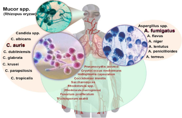
The principal fungal coinfections in patients with COVID-19.
In patients with pulmonary infections caused by Aspergillus, rhino-orbital-brain involvement is induced by Mucor, while upper gastrointestinal infections are associated with Candida, and bloodstream infections are linked to Candida dissemination (Figure 1) [19]. In addition to mucormycosis, meningoencephalitis caused by Cryptococcus neoformans has also been reported [20]. The diagnosis of histoplasmosis should be considered in endemic countries, particularly among individuals with significant tourist mobility to and from these regions; some sources even suggest its increasing prevalence [21, 22].
Pneumocystis jirovecii, although rare and infrequently reported, has been observed in pulmonary infections associated with SARS-CoV-2 [23, 24]. Infection with Saccharomyces cerevisiae has been documented in ICU patients with COVID-19 following Saccharomyces supplementation [25]. Fusarium may pose complex challenges even for patients with CoV whose immune systems are not severely compromised [26]. Rhodotorula mucilaginosa can be present either alone or in conjunction with species such as Candida glabrata [27]. Coinfection with Trichosporon asahii and COVID-19 has been reported in the form of nosocomial pneumonia or fungemia [27, 28].
3. Mucormycosis
Mucormycosis, also known as zygomycosis or “black fungus,” is a serious angioinvasive fungal infection. It is caused by a group of molds known as Mucoromycetes, which belong to the subphylum Mucoromycotina and the order Mucorales. The most common fungi responsible for mucormycosis include Rhizopus spp., Mucor spp., Rhizomucor spp., Syncephalastrum spp., Cunninghamella bertholletiae, Apophysomyces spp., and Lichtheimia (formerly Absidia) spp [29]. Notably, Rhizopus oryzae accounts for approximately 60% of cases and 90% of the rhino-orbital-cerebral mucormycosis (ROCM) variant [30].
Mucormycosis is associated with several underlying conditions that increase susceptibility to infection, including prolonged neutropenia, hematologic malignancies, uncontrolled diabetes, iron chelation therapy, and skin breaches. These factors significantly elevate the risk of invasive disease, particularly in non-COVID-19 patients [31]. The emergence of mucormycosis has complicated the management of COVID-19, especially among critically ill patients on mechanical ventilation, who are at heightened risk for fungal infections. In March 2021, 41 cases of COVID-19-associated mucormycosis (CAM) were reported globally, with India accounting for 70% of these cases. The second wave in India saw a surge, with Maharashtra reporting 2245 cases and 120 fatalities, and Rajasthan documenting 2651 cases by June 2021 [32]. A study of 102 CAM cases indicated that 69.6% of patients were male, and 70.5% had concurrent COVID-19. Steroid use was prevalent in 68.6% of cases, while diabetes mellitus affected 88.2% of patients. Mucormycosis primarily impacted the sino-nasal area (72.5%), with a recorded mortality rate of 23.5% [33]. The incidence of CAM is particularly high among diabetic patients, with approximately 95% of severe COVID-19 cases affected [32]. The predominant causative agent in India is Rhizopus arrhizus, along with other species such as Mucor irregularis and Apophysomyces variabilis [34–36]. The number of countries reporting CAM has been discussed in a previous study (Figure 2) [37].
Figure 2.
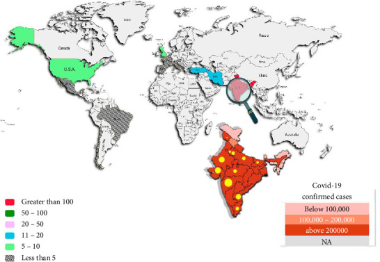
Global distribution of COVID-19-associated mucormycosis.
In India, the prevalence of mucormycosis is significantly higher than in developed countries, exacerbated by the widespread use of immunosuppressants and corticosteroids for COVID-19 treatment [29, 38]. Additionally, both hyperglycemia and COVID-19 can disrupt iron metabolism, further increasing the risk of mucormycosis [39]. Diagnosing and treating mucormycosis in COVID-19 patients poses significant challenges, necessitating faster diagnostic methods and alternative treatment options [40, 41]. Excessive reliance on conventional precautions may disrupt nasal microbiomes, thereby increasing the risk of fungal infections [42]. Interestingly, increased zinc consumption, aimed at preventing viral infections, may inadvertently inhibit fungal growth, suggesting a potential therapeutic avenue [43]. The diversity of fungal species involved, such as Rhizopus microsporus and Lichtheimia corymbifera, highlights the complexities of managing mucormycosis [44]. Recognizing these factors enables clinicians to effectively address the challenges posed by mucormycosis during the COVID-19 pandemic.
4. Aspergillosis
Aspergillosis encompasses numerous mold species that are prevalent in diverse climates globally, frequently isolated from both outdoor and indoor environments, including tertiary care centers. It is a leading cause of life-threatening fungal infections. Despite significant advancements in diagnosing and treating aspergillosis, effectively preventing and managing severe hospital-acquired fungal diseases remains challenging, with high mortality rates, especially among immunocompromised patients affected by invasive infections [45–47].
Infections are typically caused by Aspergillus fumigatus (roughly 90%), followed by A. flavus, A. nidulans, A. niger, A. terreus, and A. versicolor [48]. The prevalence of aspergillosis has increased due to modern medical practices and a rising number of individuals with compromised immune systems from cancer treatments, organ transplants, and prolonged immunosuppressive therapy. It manifests in several forms: invasive pulmonary aspergillosis (IPA), chronic pulmonary aspergillosis (CPA), allergic bronchopulmonary aspergillosis (ABPA), chronic rhinosinusitis (CRC), fungal asthma, and Aspergillus bronchitis [49]. Notably, IPA incidence has risen 10-fold in the past two decades [50].
IPA is a recognized complication in severely immunosuppressed individuals, such as those undergoing hematopoietic transplantation or chemotherapy, and is associated with high mortality rates [49]. Studies indicate that patients with severe acute respiratory distress syndrome (ARDS) from influenza are also at risk for rapid progression to IPA, linked to prolonged hospital stays and higher mortality [51]. Other risk factors include liver cirrhosis, systemic connective tissue diseases, chronic kidney disease (CKD) or renal replacement therapy (RRT), influenza-associated aspergillosis (IAA) [51, 52], advanced solid cancers, and diabetes mellitus [53].
Recent diagnostic advancements show that approximately 50% of IPA cases occur in ICUs, often in patients not experiencing neutropenia [49, 51]. The association between ARDS and IPA suggests that alveolar damage may facilitate fungal invasion [54]. COVID-19-associated pulmonary aspergillosis (CAPA) is a significant concern, with A. fumigatus identified as the primary coinfecting species in patients with COVID-19, alongside A. flavus and A. terreus [55].
Predisposing factors for CAPA include diabetes, immunomodulatory therapies, prior respiratory diseases, cardiovascular disease, hepatic disease, renal failure, prior use of antibiotics and antifungal treatments, as well as environmental and logistical factors. Similar to other forms of pulmonary aspergillosis, CAPA requires underlying host factors such as allergic conditions (e.g., asthma), airway diseases (e.g., bronchial dilation, cystic fibrosis), chronic lung cavities (e.g., tuberculosis, sarcoidosis), or immune deficiency [53].
5. Candidiasis
Candida is a genus of yeast comprising approximately 200 species, including Candida albicans, C. glabrata, C. parapsilosis, C. tropicalis, and C. krusei, commonly found on mucosal surfaces [56, 57]. While Candida can infect both immunocompetent and immunocompromised individuals, infections are more prevalent in those with weakened immune systems, leading to candidiasis being referred to as the “disease of the diseased” [58]. Candida species can cause a range of clinical symptoms, from mucocutaneous overgrowth to severe disseminated infections such as candidemia. Although Candida albicans is frequently found in humans, it does not always cause symptoms. However, it can lead to various chronic and acute opportunistic infections in immunocompromised individuals, including diabetics, organ transplant recipients, and people living with human immunodeficiency virus (HIV), manifesting as thrush, vaginitis, cutaneous infections, and nail infections [59]. Notably, the incidence of infections caused by non-albicans Candida (NAC) species is increasing, with some exhibiting resistance to standard antifungal treatments. Several studies have reported on C. albicans, C. parapsilosis, C. tropicalis, C. glabrata, C. guilliermondii, and C. krusei, noting their occasional resistance to conventional antifungal treatments [59, 60]. One study at Siriraj Hospital in Thailand analyzed data from 156 candidemia patients between January 2016 and December 2017, finding that 71.2% had candidemia caused by NAC species, reflecting an increasing prevalence in the region [61]. Additionally, research indicated that Pichia kudriavzevii, C. dubliniensis, and C. glabrata exhibited decreased susceptibility to azoles [62].
Various virulence mechanisms enable Candida species to transition from harmless commensals to pathogens, including adherence to host tissues, biofilm formation, and the production of extracellular hydrolytic enzymes [63]. Factors contributing to this transition include severe immunosuppression, prematurity, broad-spectrum antibiotic use, empirical antifungal therapy, diabetes, renal failure, and the presence of catheters, as well as major surgery, and burns [64]. While clinical symptoms of infections caused by NAC spp. are often similar, some NAC spp. exhibit inherent resistance to antifungal medications or can develop resistance [65]. Candida spp., especially from NAC species, is a common ICU pathogen, infecting 6% to 10% of patients. The incidence of candidemia is increasing, with invasive candidiasis responsible for 19% to 40% of all deaths and a mortality rate exceeding 70% among ICU patients [66]. Candida auris has emerged as a significant NAC pathogen since 2009. It belongs to the Clavispora clade of the Metschnikowiaceae family [67, 68]. The phylogenetic relationship of C. auris with other Candida species remains unclear due to the rarity of some related species; however, five clades have been identified, linked to species such as C. haemulonii, C. pseudohaemulonii, C. duobushaemulonii, and more recently, C. heveicola [69, 70].
C. auris infections have been reported in over 40 countries, affecting 5% to 10% of infected individuals, ranging from mild infections like otitis media to severe invasive candidiasis. Its epidemiology is similar to that of other Candida species [71]. Initially, Aspergillus fumigatus was seen as the main cause of fungal superinfections in critically ill COVID-19 patients, but candidiasis has become increasingly significant. This shift is due to factors like antibiotic and corticosteroid use, central venous catheters, and SARS-CoV-2 damage in ARDS patients. Candida albicans is the most common yeast in invasive infections among these patients, followed by C. auris, C. glabrata, C. parapsilosis, C. tropicalis, S. cerevisiae, and Rhodotorula spp [72]. In New Delhi, C. auris was responsible for 67% of candidiasis cases [73]. It was first reported in Salvador, Brazil, in December 2020 [74], with subsequent cases in Mexico [75]. The role of C. auris in patients and its transmission via respiratory equipment has been highlighted in several studies [76]. Other species, including C. glabrata, C. dubliniensis, C. parapsilosis, C. tropicalis, and C. krusei, have also been identified. In COVID-19 cases, various Candida species have been isolated from candidemia [77, 78].
Early and accurate identification is crucial for effective treatment of opportunistic fungal infections, but diagnostic challenges complicate detection for healthcare professionals.
6. Challenges in COVID-19-Associated Fungal Disease
Diagnosing fungal infections based solely on clinical signs, symptoms, and nonspecific radiographic manifestations can be challenging. Laboratory diagnosis of fungal diseases typically involves microscopic examination, culture, antigen or antibody testing, and molecular assays [79–81]. Direct microscopic examination requires sampling from the site of infection; however, identifying the genus or species based solely on microscopic features may not always be feasible or sufficient. Additionally, culture techniques can be insensitive and time-consuming. Fungi are also part of the human microbiome, which limits the effectiveness of antigen- and antibody-based assays, as fungal colonization can lead to false-positive results [82].
Moreover, routine serologic tests for emerging fungi are often unavailable, and current diagnostic methods require specialized mycological expertise. As the diversity of fungi that clinical mycologists must identify increases, there is a pressing need for innovative identification approaches beyond traditional phenotypic methods. Consequently, molecular techniques, particularly polymerase chain reaction (PCR) and antigen detection, are gaining prominence as alternatives to culture-based diagnostics for fungal infections [81, 83–85]. PCR, which targets the highly variable internal transcribed spacer (ITS) regions of fungal ribosomal DNA (rDNA), is a valuable tool for distinguishing fungal species [86]. However, challenges persist, especially in diagnosing IFIs, which can remain undetected until postmortem in some cases [87].
This review will address the diagnostic challenges associated with opportunistic fungal infections frequently observed during the COVID-19 pandemic.
7. Diagnosis of CAM
Diagnosis of CAM, due to its rapid invasion and spread throughout the body, affecting most vital organs, should begin at the time of hospitalization [88, 89]. This process should take into account the patient's history, including underlying conditions and other risk factors. Mucormycosis can be categorized based on its location: pulmonary, gastrointestinal, cutaneous, disseminated, and especially ROCM [29]. ROCM typically manifests as sinusitis in the ethmoidal or sphenoidal sinuses and can progress to cavernous sinus syndrome or thrombosis of the internal carotid artery [90].
Necrotic ulcers resulting from osteomyelitis of the facial bones can impair the optic and cranial nerves, leading to headaches and facial discomfort. Diagnostic samples, including scrapings from the upper turbinates, aspirated sinus material, sputum, and biopsy specimens, can be examined using 10% potassium hydroxide (KOH) or optical brighteners like Blankophor and Calcofluor [85, 91, 92]. Mucorales are characterized by thick-walled, irregular, aseptate, and refractile hyphae measuring 6 to 25 μm in diameter, often exhibiting swollen and twisted forms with common 90° bifurcations [93, 94]. Histological analysis may reveal acute suppurative and localized granulomatous inflammation. Fungal elements are readily visible on hematoxylin and eosin-stained sections, while periodic acid-Schiff or Grocott's methenamine silver staining enhances the visibility of hyphae, particularly in cases of vascular invasion leading to thrombosis and infarction [85, 92].
Direct microscopic analysis and histopathological examination for Mucorales detection can present diagnostic challenges, as these fungi may be difficult to differentiate from Aspergillus hyphae. Mucorales grow rapidly (3 to 7 days) on standard fungal culture media (e.g., potato dextrose agar and Sabouraud agar) at 25°C to 30°C; however, only about 50% of cases may yield positive cultures despite positive microscopy findings [95, 96]. Coinfection of the lungs by Rhizomucor spp., and SARS-CoV-2 can occur, resulting in symptoms like dyspnea, cough, and airway bleeding [37].
Pulmonary mucormycosis (PM) primarily affects the bronchial airways and lung parenchyma, with potential extension into the chest wall, leading to complications such as cavitation and pericarditis [97]. An early diagnostic feature of PM is the reverse-halo sign on computed tomography (CT) scans, although this finding is rare in COVID-19 cases. Magnetic resonance imaging (MRI) effectively captures soft tissue and evaluates disease severity [98–100].
Some cases may show subtle thickening of the sinus mucosa or extraocular muscles without visible abnormalities in the sinus bones, despite clinical symptoms. A hypointense rim around lesions may indicate the accumulation of blood products or metals like iron and magnesium due to fungal activity [101].
Rhino-cerebral mucormycosis often progresses with orbital invasion, increasing the risk of cavernous sinus infections and internal carotid thrombosis, which can elevate mortality rates. A “black hole” sign, representing a lack of contrast uptake by lesions, has been noted [102]. In patients with COVID-19, whether alone or in conjunction with mucormycosis, abnormal levels of nonspecific laboratory markers associated with COVID-19-related sepsis have been observed, including elevated lactate dehydrogenase, C-reactive protein, D-dimer, and renal profile derangements, along with cytopenia [98, 103–105].
For bacterial and yeast infections, matrix-assisted laser desorption/ionization time-of-flight mass spectrometry (MALDI-TOF MS) is a rapid and reliable method for detecting culture isolates. However, the handling of filamentous fungi presents greater challenges, necessitating several pre-analytical procedures. Despite these challenges, researchers have successfully employed MALDI-TOF MS technologies for the identification of Mucorales isolates [106–109].
Currently, molecular detection of Mucorales primarily relies on DNA detection, as β-D-glucan and galactomannan (GM) assays are insufficient for identifying this fungus [80, 110]. Initially, the detection of Mucorales DNA (specifically 18S rRNA) from clinical specimens served as a complementary tool to histopathological and microbiological diagnoses [111, 112]. Assays have been developed to detect Mucorales DNA in blood and urine samples [113, 114]. Notably, the levels of circulating Mucorales DNA in serum are significantly elevated compared to those in invasive aspergillosis (IA), likely due to the angioinvasive nature of Mucorales infections [113, 115].
The quantity and extractability of fungal DNA in clinical samples, influenced by sample type, greatly impact the sensitivity of DNA-based diagnostic methods. Formalin-fixed paraffin-embedded tissue (FFPET) exhibits lower DNA extractability compared to fresh tissue due to the detrimental effects of formalin on DNA [116]. This partial fragmentation of DNA reduces the sensitivity of PCR detection [80, 117]. Most DNA-based detection technologies utilize PCR to amplify target genetic material, resulting in high analytical sensitivity, which also increases the risk of infection by common environmental fungi [79, 80, 118]. Researchers and medical professionals have employed various diagnostic methods, including quantitative PCR, reverse transcription PCR (RT-PCR), and real-time PCR, to evaluate the performance of CAM diagnostics. These approaches have yielded both successful outcomes and occasional limitations or failures [113, 119].
8. Diagnosis of CAPA
The diagnosis of CAPA relies on the patient's medical history and clinical findings. Symptoms of IA are often nonspecific, with a lack of response to empiric antibiotic treatment being a common indicator. C-reactive protein (CRP) and procalcitonin (PCT) are nonspecific inflammatory markers useful for assessing treatment efficacy post-diagnosis [120, 121]. Diagnostic methods for IA include imaging, microscopy, histopathology, culture, antigen detection, and DNA detection [122] (Figure 3).
Figure 3.
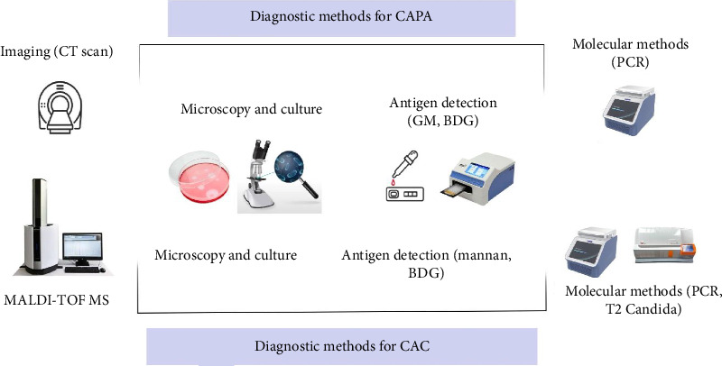
Diagnostic methods for COVID-19-associated fungal infections: CAPA and CAC.
Microscopic examination is a straightforward method for detecting IA, but Aspergillus species rarely sporulate in vivo, leading to potential misidentification. Sensitivity varies widely (0%–90%) due to differences in techniques and mycologist expertise [123–126]. A positive culture from the same specimen is required for confirmation, as visible hyphae alone are insufficient. Obtaining biopsy samples for pulmonary IA can be risky due to low platelet counts. Aspergillus hyphae are thin (1–3 μm) and septate, complicating differentiation from other fungi such as Scedosporium and Fusarium species [122].
Historically, culture was the standard diagnostic method before the introduction of GM and PCR assays [127]. Aspergillus typically grows rapidly (within 48 h) on various media, but culture sensitivity is limited regarding infection versus colonization [122]. Characteristic CT findings for IA include reticular opacities and halo signs, but these are not specific and can overlap with other infections (such as Scedosporium spp. or Cryptococcus spp.), including COVID-19-associated PM [128].
Immunosuppressed patients receiving glucocorticoids may present with Aspergillus bronchopneumonia and varied CT abnormalities, including multiple nodules, lobar infiltrates, and widespread ground-glass opacities. The radiographic signs of these peribronchial zones of consolidation are non-specific and can resemble findings from other infections [129]. CT, particularly when combined with high-resolution CT (HRCT), alongside other diagnostic modalities, is valuable for diagnosing IPA. However, CT imaging can obscure parenchymal abnormalities indicative of CAPA. Detecting IA in mechanically ventilated COVID-19 patients can be challenging due to nodular infiltrates that complicate the identification of surrounding halos [130]. This challenge is exacerbated in severe COVID-19 pneumonia, where CT typically reveals bilateral peripheral ground-glass opacities, consolidation, and bronchovascular thickening. Consequently, distinguishing IA radiological signs from those associated with severe COVID-19 pneumonia is significant [131]. Patti et al. described a mechanically ventilated COVID-19 patient with newly developed thin-walled cavitary lesions populated by fungal ball-like lesions on CT [131]. Additionally, the transport of critically ill patients poses risks, making CT scans not always feasible [132].
GM assays, similar to those for diagnosing mucormycosis, are also unreliable. Other fungi that produce GM include Penicillium, Fusarium, Alternaria, Histoplasma, and Cryptococcus [122]. Swanink et al. noted that Candida spp. can yield low GM assay responses, possibly due to hydroxychloroquine (HCQ) use in COVID-19 treatment protocols [133]. The percentage of neutropenic individuals with IPA who test positive by culture ranges from 10% to 58% [122, 134]. Aspergillus species exhibit a high glucan content in their cell walls, particularly β-D-1,3-glucan (BDG) [135]. A study by Digby et al. found elevated BDG levels in ICU patients with bacterial and fungal infections compared to those without infections [136].
Some studies suggest that early BDG detection during infection progression is more likely than GM detection [137]. The BDG assay has traditionally been used in hematology patients at risk for IPA due to neutrophil deficiency, but it has also shown utility in critical care [138] and COPD patients [139]. In an animal model of IA, the BDG assay demonstrated earlier positivity in bronchoalveolar lavage (BAL) samples compared to serum, with greater overall sensitivity [140]. However, serum BDG also plays a role. Due to limited literature, uncertainty remains regarding its utility in diagnosing CAPA [141]. Despite these challenges, GM measurement techniques are still regarded as the gold standard in many laboratories, including at the PCR level.
9. COVID-19-Associated Candidiasis (CAC) Diagnosis
Physical symptoms, microscopic inspection, and blood cultures are the most common methods for diagnosing candidiasis and candidemia. Recently, multiple instances of oral candidiasis have been documented in COVID-19 patients, raising concerns about increased morbidity and mortality. Early detection of oral candidiasis in these individuals is critical for effective treatment [142]. However, relying solely on clinical criteria is insufficient, as many symptoms are not pathognomonic and are often identified only when treatment becomes difficult. A negative result does not exclude the possibility of infection. Microscopic inspection is a rapid and beneficial diagnostic tool [81, 143]. Blood cultures are considered the gold standard for diagnosing candidiasis, particularly in severe cases of candidemia or invasive candidiasis [144] (Figure 3).
However, fungal cell counts in infected tissues or circulating in the bloodstream are usually low, which can hinder the detection of Candida spp. using simple blood culture tests. As a result, more invasive procedures may be required [17, 27, 72]. Notably, only 50%–70% of patients with Candida bloodstream infections yield positive blood cultures [145]. Additionally, blood cultures may be negative in some COVID-19 patients, especially if drawn after the onset of C. auris fungemia [76]. Traditional phenotypic techniques have led to misidentification of C. auris, commonly mistaking it for C. sake, C. duobushaemulonii, Rhodotorula glutinis, C. haemulonii, or other Candida species [146]. C. auris can thrive in specific media, such as Sabouraud broth, at elevated temperatures (40°C–42°C) or in high salt concentrations (10%) [147–149].
On CHROMagar Candida medium, C. auris typically forms pink colonies, making it challenging to differentiate from C. glabrata and various other yeast species, including those in the C. haemulonii complex [150, 151]. Importantly, C. auris can switch between different colony phenotypes, such as pink, white, and dark purple [147, 151]. A novel chromogenic selective medium, CHROMagar™ Candida Plus, has been developed that allows for the differentiation of C. auris, which forms distinct cream-colored colonies with a blue halo after 48 h of incubation at 37°C [152]. CHROMagar supplemented with Pal's medium has also proven useful for distinguishing C. auris from C. haemulonii [153]. Automated yeast identification systems commonly misidentify C. auris isolates as C. haemulonii or Rhodotorula glutinis [47, 151]. An FDA-approved automated molecular test, the T2Candida nano-diagnostic panel, utilizes magnetic resonance technology to directly detect Candida spp. in whole blood samples within 5 h. This test targets the five most prevalent pathogenic Candida spp., including C. krusei, C. glabrata, C. tropicalis, C. albicans, and C. parapsilosis, and can also detect C. auris [154, 155].
Confirmation of C. auris infection requires additional methods. MALDI-TOF MS technology can rapidly identify yeast species directly from blood cultures within minutes, eliminating the need for sub-culturing [154]. Other techniques, such as BDG assays and mannan antigen testing, are useful for detecting deep-seated infections [154, 156]. Commercial enzyme-linked immunosorbent assay (ELISA) kits can detect Candida mannan antigen (MN) in serum samples, offering relatively high specificity and sensitivity [157]. It is important to note that BDG assays are not species-specific and may yield false positives. Although PCR assays on blood samples are not FDA-approved for diagnostic use, they can identify specific Candida spp. [17, 158]. In patients suspected of having invasive candidiasis, elevated levels of MN in the blood have demonstrated a sensitivity of 58% and a specificity of 93% [157, 159]. Blood PCR has shown pooled sensitivity and specificity of 95% and 92%, respectively, for detecting proven or probable invasive candidiasis compared to at-risk controls [72, 160].
The T2Candida Panel, which amplifies the ITS2 region, directly detects Candida spp. in EDTA blood samples within 5 h and has demonstrated efficacy in diagnosing candidemia and intra-abdominal candidiasis. However, its technical requirements may pose challenges [155, 158, 161]. To enhance sensitivity, combining multiple diagnostic techniques is recommended [27, 154, 156, 159]. The ITS-PCR assay allows differentiation of C. auris from C. haemulonii based on amplicon size [162]. Another approach utilizes direct PCR targeting glycosylphosphatidylinositol (GPI) protein–encoding genes to speciate C. auris and 18 other Candida species by visualizing different-sized amplicons on a gel [163]. Evaluation of a proficiency panel of 44 C. auris and 97 other yeast isolates achieved 100% accuracy (100% sensitivity and specificity) [164]. This assay has been modified to enable direct detection of C. auris from patient swabs, demonstrating 93% sensitivity and 96% specificity [165]. Several laboratories have developed real-time PCR tests for Candida auris, with ongoing research in this area [166–169].
The challenges of fungal infections in the context of COVID-19 extend beyond previous concerns. Patients may experience multiple concurrent fungal infections, complicating the differentiation from bacterial infections and viral superinfections. This multiplicity can hinder the diagnosis of other abnormalities. For instance, coinfections like mucormycosis and oral candidiasis require skilled mycologists to accurately identify based on clinical signs, medical history, and lab findings. Proper sample collection is crucial; some require microscopic examination, while others are cultured, with additional assays like spectrometry and molecular testing. However, challenges arise, particularly with mucormycosis and candidiasis coinfection, where biopsy is necessary due to the delicate nature of mucoromycetes and the risk of spreading infection. Furthermore, corticosteroid treatment can render tissues extremely thin and fragile, complicating the diagnosis and management of these infections [142].
In another scenario, a diabetic man with COVID-19 in the ICU was diagnosed with concurrent pulmonary aspergillosis (Aspergillus fumigatus) and mucormycosis. He underwent comprehensive diagnostics and received multiple antifungal and bacterial therapies. On the ninth day, he developed methicillin-resistant Staphylococcus aureus (MRSA) and Klebsiella pneumoniae ventilator-associated pneumonia. By day 36, despite requiring ventilator support, he tolerated intravenous liposomal amphotericin B (L-AmB) therapy and was eventually transferred to a long-term acute care facility [39]. Another case involved a 78-year-old male with type 2 diabetes mellitus and ketoacidosis, who presented with facial swelling, paralysis, ptosis, and a necrotic palatal ulcer. No COVID-19 coinfection was noted. CT imaging revealed a right maxillary sinus abscess, and cultured secretions identified A. fumigatus. Pathological examination confirmed mucormycosis. The patient received voriconazole, amphotericin B deoxycholate, and surgical debridement but unfortunately did not survive. In immunocompromised individuals, the coexistence of rhino-cerebral mucormycosis and aspergillosis should be considered for early treatment and improved prognosis [170]. A 69-year-old woman with a history of hypertension and diabetes presented with cerebro-rhino-orbital mucormycosis and an aspergillosis coinfection. The challenges of early detection of invasive mucormycosis highlight the importance of considering concomitant infections like aspergillosis. Fortunately, the patient was discharged after a successful recovery [171].
10. Economic Impact of Pandemics on Global Healthcare Systems
Given the aggressive nature of fungal pathogens, particularly mucormycosis, and their resistance to antifungal therapies, timely diagnosis is crucial for effective management and related research. While we did not find a specific article addressing the importance of timely interventions in Aspergillus and Candida infections, healthcare professionals must remain vigilant about potential false negatives and employ various diagnostic techniques, including molecular approaches, to achieve more accurate results.
These factors necessitate the availability of competent healthcare personnel and well-equipped laboratory facilities, which can be particularly challenging in low- and middle-income countries (LMICs). Notably, the ongoing COVID-19 pandemic has significantly affected LMICs, especially in regions such as Asia, sub-Saharan Africa, and South America. Despite efforts to enhance healthcare in these areas, a substantial portion of the population still lacks access to adequate diagnostic and treatment services, as well as sufficient medical personnel. The economies of nations worldwide have been considerably impacted by the COVID-19 pandemic, resulting in both short-term budgetary effects and long-term economic consequences. Measures such as quarantine, health facility preparedness, isolation of infected individuals, and contact tracing require significant public health resource allocation, human resources, and implementation costs. Additionally, healthcare expenses associated with antibiotics, medical supplies, personal protective equipment, and consumables further strain resources. Pandemics can lead to fiscal stress by reducing tax revenues while increasing expenditures, particularly in LMICs, where fiscal constraints are more pronounced and tax systems require further development. For instance, during the Ebola virus outbreak, the loss of government income due to quarantine and curfews severely impacted Liberia's economy, leading to increased public health spending, economic downturns, and reduced revenue. Economic shocks are common during pandemics, exacerbated by labor shortages across various professions, including diagnostic and treatment specialists, alongside increased mortality among both the general population and healthcare workers [172, 173]. Additional challenges include transportation disruptions, workplace closures, trade and travel restrictions, and border closures.
Together, these factors lead to decreased access to specialized care and a sharp rise in the cost of diagnostic kits and research budgets aimed at combating the CoVs. In such circumstances, the risk of other infections, including opportunistic fungal infections, which can lead to significant outbreaks or complicated treatment cases, is often overlooked. These challenges may be further exacerbated in tropical and subtropical regions (e.g., CAM in India). Thus, alternative methods like MALDI-TOF MS, which has a direct cost of only $1403.20 or $35.08 per isolate, resulting in a 74.0% cost savings, can benefit not only LMICs but also numerous clinics and laboratories in more affluent countries facing budget constraints [174]. More expensive methods, such as PCR, may not be reliably feasible. It is important to note that these fungi are typically saprophytic and present in the environment. For instance, Aspergillus spores, which are ubiquitous even in controlled environments, can contaminate molecular detection processes and yield false-positive results, raising questions about the accuracy of these molecular methods.
Candida auris further complicates this issue. Since its initial identification in 2009 from the ear discharge of a 70-year-old Japanese patient, C. auris has spread rapidly worldwide [69]. This species exhibits unique characteristics, including the ability to form biofilms on various surfaces, facilitating nosocomial transmission. C. auris has been isolated from catheters, central and peripheral line tips, and neurological shunts, among other indwelling medical devices [151]. Moreover, C. auris can persist in hospital environments for months by forming “dry” biofilms on environmental surfaces and equipment, such as reusable temperature sensors. Its biofilm-forming capability contributes to its role as a persistent colonizer and a challenging pathogen to eradicate within healthcare facilities [151]. Transcriptional studies have shown that biofilm development confers protection against antifungal medications and detergents, with mature biofilms (aged 24 h) demonstrating resistance to triazoles, polyenes, and echinocandins [175, 176].
As a result, the morphological features and initial diagnosis of this fungus can present challenges for laboratory personnel. Concerning CAM, it is noteworthy that many patients receiving medical treatment, but not on oxygen therapy, have been infected and diagnosed with mucormycosis. However, there does not appear to be a definitive link between oxygen therapy and susceptibility to this infection [177]. In general, it is crucial to adhere to established protocols, maintain distance, and limit the use of supportive devices, such as mechanical ventilator. While these measures are ideal in theory, the reality of managing severe COVID-19 cases, a surge of patients requiring urgent hospitalization, and overwhelmed medical staff under significant stress makes it challenging to observe all principles for addressing fungal species that are likely to be resistant to fungicides or that may rapidly develop resistance. Timely detection and differentiation of opportunistic fungal species from other infectious agents pose significant challenges. Currently, no studies have examined the effects of corticosteroids, immunomodulators, and other risk factors on the morphology of hyphae, spores, conidia, and yeasts. However, based on the authors' experience with dermatophytic fungi, this may also present moderate challenges.
11. Conclusion
IFIs have become a significant public health issue, particularly highlighted by recent outbreaks. These infections pose serious risks to hospitalized patients, especially in ICUs, with mortality rates ranging from 5% to 70% [14]. During the COVID-19 pandemic, the incidence of IFIs among patients with severe SARS-CoV-2 infection was reported between 4% and 27.7%, primarily involving pathogens such as Mucor, Aspergillus, and Candida species [16].
Timely and accurate identification of these fungal pathogens is essential for effective treatment; however, diagnostic challenges hinder early detection. Delayed treatment, particularly for invasive candidiasis, can increase mortality rates by 20% [178, 179]. Existing diagnostic methods, including microscopy, culture, antigen or antibody testing, and molecular assays, are limited by factors such as laboratory personnel expertise and the potential for false-positive results [79–81].
Advancements in molecular techniques and antigen detection offer alternatives to traditional diagnostics. Methods like PCR and mass spectrometry show promise for rapid and accurate identification of fungal infections, yet issues such as contamination and cost-effectiveness remain significant challenges. In resource-limited settings, environmental factors further complicate the management of IFIs, underscoring the need for improved diagnostic approaches.
In humid environments, such as India, conditions favor the growth of Mucor spp., especially in overcrowded settings with limited healthcare resources. The emergence of species like Candida auris, which exhibit high resistance to various disinfectants, presents additional challenges, particularly during the COVID-19 pandemic and in the context of chronic infectious diseases.
Looking ahead, addressing the challenges and limitations associated with future viral infections will be paramount. Key obstacles include the following:
1. Resource constraints: Many healthcare systems, particularly in LMICs, face significant limitations in resources, infrastructure, and trained personnel, which can hinder timely diagnosis and treatment of viral infections.
2. Diagnostic limitations: Current diagnostic methods may not be sufficiently rapid or sensitive to detect emerging viral pathogens early, leading to delays in intervention and increased transmission rates.
3. Viral variability: The ability of viruses to mutate rapidly poses a challenge for vaccine development and effectiveness, as well as for diagnostic accuracy.
4. Public health infrastructure: Insufficient public health infrastructure can impede effective surveillance and response to outbreaks, making it difficult to implement timely interventions.
5. Antiviral resistance: Just as with fungal pathogens, the emergence of resistant viral strains can complicate treatment options and necessitate ongoing research and development of new antiviral agents.
Acknowledging these limitations and challenges is crucial for developing effective strategies to combat future viral infections. Further research and development are necessary to enhance the accuracy and accessibility of diagnostics, ultimately improving patient outcomes in the face of these growing threats.
Acknowledgments
We are grateful to Rasoul Soleimani-Estyar and Danial Astaraki for their assistance in conducting this research.
Contributor Information
Amir Sasan Mozaffari Nejad, Email: asmozafarinejad@yahoo.in.
Saeme Asgari, Email: saeme.asgari@iau.ac.ir.
Data Availability Statement
Data sharing is not applicable to this article as no new data were created or analyzed in this study.
Conflicts of Interest
The authors declare no conflicts of interest.
Funding
No funding was received for this research.
References
- 1.Huremović D. Brief History of Pandemics (Pandemics Throughout History) Psychiatry of Pandemics . 2019:7–35. doi: 10.1007/978-3-030-15346-5_2. [DOI] [Google Scholar]
- 2.Li F. Structure, Function, and Evolution of Coronavirus Spike Proteins. Annual Review of Virology . 2016;3(1):237–261. doi: 10.1146/annurev-virology-110615-042301. [DOI] [PMC free article] [PubMed] [Google Scholar]
- 3.Shahgolzari M., Yavari A., Arjeini Y., et al. Immunopathology and Immunopathogenesis of COVID-19, What We Know and What We Should Learn. Gene Reports . 2021;25 doi: 10.1016/j.genrep.2021.101417. [DOI] [PMC free article] [PubMed] [Google Scholar]
- 4.Cui J., Li F., Shi Z. L. Origin and Evolution of Pathogenic Coronaviruses. Nature Reviews Microbiology . 2019;17(3):181–192. doi: 10.1038/s41579-018-0118-9. [DOI] [PMC free article] [PubMed] [Google Scholar]
- 5.Hu B., Guo H., Zhou P., Shi Z.-L. Characteristics of SARS-CoV-2 and COVID-19. Nature Reviews Microbiology . 2021;19(3):141–154. doi: 10.1038/s41579-020-00459-7. [DOI] [PMC free article] [PubMed] [Google Scholar]
- 6.Zandi M., Shafaati M., Kalantar-Neyestanaki D., et al. The Role of SARS-CoV-2 Accessory Proteins in Immune Evasion. Biomedicine & Pharmacotherapy . 2022;156 doi: 10.1016/j.biopha.2022.113889. [DOI] [PMC free article] [PubMed] [Google Scholar]
- 7.Kesheh M. M., Hosseini P., Soltani S., Zandi M. An Overview on the Seven Pathogenic Human Coronaviruses. Reviews in Medical Virology . 2022;32(2):p. e2282. doi: 10.1002/rmv.2282. [DOI] [PubMed] [Google Scholar]
- 8.Hui D. S., I Azhar E., Madani T., et al. The Continuing 2019-nCoV Epidemic Threat of Novel Coronaviruses to Global Health—The Latest 2019 Novel Coronavirus Outbreak in Wuhan, China. International Journal of Infectious Diseases . 2020;91:264–266. doi: 10.1016/j.ijid.2020.01.009. [DOI] [PMC free article] [PubMed] [Google Scholar]
- 9.Wu J. T., Leung K., Leung G. M. Nowcasting and Forecasting the Potential Domestic and International Spread of the 2019-nCoV Outbreak Originating in Wuhan, China: A Modelling Study. The Lancet . 2020;395(10225):689–697. doi: 10.1016/s0140-6736(20)30260-9. [DOI] [PMC free article] [PubMed] [Google Scholar]
- 10.Deng S.-Q., Peng H.-J. Characteristics of and Public Health Responses to the Coronavirus Disease 2019 Outbreak in China. Journal of Clinical Medicine . 2020;9(2):p. 575. doi: 10.3390/jcm9020575. [DOI] [PMC free article] [PubMed] [Google Scholar]
- 11.Han Q., Lin Q., Jin S., You L. Coronavirus 2019-nCoV: A Brief Perspective From the Front Line. Journal of Infection . 2020;80(4):373–377. doi: 10.1016/j.jinf.2020.02.010. [DOI] [PMC free article] [PubMed] [Google Scholar]
- 12.Al-Hatmi A. M., Mohsin J., Al-Huraizi A., Khamis F. COVID-19 Associated Invasive Candidiasis. Journal of Infection . 2021;82(2):e45–e46. doi: 10.1016/j.jinf.2020.08.005. [DOI] [PMC free article] [PubMed] [Google Scholar]
- 13.Feldman C., Anderson R. The Role of Co-Infections and Secondary Infections in Patients with COVID-19. Pneumonia (Nathan Qld) . 2021;13(1) doi: 10.1186/s41479-021-00083-w. [DOI] [PMC free article] [PubMed] [Google Scholar]
- 14.Coşkun A. S., Durmaz S. Fungal Infections in COVID-19 Intensive Care Patients. Polish Journal of Microbiology . 2021;70(3):395–400. doi: 10.33073/pjm-2021-039. [DOI] [PMC free article] [PubMed] [Google Scholar]
- 15.Litvintseva A. P., Brandt M. E., Mody R. K., Lockhart S. R. Investigating Fungal Outbreaks in the 21st Century. PLoS Pathogens . 2015;11(5):p. e1004804. doi: 10.1371/journal.ppat.1004804. [DOI] [PMC free article] [PubMed] [Google Scholar]
- 16.Segrelles-Calvo G., de S Araújo G. R., Llopis-Pastor E., et al. Candida Spp. Co-Infection in COVID-19 Patients With Severe Pneumonia: Prevalence Study and Associated Risk Factors. Respiratory Medicine . 2021;188 doi: 10.1016/j.rmed.2021.106619. [DOI] [PMC free article] [PubMed] [Google Scholar]
- 17.Ghosh A., Sarkar A., Paul P., Patel P. The Rise in Cases of Mucormycosis, Candidiasis and Aspergillosis Amidst COVID19. Fungal Biology Reviews . 2021;38:67–91. doi: 10.1016/j.fbr.2021.09.003. [DOI] [PMC free article] [PubMed] [Google Scholar]
- 18.Song G., Liang G., Liu W. Fungal Co-Infections Associated With Global COVID-19 Pandemic: A Clinical and Diagnostic Perspective From China. Mycopathologia . 2020;185(4):599–606. doi: 10.1007/s11046-020-00462-9. [DOI] [PMC free article] [PubMed] [Google Scholar]
- 19.Silva L. N., de Mello T. P., de Souza Ramos L., Branquinha M. H., Roudbary M., Dos Santos A. L. S. Fungal Infections in COVID-19-Positive Patients: A Lack of Optimal Treatment Options. Current Topics in Medicinal Chemistry . 2020;20(22):1951–1957. doi: 10.2174/156802662022200917110102. [DOI] [PubMed] [Google Scholar]
- 20.Ghanem H., Sivasubramanian G. Cryptococcus Neoformans Meningoencephalitis in an Immunocompetent Patient after COVID-19 Infection . Case Reports in Infectious Diseases; 2021. [DOI] [PMC free article] [PubMed] [Google Scholar]
- 21.Pemán J., Ruiz-Gaitán A., García-Vidal C., et al. Fungal Co-Infection in COVID-19 Patients: Should We Be Concerned? Revista Iberoamericana De Micologia . 2020;37(2):41–46. doi: 10.1016/j.riam.2020.07.001. [DOI] [PMC free article] [PubMed] [Google Scholar]
- 22.Roudbary M., Kumar S., Kumar A., Černáková L., Nikoomanesh F., Rodrigues C. F. Overview on the Prevalence of Fungal Infections, Immune Response, and Microbiome Role in COVID-19 Patients. Journal of Fungi . 2021;7(9):p. 720. doi: 10.3390/jof7090720. [DOI] [PMC free article] [PubMed] [Google Scholar]
- 23.Abdoli A., Falahi S., Kenarkoohi A. COVID-19-Associated Opportunistic Infections: A Snapshot on the Current Reports. Clinical and Experimental Medicine . 2021;22(3):327–346. doi: 10.1007/s10238-021-00751-7. [DOI] [PMC free article] [PubMed] [Google Scholar]
- 24.Chang C. C., Senining R., Kim J., Goyal R. An Acute Pulmonary Coccidioidomycosis Coinfection in a Patient Presenting With Multifocal Pneumonia with COVID-19. Journal of Investigative Medicine High Impact Case Reports . 2020;8 doi: 10.1177/2324709620972244. [DOI] [PMC free article] [PubMed] [Google Scholar]
- 25.Ventoulis I., Sarmourli T., Amoiridou P., et al. Bloodstream Infection by Saccharomyces cerevisiae in Two COVID-19 Patients After Receiving Supplementation of Saccharomyces in the ICU. Journal of Fungi . 2020;6(3):p. 98. doi: 10.3390/jof6030098. [DOI] [PMC free article] [PubMed] [Google Scholar]
- 26.Poignon C., Blaize M., Vezinet C., Lampros A., Monsel A., Fekkar A. Invasive Pulmonary Fusariosis in an Immunocompetent Critically Ill Patient With Severe COVID-19. Clinical Microbiology and Infection . 2020;26(11):1582–1584. doi: 10.1016/j.cmi.2020.06.026. [DOI] [PMC free article] [PubMed] [Google Scholar]
- 27.Arastehfar A., Shaban T., Zarrinfar H., et al. Candidemia Among Iranian Patients With Severe COVID-19 Admitted to ICUs. Journal of Fungi . 2021;7(4):p. 280. doi: 10.3390/jof7040280. [DOI] [PMC free article] [PubMed] [Google Scholar]
- 28.Segrelles-Calvo G., Araújo G. R. D. S., Llopis-Pastor E., Frasés S. Trichosporon Asahii as Cause of Nosocomial Pneumonia in Patient With COVID-19: A Triple Co-Infection. Archivos de Bronconeumología . 2021;57:46–48. doi: 10.1016/j.arbres.2020.11.007. [DOI] [PMC free article] [PubMed] [Google Scholar]
- 29.Kottarathil M., Thayanidhi P., Sathyamurthy P., Jyoti Kindo A. Rise of Mucormycosis during the COVID-19 Pandemic and the Challenges Faced. Current Medical Mycology . 2023;9(1):44–55. doi: 10.18502/cmm.2023.345032.1400. [DOI] [PMC free article] [PubMed] [Google Scholar]
- 30.Pippal S. K., Kumar D., Ukawat L. Management Challenge of Rhino-Orbito-Cerebral Mucormycosis in Covid 19 Era: A Prospective Observational Study. Indian Journal of Otolaryngology and Head & Neck Surgery . 2021;74(S2):3485–3491. doi: 10.1007/s12070-021-02947-5. [DOI] [PMC free article] [PubMed] [Google Scholar]
- 31.Jeong W., Keighley C., Wolfe R., et al. The Epidemiology and Clinical Manifestations of Mucormycosis: A Systematic Review and Meta-Analysis of Case Reports. Clinical Microbiology and Infection . 2019;25(1):26–34. doi: 10.1016/j.cmi.2018.07.011. [DOI] [PubMed] [Google Scholar]
- 32.Mahalaxmi I., Jayaramayya K., Venkatesan D., et al. Mucormycosis: An Opportunistic Pathogen During COVID-19. Environmental Research . 2021;201 doi: 10.1016/j.envres.2021.111643. [DOI] [PMC free article] [PubMed] [Google Scholar]
- 33.Singh K., Kumar S., Shastri S., Sudershan A., Mansotra V. Black Fungus Immunosuppressive Epidemic with Covid-19 Associated Mucormycosis (Zygomycosis): A Clinical and Diagnostic Perspective From India. Immunogenetics . 2021;74(2):197–206. doi: 10.1007/s00251-021-01226-5. [DOI] [PMC free article] [PubMed] [Google Scholar]
- 34.Prakash H., Chakrabarti A. Epidemiology of Mucormycosis in India. Microorganisms . 2021;9(3):p. 523. doi: 10.3390/microorganisms9030523. [DOI] [PMC free article] [PubMed] [Google Scholar]
- 35.Sarkar S., Gokhale T., Choudhury S. S., Deb A. K. COVID-19 and Orbital Mucormycosis. Indian Journal of Ophthalmology . 2021;69(4):1002–1004. doi: 10.4103/ijo.ijo_3763_20. [DOI] [PMC free article] [PubMed] [Google Scholar]
- 36.Zurl C., Hoenigl M., Schulz E., et al. Autopsy Proven Pulmonary Mucormycosis Due to Rhizopus Microsporus in a Critically Ill COVID-19 Patient With Underlying Hematological Malignancy. Journal of Fungi . 2021;7(2):p. 88. doi: 10.3390/jof7020088. [DOI] [PMC free article] [PubMed] [Google Scholar]
- 37.Yasmin F., Najeeb H., Naeem A., et al. COVID-19 Associated Mucormycosis: A Systematic Review From Diagnostic Challenges to Management. Diseases . 2021;9(4):p. 65. doi: 10.3390/diseases9040065. [DOI] [PMC free article] [PubMed] [Google Scholar]
- 38.Chandley P., Subba P., Rohatgi S. COVID-19-Associated Mucormycosis: A Matter of Concern Amid the SARS-CoV-2 Pandemic. Vaccines . 2022;10(8):p. 1266. doi: 10.3390/vaccines10081266. [DOI] [PMC free article] [PubMed] [Google Scholar]
- 39.John T. M., Jacob C. N., Kontoyiannis D. P. When Uncontrolled Diabetes Mellitus and Severe COVID-19 Converge: The Perfect Storm for Mucormycosis. Journal of Fungi . 2021;7(4):p. 298. doi: 10.3390/jof7040298. [DOI] [PMC free article] [PubMed] [Google Scholar]
- 40.Johnson A. K., Ghazarian Z., Cendrowski K. D., Persichino J. G. Pulmonary Aspergillosis and Mucormycosis in a Patient With COVID-19. Medical mycology case reports . 2021;32:64–67. doi: 10.1016/j.mmcr.2021.03.006. [DOI] [PMC free article] [PubMed] [Google Scholar]
- 41.Moorthy A., Gaikwad R., Krishna S., et al. SARS-CoV-2, Uncontrolled Diabetes and Corticosteroids—An Unholy Trinity in Invasive Fungal Infections of the Maxillofacial Region? A Retrospective, Multi-Centric Analysis. Journal of Maxillofacial and Oral Surgery . 2021;20(3):418–425. doi: 10.1007/s12663-021-01532-1. [DOI] [PMC free article] [PubMed] [Google Scholar]
- 42.Staats C. C., Kmetzsch L., Schrank A., Vainstein M. H. Fungal Zinc Metabolism and Its Connections to Virulence. Frontiers in Cellular and Infection Microbiology . 2013;3:p. 65. doi: 10.3389/fcimb.2013.00065. [DOI] [PMC free article] [PubMed] [Google Scholar]
- 43.Leonardelli F., Macedo D., Dudiuk C., et al. In Vitro Activity of Combinations of Zinc Chelators With Amphotericin B and Posaconazole Against Six Mucorales Species. Antimicrobial Agents and Chemotherapy . 2019;63(5) doi: 10.1128/aac.00266-19. [DOI] [PMC free article] [PubMed] [Google Scholar]
- 44.Fernández-García O., Guerrero-Torres L., Roman-Montes C. M., et al. Isolation of Rhizopus Microsporus and Lichtheimia Corymbifera From Tracheal Aspirates of Two Immunocompetent Critically Ill Patients With COVID-19. Medical Mycology Case Reports . 2021;33:32–37. doi: 10.1016/j.mmcr.2021.07.001. [DOI] [PMC free article] [PubMed] [Google Scholar]
- 45.Kamkar A., Fallah A. A., Mozaffari Nejad A. S. The Review of Aflatoxin M1 Contamination in Milk and Dairy Products Produced in Iran. Toxin Reviews . 2014;33(4):160–168. doi: 10.3109/15569543.2014.922580. [DOI] [Google Scholar]
- 46.Mozaffarinejad A. S., Giri A. The Measurement of Aflatoxin B1 in Chilli and Black Peppers of Qaemshahr, Iran. Journal of Kerman University of Medical Sciences . 2015;22(2):185–193. [Google Scholar]
- 47.Chowdhary A., Sharma C., Meis J. F. Azole-Resistant Aspergillosis: Epidemiology, Molecular Mechanisms, and Treatment. The Journal of Infectious Diseases . 2017;216(suppl_3):S436–S444. doi: 10.1093/infdis/jix210. [DOI] [PubMed] [Google Scholar]
- 48.Paulussen C., Hallsworth J. E., Álvarez‐Pérez S., et al. Ecology of Aspergillosis: Insights Into the Pathogenic Potency of Aspergillus fumigatus and Some Other Aspergillus Species. Microbial Biotechnology . 2017;10(2):296–322. doi: 10.1111/1751-7915.12367. [DOI] [PMC free article] [PubMed] [Google Scholar]
- 49.Kosmidis C., Denning D. W. The Clinical Spectrum of Pulmonary Aspergillosis. Thorax . 2015;70(3):270–277. doi: 10.1136/thoraxjnl-2014-206291. [DOI] [PubMed] [Google Scholar]
- 50.Ashtiani N. M., Kachuei R., Yalfani R., Harchegani A. B., Nosratabadi M. Identification of Aspergillus Sections Flavi, Nigri, and Fumigati and Their Differentiation Using Specific Primers. Informe Medico . 2017;25(2):127–132. [PubMed] [Google Scholar]
- 51.Schauwvlieghe A. F., Rijnders B. J., Philips N., et al. Invasive Aspergillosis in Patients Admitted to the Intensive Care Unit With Severe Influenza: A Retrospective Cohort Study. The Lancet Respiratory Medicine . 2018;6(10):782–792. doi: 10.1016/s2213-2600(18)30274-1. [DOI] [PubMed] [Google Scholar]
- 52.Vanderbeke L., Spriet I., Breynaert C., Rijnders B. J., Verweij P. E., Wauters J. Invasive Pulmonary Aspergillosis Complicating Severe Influenza: Epidemiology, Diagnosis and Treatment. Current Opinion in Infectious Diseases . 2018;31(6):471–480. doi: 10.1097/qco.0000000000000504. [DOI] [PubMed] [Google Scholar]
- 53.Montrucchio G., Lupia T., Lombardo D., et al. Risk Factors for Invasive Aspergillosis in ICU Patients With COVID-19: Current Insights and New Key Elements. Annals of Intensive Care . 2021;11(1):136–211. doi: 10.1186/s13613-021-00923-4. [DOI] [PMC free article] [PubMed] [Google Scholar]
- 54.Blot S., Rello J., Koulenti D. Diagnosing Invasive Pulmonary Aspergillosis in ICU Patients: Putting the Puzzle Together. Current Opinion in Critical Care . 2019;25(5):430–437. doi: 10.1097/mcc.0000000000000637. [DOI] [PubMed] [Google Scholar]
- 55.Cocio T. A., Siqueira L. P. M., Riciluca K. C. T., et al. Significance of Aspergillus Spp. Isolation in Defining Cases of COVID-19 Associated Pulmonary Aspergillosis–CAPA. Brazilian Journal of Infectious Diseases . 2023;27(4) doi: 10.1016/j.bjid.2023.102793. [DOI] [PMC free article] [PubMed] [Google Scholar]
- 56.Hallen-Adams H. E., Suhr M. J. Fungi in the Healthy Human Gastrointestinal Tract. Virulence . 2017;8(3):352–358. doi: 10.1080/21505594.2016.1247140. [DOI] [PMC free article] [PubMed] [Google Scholar]
- 57.Rolling T., Hohl T. M., Zhai B. Minority Report: The Intestinal Mycobiota in Systemic Infections. Current Opinion in Microbiology . 2020;56:1–6. doi: 10.1016/j.mib.2020.05.004. [DOI] [PMC free article] [PubMed] [Google Scholar]
- 58.Deorukhkar S. C., Saini S., Mathew S. Non-Albicans Candida Infection: an Emerging Threat. Interdisciplinary Perspectives on Infectious Diseases . 2014;2014:1–7. doi: 10.1155/2014/615958. [DOI] [PMC free article] [PubMed] [Google Scholar]
- 59.Shahin-Jafari A., Bayat M., Shahhosseiny M. H., Tajik P., Roudbar-Mohammadi S. Effect of Long-Term Exposure to Mobile Phone Radiation on Alpha-Int1 Gene Sequence of Candida Albicans. Saudi Journal of Biological Sciences . 2016;23(3):426–433. doi: 10.1016/j.sjbs.2015.05.001. [DOI] [PMC free article] [PubMed] [Google Scholar]
- 60.Taei M., Chadeganipour M., Mohammadi R. An Alarming Rise of Non-Albicans Candida Species and Uncommon Yeasts in the Clinical Samples; a Combination of Various Molecular Techniques for Identification of Etiologic Agents. BMC Research Notes . 2019;12(1):779–787. doi: 10.1186/s13104-019-4811-1. [DOI] [PMC free article] [PubMed] [Google Scholar]
- 61.Ngamchokwathana C., Chongtrakool P., Waesamaae A., Chayakulkeeree M. Risk Factors and Outcomes of Non-Albicans Candida Bloodstream Infection in Patients With Candidemia at Siriraj Hospital—Thailand’s Largest National Tertiary Referral Hospital. Journal of Fungi . 2021;7(4):p. 269. doi: 10.3390/jof7040269. [DOI] [PMC free article] [PubMed] [Google Scholar]
- 62.Nadagir S. D., Chunchanur S. K., Halesh L., Yasmeen K., Chandrasekhar M., Patil B. Significance of Isolation and Drug Susceptibility Testing of Non-Candida Albicans Species Causing Oropharyngeal Candidiasis in HIV Patients. Southeast Asian Journal of Tropical Medicine & Public Health . 2008;39(3):492–495. [PubMed] [Google Scholar]
- 63.Sardi J., Scorzoni L., Bernardi T., Fusco-Almeida A., Mendes Giannini M. J. S. Candida Species: Current Epidemiology, Pathogenicity, Biofilm Formation, Natural Antifungal Products and New Therapeutic Options. Journal of Medical Microbiology . 2013;62(1):10–24. doi: 10.1099/jmm.0.045054-0. [DOI] [PubMed] [Google Scholar]
- 64.Hankovszky P., Társy D., Öveges N., Molnár Z. Invasive Candida Infections in the ICU: Diagnosis and Therapy. The Journal of Critical Care Medicine. . 2015;1(4):129–139. doi: 10.1515/jccm-2015-0025. [DOI] [PMC free article] [PubMed] [Google Scholar]
- 65.Sullivan D., Henman M., Moran G., et al. Molecular Genetic Approaches to Identification, Epidemiology and Taxonomy of Non-Albicans Candida Species. Journal of Medical Microbiology . 1996;44(6):399–408. doi: 10.1099/00222615-44-6-399. [DOI] [PubMed] [Google Scholar]
- 66.Marra A. R., Camargo L. F. A., Pignatari A. C. C., et al. Nosocomial Bloodstream Infections in Brazilian Hospitals: Analysis of 2,563 Cases From a Prospective Nationwide Surveillance Study. Journal of Clinical Microbiology . 2011;49(5):1866–1871. doi: 10.1128/jcm.00376-11. [DOI] [PMC free article] [PubMed] [Google Scholar]
- 67.Chybowska A. D., Childers D. S., Farrer R. A. Nine Things Genomics Can Tell Us About Candida Auris. Frontiers in Genetics . 2020;11:p. 351. doi: 10.3389/fgene.2020.00351. [DOI] [PMC free article] [PubMed] [Google Scholar]
- 68.Garcia-Bustos V., Cabanero-Navalon M. D., Ruiz-Saurí A., et al. What Do We Know about Candida Auris? State of the Art, Knowledge Gaps, and Future Directions. Microorganisms . 2021;9(10):p. 2177. doi: 10.3390/microorganisms9102177. [DOI] [PMC free article] [PubMed] [Google Scholar]
- 69.Satoh K., Makimura K., Hasumi Y., Nishiyama Y., Uchida K., Yamaguchi H. Candida Auris Sp a Novel Ascomycetous Yeast Isolated From the External Ear Canal of an Inpatient in a Japanese Hospital. Microbiology and Immunology . 2009;53(1):41–44. doi: 10.1111/j.1348-0421.2008.00083.x. [DOI] [PubMed] [Google Scholar]
- 70.Cendejas-Bueno E., Kolecka A., Alastruey-Izquierdo A., et al. Reclassification of the Candida Haemulonii Complex as Candida Haemulonii (C. Haemulonii Group I), C. Duobushaemulonii Sp. nov.(C. Haemulonii Group II), and C. Haemulonii Var. Vulnera Var. nov.: Three Multiresistant Human Pathogenic Yeasts. Journal of Clinical Microbiology . 2012;50(11):3641–3651. doi: 10.1128/jcm.02248-12. [DOI] [PMC free article] [PubMed] [Google Scholar]
- 71.Keighley C., Garnham K., Harch S., et al. Candida Auris: Diagnostic Challenges and Emerging Opportunities for the Clinical Microbiology Laboratory. Current fungal infection reports . 2021;15(3):116–126. doi: 10.1007/s12281-021-00420-y. [DOI] [PMC free article] [PubMed] [Google Scholar]
- 72.Arastehfar A., Carvalho A., van de Veerdonk F. L., et al. COVID-19 Associated Pulmonary Aspergillosis (CAPA)—From Immunology to Treatment. Journal of Fungi . 2020;6(2):p. 91. doi: 10.3390/jof6020091. [DOI] [PMC free article] [PubMed] [Google Scholar]
- 73.Chowdhary A., Tarai B., Singh A., Sharma A. Multidrug-Resistant Candida Auris Infections in Critically Ill Coronavirus Disease Patients, India, April–July 2020. Emerging Infectious Diseases . 2020;26(11):2694–2696. doi: 10.3201/eid2611.203504. [DOI] [PMC free article] [PubMed] [Google Scholar]
- 74.de Almeida J. N., Jr, Francisco E. C., Hagen F., et al. Emergence of Candida Auris in Brazil in a COVID-19 Intensive Care Unit. Journal of Fungi . 2021;7(3):p. 220. doi: 10.3390/jof7030220. [DOI] [PMC free article] [PubMed] [Google Scholar]
- 75.Villanueva-Lozano H., Treviño-Rangel R. d J., González G. M., et al. Outbreak of Candida Auris Infection in a COVID-19 Hospital in Mexico. Clinical Microbiology and Infection . 2021;27(5):813–816. doi: 10.1016/j.cmi.2020.12.030. [DOI] [PMC free article] [PubMed] [Google Scholar]
- 76.Goravey W., Ali G. A., Ali M., Ibrahim E. B., Al Maslamani M., Abdel Hadi H. Ominous Combination: COVID‐19 Disease and Candida Auris Fungemia—Case Report and Review of the Literature. Clinical Case Reports . 2021;9(9) doi: 10.1002/ccr3.4827. [DOI] [PMC free article] [PubMed] [Google Scholar]
- 77.Salehi M., Ahmadikia K., Mahmoudi S., et al. Oropharyngeal Candidiasis in Hospitalised COVID‐19 Patients From Iran: Species Identification and Antifungal Susceptibility Pattern. Mycoses . 2020;63(8):771–778. doi: 10.1111/myc.13137. [DOI] [PMC free article] [PubMed] [Google Scholar]
- 78.Awada B., Alam W., Chalfoun M., Araj G., Bizri A. R. COVID-19 and Candida Duobushaemulonii Superinfection: A Case Report. Journal of Medical Mycology . 2021;31(3) doi: 10.1016/j.mycmed.2021.101168. [DOI] [PMC free article] [PubMed] [Google Scholar]
- 79.Lackner M., Lass-Florl C. Up-date on Diagnostic Strategies of Invasive Aspergillosis. Current Pharmaceutical Design . 2013;19(20):3595–3614. doi: 10.2174/13816128113199990323. [DOI] [PubMed] [Google Scholar]
- 80.Lackner N., Posch W., Lass-Flörl C. Microbiological and Molecular Diagnosis of Mucormycosis: From Old to New. Microorganisms . 2021;9(7):p. 1518. doi: 10.3390/microorganisms9071518. [DOI] [PMC free article] [PubMed] [Google Scholar]
- 81.Schelenz S., Barnes R. A., Barton R. C., et al. British Society for Medical Mycology Best Practice Recommendations for the Diagnosis of Serious Fungal Diseases. The Lancet Infectious Diseases . 2015;15(4):461–474. doi: 10.1016/s1473-3099(15)70006-x. [DOI] [PubMed] [Google Scholar]
- 82.Singh V. Fungal Rhinosinusitis: Unravelling the Disease Spectrum. Journal of Maxillofacial and Oral Surgery . 2019;18(2):164–179. doi: 10.1007/s12663-018-01182-w. [DOI] [PMC free article] [PubMed] [Google Scholar]
- 83.Kozel T. R., Wickes B. Fungal Diagnostics. Cold Spring Harbor Perspectives in medicine . 2014;4(4):p. a019299. doi: 10.1101/cshperspect.a019299. [DOI] [PMC free article] [PubMed] [Google Scholar]
- 84.Lass-Flörl C. Current Challenges in the Diagnosis of Fungal Infections. Methods in Molecular Biology . 2017;1508:3–15. doi: 10.1007/978-1-4939-6515-1_1. [DOI] [PubMed] [Google Scholar]
- 85.Lass-Flörl C., Mayr A. Diagnosing Invasive Fungal Diseases–Limitations of Microbiological Diagnostic Methods. Expert Opinion on Medical Diagnostics . 2009;3(4):461–470. doi: 10.1517/17530050902878031. [DOI] [PubMed] [Google Scholar]
- 86.Martin K. J., Rygiewicz P. T. Fungal-Specific PCR Primers Developed for Analysis of the ITS Region of Environmental DNA Extracts. BMC microbiology . 2005;5(1):28–11. doi: 10.1186/1471-2180-5-28. [DOI] [PMC free article] [PubMed] [Google Scholar]
- 87.Miceli M. H., Maertens J., editors. Seminars in Respiratory and Critical Care Medicine . Thieme Medical Publishers; 2015. Role of Non-Culture-Based Tests, With an Emphasis on Galactomannan Testing for the Diagnosis of Invasive Aspergillosis. [DOI] [PubMed] [Google Scholar]
- 88.Hoenigl M., Seidel D., Carvalho A., et al. The Emergence of COVID-19 Associated Mucormycosis: A Review of Cases from 18 Countries. The Lancet Microbe . 2022;3(7):e543–e552. doi: 10.1016/s2666-5247(21)00237-8. [DOI] [PMC free article] [PubMed] [Google Scholar]
- 89.Riad A., Shabaan A. A., Issa J., et al. COVID-19-Associated Mucormycosis (CAM): Case-Series and Global Analysis of Mortality Risk Factors. Journal of Fungi . 2021;7(10):p. 837. doi: 10.3390/jof7100837. [DOI] [PMC free article] [PubMed] [Google Scholar]
- 90.Manjunath K. S., Shivaswamy S., Kulkarni J. D., Kenkare Venkatachalaiah R. Rhino-Orbito-Cerebral Mucormycosis (ROCM) With Internal Carotid Artery Stenosis in a Diabetic Patient With Caries Tooth and Oroantral Fistula. BJR Case Reports . 2016;2(2) doi: 10.1259/bjrcr.20150447. [DOI] [PMC free article] [PubMed] [Google Scholar]
- 91.Frater J. L., Hall G. S., Procop G. W. Histologic Features of Zygomycosis: Emphasis on Perineural Invasion and Fungal Morphology. Archives of Pathology and Laboratory Medicine . 2001;125(3):375–378. doi: 10.5858/2001-125-0375-hfoz. [DOI] [PubMed] [Google Scholar]
- 92.Lass-Flörl C. Zygomycosis: Conventional Laboratory Diagnosis. Clinical Microbiology and Infection . 2009;15:60–65. doi: 10.1111/j.1469-0691.2009.02999.x. [DOI] [PubMed] [Google Scholar]
- 93.Monheit J., Cowan D., Moore D. Rapid Detection of Fungi in Tissues Using Calcofluor White and Fluorescence Microscopy. Archives of Pathology and Laboratory Medicine . 1984;108(8):616–618. [PubMed] [Google Scholar]
- 94.Ribes J. A., Vanover-Sams C. L., Baker D. J. Zygomycetes in Human Disease. Clinical Microbiology Reviews . 2000;13(2):236–301. doi: 10.1128/cmr.13.2.236. [DOI] [PMC free article] [PubMed] [Google Scholar]
- 95.Skiada A., Lass-Floerl C., Klimko N., Ibrahim A., Roilides E., Petrikkos G. Challenges in the Diagnosis and Treatment of Mucormycosis. Medical Mycology . 2018;56(suppl_1):S93–S101. doi: 10.1093/mmy/myx101. [DOI] [PMC free article] [PubMed] [Google Scholar]
- 96.Skiada A., Pavleas I., Drogari-Apiranthitou M. Epidemiology and Diagnosis of Mucormycosis: An Update. Journal of Fungi . 2020;6(4):p. 265. doi: 10.3390/jof6040265. [DOI] [PMC free article] [PubMed] [Google Scholar]
- 97.Hayden R., Qian X., Procop G., Roberts G., Lloyd R. In Situ Hybridization for the Identification of Filamentous Fungi in Tissue Section. Diagnostic Molecular Pathology . 2002;11(2):119–126. doi: 10.1097/00019606-200206000-00009. [DOI] [PubMed] [Google Scholar]
- 98.Abdelsalam M., Althaqafi R. M. M., Assiri S. A., et al. Clinical and Laboratory Findings of COVID-19 in High-Altitude Inhabitants of Saudi Arabia. Frontiers of Medicine . 2021;8 doi: 10.3389/fmed.2021.670195. [DOI] [PMC free article] [PubMed] [Google Scholar]
- 99.Francone M., Iafrate F., Masci G. M., et al. Chest CT Score in COVID-19 Patients: Correlation With Disease Severity and Short-Term Prognosis. European Radiology . 2020;30(12):6808–6817. doi: 10.1007/s00330-020-07033-y. [DOI] [PMC free article] [PubMed] [Google Scholar]
- 100.Al-Smadi A. S., Bhatnagar A., Ali R., Lewis N., Johnson S. Correlation of Chest Radiography Findings With the Severity and Progression of COVID-19 Pneumonia. Clinical Imaging . 2021;71:17–23. doi: 10.1016/j.clinimag.2020.11.004. [DOI] [PMC free article] [PubMed] [Google Scholar]
- 101.Spellberg B., Edwards Jr J., Ibrahim A. Novel Perspectives on Mucormycosis: Pathophysiology, Presentation, and Management. Clinical Microbiology Reviews . 2005;18(3):556–569. doi: 10.1128/cmr.18.3.556-569.2005. [DOI] [PMC free article] [PubMed] [Google Scholar]
- 102.Hammer M. M., Madan R., Hatabu H. Pulmonary Mucormycosis: Radiologic Features at Presentation and Over Time. American Journal of Roentgenology . 2018;210(4):742–747. doi: 10.2214/ajr.17.18792. [DOI] [PubMed] [Google Scholar]
- 103.Almyroudis N. G., Sutton D. A., Fothergill A. W., Rinaldi M. G., Kusne S. In Vitro Susceptibilities of 217 Clinical Isolates of Zygomycetes to Conventional and New Antifungal Agents. Antimicrobial Agents and Chemotherapy . 2007;51(7):2587–2590. doi: 10.1128/aac.00452-07. [DOI] [PMC free article] [PubMed] [Google Scholar]
- 104.Marty F. M., Ostrosky-Zeichner L., Cornely O. A., et al. Isavuconazole Treatment for Mucormycosis: A Single-Arm Open-Label Trial and Case-Control Analysis. The Lancet Infectious Diseases . 2016;16(7):828–837. doi: 10.1016/s1473-3099(16)00071-2. [DOI] [PubMed] [Google Scholar]
- 105.Fathalla L. A., Kamal L. M., Salaheldin O., et al. Laboratory Biomarker Predictors for Disease Progression and Outcome Among Egyptian COVID-19 Patients. International Journal of Immunopathology and Pharmacology . 2022;36 doi: 10.1177/03946320221096207. [DOI] [PMC free article] [PubMed] [Google Scholar]
- 106.Shao J., Wan Z., Li R., Yu J. Species Identification and Delineation of Pathogenic Mucorales by Matrix-Assisted Laser Desorption Ionization–Time of Flight Mass Spectrometry. Journal of Clinical Microbiology . 2018;56(4):018866-17–e1917. doi: 10.1128/jcm.01886-17. [DOI] [PMC free article] [PubMed] [Google Scholar]
- 107.De Carolis E., Posteraro B., Lass-Flörl C., et al. Species Identification of Aspergillus, Fusarium and Mucorales With Direct Surface Analysis by Matrix-Assisted Laser Desorption Ionization Time-Of-Flight Mass Spectrometry. Clinical Microbiology and Infection . 2012;18(5):475–484. doi: 10.1111/j.1469-0691.2011.03599.x. [DOI] [PubMed] [Google Scholar]
- 108.Zvezdanova M., Escribano P., Ruiz A., et al. Increased Species-Assignment of Filamentous Fungi Using MALDI-TOF MS Coupled With a Simplified Sample Processing and an In-House Library. Medical Mycology . 2019;57(1):63–70. doi: 10.1093/mmy/myx154. [DOI] [PubMed] [Google Scholar]
- 109.Schwarz P., Guedouar H., Laouiti F., Grenouillet F., Dannaoui E. Identification of Mucorales by Matrix-Assisted Laser Desorption Ionization Time-Of-Flight Mass Spectrometry. Journal of Fungi . 2019;5(3):p. 56. doi: 10.3390/jof5030056. [DOI] [PMC free article] [PubMed] [Google Scholar]
- 110.Kontoyiannis D. P., Lewis R. E. How I Treat Mucormycosis. Blood . 2011;118(5):1216–1224. doi: 10.1182/blood-2011-03-316430. [DOI] [PMC free article] [PubMed] [Google Scholar]
- 111.Bialek R., Konrad F., Kern J., et al. PCR Based Identification and Discrimination of Agents of Mucormycosis and Aspergillosis in Paraffin Wax Embedded Tissue. Journal of Clinical Pathology . 2005;58(11):1180–1184. doi: 10.1136/jcp.2004.024703. [DOI] [PMC free article] [PubMed] [Google Scholar]
- 112.Rickerts V., Just-Nübling G., Konrad F., et al. Diagnosis of Invasive Aspergillosis and Mucormycosis in Immunocompromised Patients by Seminested PCR Assay of Tissue Samples. European Journal of Clinical Microbiology & Infectious Diseases . 2006;25(1):8–13. doi: 10.1007/s10096-005-0078-7. [DOI] [PubMed] [Google Scholar]
- 113.Millon L., Herbrecht R., Grenouillet F., et al. Early Diagnosis and Monitoring of Mucormycosis by Detection of Circulating DNA in Serum: Retrospective Analysis of 44 Cases Collected Through the French Surveillance Network of Invasive Fungal Infections (RESSIF) Clinical Microbiology and Infection . 2016;22(9):810.e1–810.e8. doi: 10.1016/j.cmi.2015.12.006. [DOI] [PubMed] [Google Scholar]
- 114.Baldin C., Soliman S. S., Jeon H. H., et al. PCR-Based Approach Targeting Mucorales-Specific Gene Family for Diagnosis of Mucormycosis. Journal of Clinical Microbiology . 2018;56(10):007466-18–e818. doi: 10.1128/jcm.00746-18. [DOI] [PMC free article] [PubMed] [Google Scholar]
- 115.Ibrahim A. S., Spellberg B., Walsh T. J., Kontoyiannis D. P. Pathogenesis of Mucormycosis. Clinical Infectious Diseases . 2012;54(suppl_1):S16–S22. doi: 10.1093/cid/cir865. [DOI] [PMC free article] [PubMed] [Google Scholar]
- 116.Bialek R., Zelck U. PCR-Diagnostik von Mukormykosen Aus Gewebeschnitten. Pathologe . 2013;34(6):511–518. doi: 10.1007/s00292-013-1831-9. [DOI] [PubMed] [Google Scholar]
- 117.Cornely O. A., Alastruey-Izquierdo A., Arenz D., et al. Global Guideline for the Diagnosis and Management of Mucormycosis: An Initiative of the European Confederation of Medical Mycology in Cooperation With the Mycoses Study Group Education and Research Consortium. The Lancet Infectious Diseases . 2019;19(12):e405–e421. doi: 10.1016/s1473-3099(19)30312-3. [DOI] [PMC free article] [PubMed] [Google Scholar]
- 118.Frickmann H., Loderstaedt U., Racz P., et al. Detection of Tropical Fungi in Formalin-Fixed, Paraffin-Embedded Tissue: Still an Indication for Microscopy in Times of Sequence-Based Diagnosis? BioMed Research International . 2015;2015:1–11. doi: 10.1155/2015/938721. [DOI] [PMC free article] [PubMed] [Google Scholar]
- 119.Seo H., Kim J. Y., Son H. J., et al. Diagnostic Performance of Real‐Time Polymerase Chain Reaction Assay on Blood for Invasive Aspergillosis and Mucormycosis. Mycoses . 2021;64(12):1554–1562. doi: 10.1111/myc.13319. [DOI] [PubMed] [Google Scholar]
- 120.Chai L. Y., Netea M. G., Teerenstra S., et al. Early Proinflammatory Cytokines and C-Reactive Protein Trends as Predictors of Outcome in Invasive Aspergillosis. The Journal of Infectious Diseases . 2010;202(9):1454–1462. doi: 10.1086/656527. [DOI] [PubMed] [Google Scholar]
- 121.Chen S. C., Kontoyiannis D. P. New Molecular and Surrogate Biomarker-Based Tests in the Diagnosis of Bacterial and Fungal Infection in Febrile Neutropenic Patients. Current Opinion in Infectious Diseases . 2010;23(6):567–577. doi: 10.1097/qco.0b013e32833ef7d1. [DOI] [PubMed] [Google Scholar]
- 122.Barton R. C. Laboratory Diagnosis of Invasive Aspergillosis: From Diagnosis to Prediction of Outcome. Scientifica . 2013;2013:1–29. doi: 10.1155/2013/459405. [DOI] [PMC free article] [PubMed] [Google Scholar]
- 123.Paugam A., Baixench M.-T., Lebuisson A., Dupouy-Camet J. Diagnostic de l’aspergillose bronchopulmonaire invasive: valeur du dosage du galactomannane dans le lavage bronchoalvéolaire de patients immunodéprimés. Pathologie Biologie . 2010;58(1):100–103. doi: 10.1016/j.patbio.2009.07.011. [DOI] [PubMed] [Google Scholar]
- 124.Denning D. W., Park S., Lass-Florl C., et al. High-Frequency Triazole Resistance Found in Nonculturable Aspergillus fumigatus From Lungs of Patients With Chronic Fungal Disease. Clinical Infectious Diseases . 2011;52(9):1123–1129. doi: 10.1093/cid/cir179. [DOI] [PMC free article] [PubMed] [Google Scholar]
- 125.Nguyen M. H., Leather H., Clancy C. J., et al. Galactomannan Testing in Bronchoalveolar Lavage Fluid Facilitates the Diagnosis of Invasive Pulmonary Aspergillosis in Patients With Hematologic Malignancies and Stem Cell Transplant Recipients. Biology of Blood and Marrow Transplantation . 2011;17(7):1043–1050. doi: 10.1016/j.bbmt.2010.11.013. [DOI] [PubMed] [Google Scholar]
- 126.Rüchel R., Schaffrinski M. Versatile Fluorescent Staining of Fungi in Clinical Specimens by Using the Optical Brightener Blankophor. Journal of Clinical Microbiology . 1999;37(8):2694–2696. doi: 10.1128/jcm.37.8.2694-2696.1999. [DOI] [PMC free article] [PubMed] [Google Scholar]
- 127.Horvath J. A., Dummer S. The Use of Respiratory-Tract Cultures in the Diagnosis of Invasive Pulmonary Aspergillosis. The American Journal of Medicine . 1996;100(2):171–178. doi: 10.1016/s0002-9343(97)89455-7. [DOI] [PubMed] [Google Scholar]
- 128.Arvanitis M., Mylonakis E. Diagnosis of Invasive Aspergillosis: Recent Developments and Ongoing Challenges. European Journal of Clinical Investigation . 2015;45(6):646–652. doi: 10.1111/eci.12448. [DOI] [PubMed] [Google Scholar]
- 129.Lewis R. E., Kontoyiannis D. P. Invasive Aspergillosis in Glucocorticoid-Treated Patients. Medical Mycology . 2009;47(s1):S271–S281. doi: 10.1080/13693780802227159. [DOI] [PubMed] [Google Scholar]
- 130.Paramythiotou E., Dimopoulos G., Koliakos N., et al. Epidemiology and Incidence of COVID-19-Associated Pulmonary Aspergillosis (CAPA) in a Greek Tertiary Care Academic Reference Hospital. Infectious diseases and therapy . 2021;10(3):1779–1792. doi: 10.1007/s40121-021-00486-8. [DOI] [PMC free article] [PubMed] [Google Scholar]
- 131.Patti R. K., Dalsania N. R., Somal N., et al. Subacute Aspergillosis “Fungal Balls” Complicating COVID-19. Journal of Investigative Medicine High Impact Case Reports . 2020;8 doi: 10.1177/2324709620966475. [DOI] [PMC free article] [PubMed] [Google Scholar]
- 132.Machado M., Valerio M., Álvarez‐Uría A., et al. Invasive Pulmonary Aspergillosis in the COVID‐19 Era: An Expected New Entity. Mycoses . 2021;64(2):132–143. doi: 10.1111/myc.13213. [DOI] [PMC free article] [PubMed] [Google Scholar]
- 133.Swanink C., Meis J., Rijs A., Donnelly J. P., Verweij P. E. Specificity of a Sandwich Enzyme-Linked Immunosorbent Assay for Detecting Aspergillus Galactomannan. Journal of Clinical Microbiology . 1997;35(1):257–260. doi: 10.1128/jcm.35.1.257-260.1997. [DOI] [PMC free article] [PubMed] [Google Scholar]
- 134.Bergeron A., Belle A., Sulahian A., et al. Contribution of Galactomannan Antigen Detection in BAL to the Diagnosis of Invasive Pulmonary Aspergillosis in Patients With Hematologic Malignancies. Chest . 2010;137(2):410–415. doi: 10.1378/chest.09-0701. [DOI] [PubMed] [Google Scholar]
- 135.Latgé J. The Cell Wall: a Carbohydrate Armour for the Fungal Cell. Molecular Microbiology . 2007;66(2):279–290. doi: 10.1111/j.1365-2958.2007.05872.x. [DOI] [PubMed] [Google Scholar]
- 136.Digby J., Kalbfleisch J., Glenn A., Larsen A., Browder W., Williams D. Serum Glucan Levels Are Not Specific for Presence of Fungal Infections in Intensive Care Unit Patients. Clinical and Vaccine Immunology . 2003;10(5):882–885. doi: 10.1128/cdli.10.5.882-885.2003. [DOI] [PMC free article] [PubMed] [Google Scholar]
- 137.Bellanger A. P., Grenouillet F., Henon T., et al. Retrospective Assessment of β‐d‐(1, 3)‐glucan for Presumptive Diagnosis of Fungal Infections. Apmis . 2011;119(4-5):280–286. doi: 10.1111/j.1600-0463.2011.02728.x. [DOI] [PubMed] [Google Scholar]
- 138.Acosta J., Catalan M., Palacio-Peréz-Medel A. D., et al. A Prospective Comparison of Galactomannan in Bronchoalveolar Lavage Fluid for the Diagnosis of Pulmonary Invasive Aspergillosis in Medical Patients Under Intensive Care: Comparison With the Diagnostic Performance of Galactomannan and of (1⟶ 3)–β–D-Glucan Chromogenic Assay in Serum Samples. Clinical Microbiology and Infection . 2011;17(7):1053–1060. doi: 10.1111/j.1469-0691.2010.03357.x. [DOI] [PubMed] [Google Scholar]
- 139.Gao X., Chen L., Hu G., Mei H. Invasive Pulmonary Aspergillosis in Acute Exacerbation of Chronic Obstructive Pulmonary Disease and the Diagnostic Value of Combined Serological Tests. Annals of Saudi Medicine . 2010;30(3):193–197. doi: 10.4103/0256-4947.62828. [DOI] [PMC free article] [PubMed] [Google Scholar]
- 140.Lengerova M., Kocmanova I., Racil Z., et al. Detection and Measurement of Fungal Burden in a guinea Pig Model of Invasive Pulmonary Aspergillosis by Novel Quantitative Nested Real-Time PCR Compared with Galactomannan and (1, 3)-β-D-Glucan Detection. Journal of Clinical Microbiology . 2012;50(3):602–608. doi: 10.1128/jcm.05356-11. [DOI] [PMC free article] [PubMed] [Google Scholar]
- 141.Chong W. H., Neu K. P. Incidence, Diagnosis and Outcomes of COVID-19-Associated Pulmonary Aspergillosis (CAPA): A Systematic Review. Journal of Hospital Infection . 2021;113:115–129. doi: 10.1016/j.jhin.2021.04.012. [DOI] [PMC free article] [PubMed] [Google Scholar]
- 142.Nambiar M., Varma S. R., Jaber M., Sreelatha S., Thomas B., Nair A. S. Mycotic Infections–Mucormycosis and Oral Candidiasis Associated With Covid-19: A Significant and Challenging Association. Journal of Oral Microbiology . 2021;13(1) doi: 10.1080/20002297.2021.1967699. [DOI] [PMC free article] [PubMed] [Google Scholar]
- 143.Morace G., Borghi E. Fungal Infections in ICU Patients: Epidemiology and the Role of Diagnostics. Minerva Anestesiologica . 2010;76(11):950–956. [PubMed] [Google Scholar]
- 144.Nieto M., Robles J. C., Causse M., et al. Polymerase Chain Reaction versus Blood Culture to Detect Candida Species in High-Risk Patients With Suspected Invasive Candidiasis: The MICAFEM Study. Infectious diseases and therapy . 2019;8(3):429–444. doi: 10.1007/s40121-019-0248-z. [DOI] [PMC free article] [PubMed] [Google Scholar]
- 145.Clancy C. J., Nguyen M. H. Finding the “Missing 50%” of Invasive Candidiasis: How Nonculture Diagnostics Will Improve Understanding of Disease Spectrum and Transform Patient Care. Clinical Infectious Diseases . 2013;56(9):1284–1292. doi: 10.1093/cid/cit006. [DOI] [PubMed] [Google Scholar]
- 146.Lockhart S. R., Etienne K. A., Vallabhaneni S., et al. Simultaneous Emergence of Multidrug-Resistant Candida Auris on 3 Continents Confirmed by Whole-Genome Sequencing and Epidemiological Analyses. Clinical Infectious Diseases . 2017;64(2):134–140. doi: 10.1093/cid/ciw691. [DOI] [PMC free article] [PubMed] [Google Scholar]
- 147.Welsh R. M., Bentz M. L., Shams A., et al. Survival, Persistence, and Isolation of the Emerging Multidrug-Resistant Pathogenic Yeast Candida Auris on a Plastic Health Care Surface. Journal of Clinical Microbiology . 2017;55(10):2996–3005. doi: 10.1128/jcm.00921-17. [DOI] [PMC free article] [PubMed] [Google Scholar]
- 148.Adams E., Quinn M., Tsay S., et al. Candida Auris in Healthcare Facilities, New York, USA, 2013–2017. Emerging Infectious Diseases . 2018;24(10):1816–1824. doi: 10.3201/eid2410.180649. [DOI] [PMC free article] [PubMed] [Google Scholar]
- 149.Ahmad S., Khan Z. Invasive Candidiasis: A Review of Nonculture-Based Laboratory Diagnostic Methods. Indian Journal of Medical Microbiology . 2012;30(3):264–269. doi: 10.4103/0255-0857.99482. [DOI] [PubMed] [Google Scholar]
- 150.Khan Z., Ahmad S., Al-Sweih N., Joseph L., Alfouzan W., Asadzadeh M. Increasing Prevalence, Molecular Characterization and Antifungal Drug Susceptibility of Serial Candida Auris Isolates in Kuwait. PLoS One . 2018;13(4):p. e0195743. doi: 10.1371/journal.pone.0195743. [DOI] [PMC free article] [PubMed] [Google Scholar]
- 151.Ahmad S., Alfouzan W. Candida Auris: Epidemiology, Diagnosis, Pathogenesis, Antifungal Susceptibility, and Infection Control Measures to Combat the Spread of Infections in Healthcare Facilities. Microorganisms . 2021;9(4):p. 807. doi: 10.3390/microorganisms9040807. [DOI] [PMC free article] [PubMed] [Google Scholar]
- 152.Borman A. M., Fraser M., Johnson E. M. CHROMagarTM Candida Plus: A Novel Chromogenic Agar That Permits the Rapid Identification of Candida Auris. Medical Mycology . 2021;59(3):253–258. doi: 10.1093/mmy/myaa049. [DOI] [PubMed] [Google Scholar]
- 153.Kumar A., Sachu A., Mohan K., Vinod V., Dinesh K., Karim S. Simple Low Cost Differentiation of Candida Auris From Candida Haemulonii Complex Using CHROMagar Candida Medium Supplemented with Pal’s Medium. Revista Iberoamericana De Micologia . 2017;34(2):109–111. doi: 10.1016/j.riam.2016.11.004. [DOI] [PubMed] [Google Scholar]
- 154.Clancy C. J., Nguyen M. H. Diagnosing Invasive Candidiasis. Journal of Clinical Microbiology . 2018;56(5):e01909-17–e01917. doi: 10.1128/jcm.01909-17. [DOI] [PMC free article] [PubMed] [Google Scholar]
- 155.Clancy C. J., Pappas P. G., Vazquez J., et al. Detecting Infections Rapidly and Easily for Candidemia Trial, Part 2 (DIRECT2): A Prospective, Multicenter Study of the T2Candida Panel. Clinical Infectious Diseases . 2018;66(11):1678–1686. doi: 10.1093/cid/cix1095. [DOI] [PubMed] [Google Scholar]
- 156.Nguyen M. H., Wissel M. C., Shields R. K., et al. Performance of Candida Real-Time Polymerase Chain Reaction, β-D-glucan Assay, and Blood Cultures in the Diagnosis of Invasive Candidiasis. Clinical Infectious Diseases . 2012;54(9):1240–1248. doi: 10.1093/cid/cis200. [DOI] [PubMed] [Google Scholar]
- 157.Mikulska M., Calandra T., Sanguinetti M., Poulain D., Viscoli C. The Use of Mannan Antigen and Anti-mannan Antibodies in the Diagnosis of Invasive Candidiasis: Recommendations From the Third European Conference on Infections in Leukemia. Critical Care . 2010;14(6) doi: 10.1186/cc9365. [DOI] [PMC free article] [PubMed] [Google Scholar]
- 158.Lamoth F. Performance of the T2Candida Panel for the Diagnosis of Intra-abdominal Candidiasis. Open Forum Infectious Diseases . Oxford University Press US; 2020. [DOI] [PMC free article] [PubMed] [Google Scholar]
- 159.Calandra T., Roberts J. A., Antonelli M., Bassetti M., Vincent J.-L. Diagnosis and Management of Invasive Candidiasis in the ICU: An Updated Approach to an Old Enemy. Critical Care . 2016;20(1):125–126. doi: 10.1186/s13054-016-1313-6. [DOI] [PMC free article] [PubMed] [Google Scholar]
- 160.Avni T., Leibovici L., Paul M. PCR Diagnosis of Invasive Candidiasis: Systematic Review and Meta-Analysis. Journal of Clinical Microbiology . 2011;49(2):665–670. doi: 10.1128/jcm.01602-10. [DOI] [PMC free article] [PubMed] [Google Scholar]
- 161.Zurl C., Prattes J., Zollner-Schwetz I., et al. T2Candida Magnetic Resonance in Patients With Invasive Candidiasis: Strengths and Limitations. Medical Mycology . 2020;58(5):632–638. doi: 10.1093/mmy/myz101. [DOI] [PubMed] [Google Scholar]
- 162.Theill L., Dudiuk C., Morales-Lopez S., et al. Single-Tube Classical PCR for Candida Auris and Candida Haemulonii Identification. Revista Iberoamericana De Micologia . 2018;35(2):110–112. doi: 10.1016/j.riam.2018.01.003. [DOI] [PubMed] [Google Scholar]
- 163.Ruiz-Gaitán A. C., Fernández-Pereira J., Valentin E., et al. Molecular Identification of Candida Auris by PCR Amplification of Species-Specific GPI Protein-Encoding Genes. International Journal of Medical Microbiology . 2018;308(7):812–818. doi: 10.1016/j.ijmm.2018.06.014. [DOI] [PubMed] [Google Scholar]
- 164.Kordalewska M., Zhao Y., Lockhart S. R., Chowdhary A., Berrio I., Perlin D. S. Rapid and Accurate Molecular Identification of the Emerging Multidrug-Resistant Pathogen Candida Auris. Journal of Clinical Microbiology . 2017;55(8):2445–2452. doi: 10.1128/jcm.00630-17. [DOI] [PMC free article] [PubMed] [Google Scholar]
- 165.Sexton D. J., Bentz M. L., Welsh R. M., Litvintseva A. P. Evaluation of a New T2 Magnetic Resonance Assay for Rapid Detection of Emergent Fungal Pathogen Candida Auris on Clinical Skin Swab Samples. Mycoses . 2018;61(10):786–790. doi: 10.1111/myc.12817. [DOI] [PubMed] [Google Scholar]
- 166.Dennis E. K., Chaturvedi S., Chaturvedi V. So Many Diagnostic Tests, So Little Time: Review and Preview of Candida Auris Testing in Clinical and Public Health Laboratories. Frontiers in Microbiology . 2021;12:p. 757835. doi: 10.3389/fmicb.2021.757835. [DOI] [PMC free article] [PubMed] [Google Scholar]
- 167.Walchak R. C., Buckwalter S. P., Zinsmaster N. M., et al. Candida Auris Direct Detection from Surveillance Swabs, Blood, and Urine Using a Laboratory-Developed PCR Method. Journal of Fungi . 2020;6(4):p. 224. doi: 10.3390/jof6040224. [DOI] [PMC free article] [PubMed] [Google Scholar]
- 168.Ibrahim A., Baron S. A., Yousfi H., et al. Development and Standardization of a Specific Real-Time PCR Assay for the Rapid Detection of Candida Auris. European Journal of Clinical Microbiology & Infectious Diseases . 2021;40(7):1547–1551. doi: 10.1007/s10096-021-04176-8. [DOI] [PubMed] [Google Scholar]
- 169.Alvarado M., Bartolome Alvarez J., Lockhart S. R., et al. Identification of Candida Auris and Related Species by Multiplex PCR Based on Unique GPI Protein‐Encoding Genes. Mycoses . 2021;64(2):194–202. doi: 10.1111/myc.13204. [DOI] [PMC free article] [PubMed] [Google Scholar]
- 170.Torres-Damas W., Yumpo-Cárdenas D., Mota-Anaya E. Coinfección de mucormicosis rinocerebral y aspergilosis sinusal. Revista Peruana de Medicina Experimental y Salud Pública . 2015;32:813–817. [PubMed] [Google Scholar]
- 171.Zayet S., Zaghdoudi A., Ammari L., Kilani B., Tiouiri Benaissa H. Cerebro-rhino-orbital Mucormycosis and Aspergillosis Coinfection in a Patient with Diabetes Mellitus: A Case Report. IDCases . 2021;23 doi: 10.1016/j.idcr.2020.e01022. [DOI] [PMC free article] [PubMed] [Google Scholar]
- 172.Flomo A. Z., Papyrakis E., Wagner N. Evaluating the Economic Effects of the Ebola Virus Disease in Liberia: A Synthetic Control Approach. Journal of International Development . 2023;35(6):1478–1504. doi: 10.1002/jid.3736. [DOI] [Google Scholar]
- 173.Shang Y., Li H., Zhang R. Effects of Pandemic Outbreak on Economies: Evidence from Business History Context. Frontiers in Public Health . 2021;9 doi: 10.3389/fpubh.2021.632043. [DOI] [PMC free article] [PubMed] [Google Scholar]
- 174.Tran A., Alby K., Kerr A., Jones M., Gilligan P. H. Cost Savings Realized by Implementation of Routine Microbiological Identification by Matrix-Assisted Laser Desorption Ionization–Time of Flight Mass Spectrometry. Journal of Clinical Microbiology . 2015;53(8):2473–2479. doi: 10.1128/jcm.00833-15. [DOI] [PMC free article] [PubMed] [Google Scholar]
- 175.Logan A., Wolfe A., Williamson J. C. Antifungal Resistance and the Role of New Therapeutic Agents. Current Infectious Disease Reports . 2022;24(9):105–116. doi: 10.1007/s11908-022-00782-5. [DOI] [PMC free article] [PubMed] [Google Scholar]
- 176.Osta-Ustarroz P., Theobald A. J., Whitehead K. A. Microbial Colonization, Biofilm Formation, and Malodour of Washing Machine Surfaces and Fabrics and the Evolution of Detergents in Response to Consumer Demands and Environmental Concerns. Antibiotics . 2024;13(12):p. 1227. doi: 10.3390/antibiotics13121227. [DOI] [PMC free article] [PubMed] [Google Scholar]
- 177.Kumar M., Sarma D. K., Shubham S., et al. Mucormycosis in COVID-19 Pandemic: Risk Factors and Linkages. Current Research in Microbial Sciences . 2021;2 doi: 10.1016/j.crmicr.2021.100057. [DOI] [PMC free article] [PubMed] [Google Scholar]
- 178.Klingspor L., Tortorano A., Peman J., et al. Invasive Candida Infections in Surgical Patients in Intensive Care Units: a Prospective, Multicentre Survey Initiated by the European Confederation of Medical Mycology (ECMM)(2006–2008) Clinical Microbiology and Infection . 2015;21(1):87.e1–87.e10. doi: 10.1016/j.cmi.2014.08.011. [DOI] [PubMed] [Google Scholar]
- 179.Coyle E. A. Invasive Candidiasis and the Utility of Antifungal Susceptibility Testing in the ICU. Journal of Pharmacy Practice . 2010;23(1):33–37. doi: 10.1177/0897190009356552. [DOI] [PubMed] [Google Scholar]
Associated Data
This section collects any data citations, data availability statements, or supplementary materials included in this article.
Data Availability Statement
Data sharing is not applicable to this article as no new data were created or analyzed in this study.