



Abstract

An advanced femtosecond laser experimental platform with high precision was developed for the reconstruction of the refractive index of intraocular lenses (IOLs), and its accuracy was rigorously evaluated. Diffraction gratings were inscribed on the surface of an acrylate sample utilizing a fiber femtosecond laser operating at a wavelength of 515 nm with a repetition rate of 40 MHz. The samples were subsequently measured using an Abbe refractometer to assess the alterations in their refractive index induced by the femtosecond laser scanning process. Scanning electron microscopy, confocal Raman microscopy, and X-ray photoelectron spectroscopy were employed to examine the morphology of the diffraction grating on the sample surface following femtosecond laser scanning. Additionally, these techniques were utilized to investigate the alterations in molecular structure within the material postlaser scanning, as well as to elucidate the underlying mechanisms responsible for changes in refractive index. Furthermore, the parameters of the femtosecond laser utilized in this study were compared with those of lasers commonly employed in clinical settings.
1. Introduction
Refractive error constitutes a primary factor contributing to patient dissatisfaction following cataract surgery,1 with studies indicating that approximately 30% of patients experience a postoperative target refraction exceeding 0.5 D.2 Currently, the predominant clinical treatments to correct residual refractive error after cataract surgery include corneal refractive surgery or intraocular lens (IOL) replacement.1,3 However, these invasive procedures can introduce additional complications for patients, such as exacerbation of dry eye symptoms and increased patient trauma.4−6 Therefore, the noninvasive correction of postoperative refractive error has emerged as a critical area of research and is urgently needed.7 The advent of light-adjustable lens (LAL) has generated considerable interest among researchers in recent years, with the ability to correct IOL diopter via targeted ultraviolet (UV) irradiation following surgery.8 In a long-term follow-up study following LAL implantation, an impressive 98% of patients achieved a postoperative target refraction of 0.5 D or less.9 Notably, this IOL is composed of specialized photosensitive silicone molecules and other materials,10 distinguishing it from the acrylic materials that are typically employed in clinical practice.11 Consequently, it is essential to investigate an innovative, noninvasive approach for rectifying postoperative residual refractive errors that can specifically target acrylic materials. Femtosecond lasers have found wide application in various fields in recent years due to their exceptionally high peak power and ultrahigh precision.12−14 Previous studies have demonstrated the application of femtosecond lasers to induce refractive index alterations in glass and hydrogel polymers,15,16 and the femtosecond laser direct writing (FLDW) has emerged as a focal point of scholarly inquiry.17,18 Considering that acrylate, the material used for IOLs, is also a transparent medium, the current research endeavor seeks to establish an experimental platform utilizing femtosecond laser for the refractive index remodeling of IOLs based on this underlying principle. This foundational work aims to facilitate the future implementation of noninvasive correction for residual refractive errors following cataract surgery using femtosecond laser technology.
2. Methodology
2.1. Theoretical Basis
In recent years, FLDW has gained widespread application across various disciplines. Furthermore, the utilization of femtosecond lasers to engrave diffraction gratings within transparent media has emerged as a prominent area of research in the domain of femtosecond laser precision processing. The femtosecond laser, characterized by its exceptionally high output peak power, exhibits an electric field strength in the localized region of up to 109 V/m when interacting with a transparent medium.19 The forbidden bandwidth of transparent media is usually much larger than the energy of a single-photon, so the femtosecond laser interaction with them usually results in two-photon or multiphoton absorption (MPA)(Figure 1).20
Figure 1.
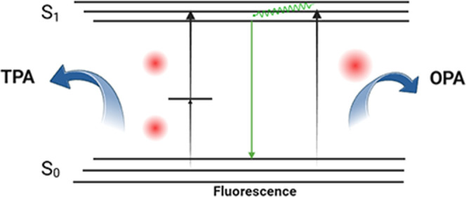
Schematic diagrams of one photon absorption and two photon absorption.
Two types of nonlinear ionization mechanisms, “nonlinear photoionization” and “avalanche ionization”, are at work in this nonlinear absorption process.21 Among them, photoionization is further divided into multiphoton ionization and electron tunneling according to the conditions of weak- and strong-field regimes, and this process, transition from one regime to the other, is controlled by the Keldysh gamma parameter
 |
1 |
where ω and E0 are the frequency and the amplitude of the driver field, I0 is the ionization potential, and e and m are the electron charge and mass.22 The conduction-band electrons can also absorb laser energy by means of free carrier absorption. Under sustained exposure to intense light, photons are absorbed by electrons in the conduction band, leading to interactions with electrons in the adjacent valence band. This process goes on and on, ultimately results in an electron avalanche.23 Following a series of nonlinear processes within the medium, a substantial number of free electron plasmas are generated, which subsequently absorb the majority of the ensuing laser energy. The accompanying effect can induce a localized refractive index change in the transparent medium. Given this, femtosecond lasers can generate a periodically distributed streak structure within a transparent medium that is optically anisotropic and demonstrates a negative change in refractive index, commonly termed a Type II structure.24,25 The reduction in refractive index can be attributed to the free carrier dispersion (FCD) effect,26 which involves the influence of free electrons and holes on the material’s refractive index. The presence of free carriers alters the material’s polarizability, thereby impacting the refractive index. In general, free carriers are capable of scattering photons, thus diminishing the material’s capacity to refract light and consequently leading to a reduction in its refractive index.27,28 This alteration arises from the periodic distribution of plasma within the molecule, occurring without inflicting damage on the material itself.
According to the theoretical basis of femtosecond laser-mediated Type II structure, we aim to utilize Two-photon direct laser writing to create diffraction gratings inside the acrylate in the hope of changing its refractive index(Figure 2). Considering the absorption spectrum characteristics of acrylate,29 we ultimately chose the green laser at 515 nm for processing. Despite acrylate exhibiting a transmittance of approximately 80% at this wavelength, our experimental setup can be equipped with an objective lens possessing a high numerical aperture and a femtosecond laser with adjustable repetition frequency, which facilitates the generation of high-power density within a specific localized region, thereby inducing a nonlinear absorption process inside the material. The utilization of a high numerical aperture objective lens enables the generation of a micron-sized spot, which, in conjunction with a high-precision displacement stage, can effectively realize the execution of high-precision micronano processing. The beam waist radius of the spot ω0 can be mathematically expressed in terms of the laser wavelength λ, the spot radius r, and the focal length of the exit lens S, as follows
 |
2 |
When processing samples with femtosecond lasers, it is also important to consider the peak energy density of the laser
 |
3 |
Where P0 is the average power of the laser and f is the repetition frequency of the laser. Given that the energy level at which the femtosecond laser generates the diffraction grating is below the material’s ablation threshold, the ablation threshold can be determined by employing the logarithmic relationship between the diameter of ablation holes and laser pulse energy30
 |
4 |
Figure 2.
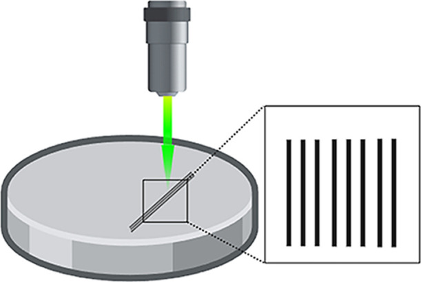
Schematic diagram of femtosecond laser engraving diffraction grating.
As a result, it will be possible to determine whether the processing energy is appropriate or not, and lay the theoretical groundwork for future experiments.
Utilizing the experimentally determined changes in the refractive index, we conducted optical simulations using Zemax(Figure 3). In these simulations, the superposition effect arises between light rays exhibiting a phase difference of (n*2π), in accordance with the Fresnel diffraction principle. Based on the Binary surface 2, the phase difference can be accrued to the pair of rays according to the following polynomial expansion
 |
5 |
Figure 3.
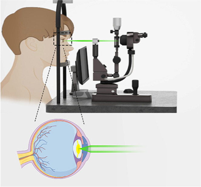
Zemax simulating femtosecond laser scanning to form a new lens within an IOL. Femtosecond lasers are incorporated into ophthalmic devices designed to emit visible light that sequentially traverses the anterior corneal surface, the posterior corneal surface, the aqueous humor, and the anterior surface of the IOL, ultimately reaching the center of the IOL, where the femtosecond laser creates a new diffraction lens.
The adjustment of IOL diopter can be achieved by setting the appropriate diffraction order and deriving the coefficient of ρ based on the phase calculation. We developed a novel diffractive lens within the monofocal IOL through phase design, enabling a reduction in the monofocal IOL diopter by 1D to 5D from its original value, without altering the single focal function. Future studies will focus on employing phase design to facilitate an increase in IOL diopter.
2.2. Femtosecond Laser System
A femtosecond laser characterized by a central wavelength of 515 nm, a repetition frequency of 40 MHz, and a pulse duration of 996 fs was employed in this study. The laser system (TCR–FS–515-20-HE, ZIMAO, China) utilizes an innovative hybrid mode-locking technology, ultrahigh-precision dispersion compensation coefficients, and a hybrid amplification mechanism incorporating both fiber and crystal elements. The femtosecond laser source, along with the half-wavelength plate, polarizer, diaphragm, and optical switch, are consolidated into a single unit. The half-wavelength plate and polarizing beam splitter (PBS) function as attenuators to regulate the output energy magnitude, while the optical switch is employed to precisely control the pulse count. The laser exhibits a beam quality factor M2 of 1.5, with optional frequency doubling modules available for 515 and 343 nm wavelengths. It demonstrates stable performance and is capable of continuous operation 24 h a day, 7 days a week, thereby fulfilling our experimental requirements. The femtosecond laser produces an output spot with a diameter of 3.6 mm. On our experimental platform, we utilize the frequency doubling module, operating at a wavelength of 515 nm.
2.3. Optical Path System
The optical path diagram of the femtosecond laser is shown in Figure 4. Following its traversal through the frequency doubling module and the beam expander (LZBE-532-1-4X, LBTEK, China), the femtosecond laser output pulse initially encounters a mirror that alters its trajectory. Subsequently, it passes through a dichroic lens (GCC-414002, Daheng Optics, China) before ultimately entering the objective lens (Olympus, Japan). Concurrently, the illumination source’s light (LB-SPL-20W, LBTEK, China) is directed through the objective lens into the CMOS camera (Basler, acA5472-5gc, Germany), facilitating real-time monitoring of the sample processing. The camera features a default resolution of 5472 × 3648 pixels and a total of 20 megapixels. Each pixel measures 2.40 × 2.40 μm, and the camera operates at a frame rate of 5 frames per second (fps) in color mode. The lens interface is a C-mount. Additionally, the color camera is equipped with an infrared cutoff filter, which is installed within a filter holder inside the lens interface. The transmittance, Tmax, exceeds 93% within the 450–620 nm wavelength range, is less than 4% in the 700–1070 nm range, and the average transmittance, Tavg, is below 1% in the 690–1070 nm range. The LED parallel light source positioned beneath the platform to provide bright field illumination. This parallel light source features a luminous area with a diameter of 20 mm and operates effectively at a working distance of 110 ± 50 mm. Light within the 400–700 nm wavelength range is permitted to enter the camera, enabling real-time monitoring of the process. The objective lens features a magnification of 40×, a numerical aperture of 0.8, and a working distance of 3.3 mm. It provides a field of view measuring 26.5 mm and has a chiaroscopic distance of 45 mm. Additionally, the lens is characterized by a postfocal plane position of −19.1 mm. A dichroic lens is an optical element engineered to selectively reflect light of a particular wavelength while transmitting light of other wavelengths. In our experimental setup, the dichroic lens employed is configured with an incidence angle of 45° and is designed to transmit light at wavelengths below 515 nm while reflecting light at wavelengths exceeding 515 nm. The beam expander matched with the objective lens, which is a 1*4× variable magnification beam expander with 2× in the optical path.
Figure 4.

Optical schematic diagram of the femtosecond laser direct writing processing system. The wavelength of 1030 nm emitted by the femtosecond laser is converted to 515 nm by the frequency doubling module for experimental processing.
2.4. Precision Machining System
The displacement stage utilized in our study was the PRO280LM(AEROTECH, USA), configured for movement along the X/Y-axis. This stage is characterized by a maximum velocity of 2 m/s, a maximum acceleration of 3 m/s2, a minimum incremental displacement of 5 nm, and a calibration accuracy of ±1 μm. The EM-LS90–20CZ (LBTEK, China) is employed along the Z-axis, featuring a stroke length of 20 mm and a resolution of 0.5 μm, which was convenient to control the lifting of the platform and precisely control the focal plane of the femtosecond laser. Together, the two sets of devices constitute a three-dimensional electrodynamic displacement stage, capable of meeting our stringent high-precision experimental requirements. Furthermore, a custom-designed fixture has been incorporated to securely hold the experimental samples in place.
2.5. Control System
The control system primarily consists of two components: the circuit and the control software. The control software employed is the Hongtuo femtosecond laser integrated processing control software, which is comprised of two main sections: the parameter setting interface and the pattern editing interface. The pattern editing interface facilitates the design of the processing path, while the parameter setting interface allows for the input of processing parameters for the femtosecond laser and the regulation of the pulse emission mode. Complex machining paths must be designed using CAD software, saved in DXF format, and subsequently imported into the relevant software. The control circuit system functions as an external auxiliary control circuit, constructed using various hardware devices to facilitate interdevice communication. This control system ensures the precise operation of the experimental platform in accordance with specified requirements.
2.6. Experimental Procedures and Methods
Our material selection is a commercially available hydrophobic acrylate IOL disc made of the same material with a diameter of 15 mm and a thickness of 2 mm. The output ratio of the femtosecond laser was systematically reduced from a value of 60% through precise manipulation of the computer software. The repetition frequency is set to 500 kHz, and the system is configured to operate in a position-synchronized output (PSO) mode, thereby effectively achieving single-pulse emission. The processing was conducted using a linear scanning pattern, with the femtosecond laser dot spacing configured at 10 μm and the line spacing at 20 μm. The operational speed was maintained at 5 mm/s, and the acceleration was set to 10 mm/s2. The surface topography of the processed sample was examined using scanning electron microscopy (SEM, JEOL, Japan). The distances between the left and right pulse points, as well as between the top and bottom pulse points, were quantified using ImageJ software to assess the precision of the motorized displacement stage, and 15 groups of data were measured independently.
With an output ratio of 25%, the scanning speed was configured to 5 mm/s, and the line spacing was set to 10 μm, the grating was engraved on the surface of the experimental sample, and a total of 10 pieces were processed. The diffraction gratings were observed with a scanning electron microscope (SEM, Zeiss Ultra Plus, Carl Zeiss AG, Germany). The alteration in refractive index of the scanned discs was quantitatively assessed using an Abbe refractometer (CJHZYG, model 2WAJ, China). The molecular organization of the scanned discs was analyzed using X-ray photoelectron spectroscopy (XPS, ESCALAB250Xi, Thermo Fisher Scientific, USA) and micro confocal Raman spectroscopy (XploRA Plus, HORIBA Jobin Yvon, France).
3. Test Results
3.1. Platform Precision
Presented below is a photograph of the processed sample following SEM (Figure 5). The Bland-Altman analysis assessed the concordance between the software-preset values and actual measured values of point and line spacing (Figure 6). The results indicate a strong agreement, validating the experimental setup’s accuracy in replicating the intended specifications, and the accuracy satisfies our experimental requirements.
Figure 5.

Surface morphology observed by SEM. Each dot represents the result of the interaction between the pulse emitted by the femtosecond laser and the material surface, and the pulse energy is set the same in the same row, and the top-down femtosecond laser energy is gradually increased.
Figure 6.

Bland–Altman plots for the comparisons between the values of point and line spacing. (a): point spacing, (b): line spacing.
3.2. Refractive Index Change
Figure 7 shows a SEM image of the diffraction grating. It was observed that the refractive index of all scanned discs exhibited a reduction. Table 1 shows the results of a paired t-test on the refractive indices of 10 samples before and after scanning. The results reveal a statistically significant difference in refractive index changes due to femtosecond laser scanning. A Cohen’s d of 142 indicates a substantial effect of the femtosecond laser on the samples’ refractive indices.
Figure 7.
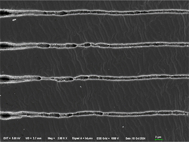
Formation of laser-induced periodic surface structure on acrylic disc surface with a line spacing of 10 μm (Top view).
Table 1. Refractive Index Change of Experimental Samples before and after Processing.
| sample size | mean ± standard deviation | mean difference | p-value | Cohen’s d value | |
|---|---|---|---|---|---|
| preprocessing | 10 | 1.5391 ± 0.004 | 0.093 | 0.000a | 142.297 |
| postprocessing | 10 | 1.4462 ± 0.004 |
P ≤ 0.05 was considered statistically significant.
3.3. Molecular Structure Detection
The spectra show strong Raman bands at 583, 699, 1001, 1051, 1250, 1493, 1625, 1748, 2899, 2946, 3033. And the Raman spectroscopy analysis indicated the absence of any novel peaks in the scanned area relative to the unscanned area. However, a reduction in fluorescence intensity was observed (Figure 8). Furthermore, the XPS spectrum revealed an increase in the content of carbonyl groups and carbon–oxygen single bonds postscanning(Figure 9a) compared to prescanning(Figure 9b), suggesting an enhancement in the hydrophilicity of the sample surface.
Figure 8.
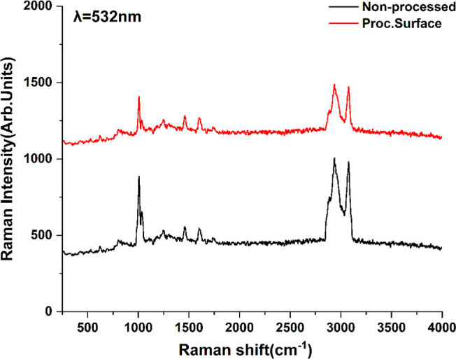
Microscopic Raman spectra of experimental samples before and after processing. The excitation wavelength was 532 nm. No new peaks appeared, indicating that the structure of the sample itself was not damaged by the femtosecond laser.
Figure 9.

X-ray photoelectron spectroscopy (XPS) of experimental samples in the processed area (a) and the unprocessed area (b) showed that the content of C=O bonds and C–O single bonds increased after processing, suggesting that the hydrophilicity of the processed area increased.
4. Discussion
With the advent of refractive cataract surgery, there is an increasing demand among patients for enhanced postoperative visual acuity.31 While the availability of diverse IOL calculation formulas enhances the surgeon’s capacity to tailor refractive outcomes to individual patient needs,32 those with a history of prior refractive surgery or other ocular surgical interventions remain at an elevated risk for residual refractive errors following surgery.1 The light-adjustable lens (LAL) is presently accessible in international markets, allowing for the adjustment of refractive errors through supplementary ultraviolet irradiation during the postoperative period. However, the long-term safety of this technology requires further investigation.7,8 Consequently, there is an urgent need in clinical practice to explore innovative methods for correcting residual refractive errors following cataract surgery. In recent years, femtosecond lasers have found extensive applications across various disciplines, with FLDW emerging as a prominent area of research within the field of femtosecond laser processing. The ultrahigh peak power of femtosecond lasers facilitates nonlinear absorption processes at the focal point within materials, subsequently inducing photochemical transformations. Consequently, we employed the FLDW to inscribe diffraction gratings on the surface of the sample, thereby altering the refractive index of the acrylate. Molecular structure of scanned samples was detected. The advantages of the platform and experimental results will be discussed.
The femtosecond laser seed source employed in this experimental platform utilizes fiber amplification technology, categorizing it as a fiber laser and thereby differentiating it from traditional solid-state lasers. Fiber lasers typically employ optical fibers doped with rare earth elements, such as erbium or ytterbium, as the gain medium.33 In contrast, solid-state lasers utilize solid materials, such as gemstone crystals or glass, as their gain medium. Common examples of solid-state laser media include Neodymium-doped YAG (yttrium–aluminum garnet) crystals and titanium-doped gemstones.34 Fiber lasers typically comprise one or more fiber amplifiers, which can be either single-mode or multimode. The inherent flexibility of the fibers facilitates a compact design that is easily integrable. However, solid-state lasers possess a comparatively complex architecture, often incorporating elements such as resonant cavities, pump sources, and devices for Q-switching or mode-locking, among others. Consequently, this complexity generally leads to larger solid-state laser systems. This structural simplicity is one of the reasons fiber lasers can be engineered to achieve a smaller size compared to solid-state lasers. Owing to the high surface area to volume ratio of the fiber, heat dissipation is highly efficient, typically necessitating only air or water cooling. Conversely, solid-state lasers often require more sophisticated cooling mechanisms, such as water or thermoelectric cooling, due to the substantial heat capacity of the solid medium. Furthermore, fiber lasers are well-suited for ultrahigh-precision micro- and nanomachining processes owing to their simplicity, reliability, and typically excellent beam quality, which is often near the diffraction limit.33 Since that the laser used in the current setup is predesigned, there is potential for substantial reduction in its size in future iterations, contingent upon specific requirements.
In this platform, the femtosecond laser operates at a wavelength of 515 nm with a repetition frequency of 500 kHz. It is noteworthy that a decrease in repetition frequency results in an increase in the pulse energy of the femtosecond laser. The pulse width is measured at 996 fs, which significantly influences the peak power of the laser. Assuming a fixed energy per pulse, a reduction in pulse width leads to an elevation in the peak power of the femtosecond laser. This increase in peak power is associated with reduced material damage, thereby enhancing safety. Femtosecond laser-induced two-photon absorption in materials necessitates exceptionally high peak power to induce modifications in the refractive index of the material while preserving the integrity of the sample. The numerical aperture of the focusing objective lens equipped with this experimental platform is as high as 0.8, because the larger the numerical aperture of the focusing objective lens, the higher the processing resolution of the experimental platform and the shorter the depth of focus. Moreover, our experimental platform is equipped with a high-precision motorized displacement stage, which can meet the needs of various processing trajectories.
The experimental platform was constructed by professional technicians to optimize the functionality of each device while maintaining the neutrality of the beam. Upon testing, the measurement results obtained under actual processing conditions align with the displacement values specified in the software. The minor discrepancies observed may be attributed to the necessity for the personnel to manually mark the center of the pulse when measuring point and line spacing. This indicates that the accuracy of the displacement platform is sufficient to meet the requirements of our experiment.
We engraved a diffraction grating inside the sample based on the fact that the femtosecond laser can induce a nonlinear absorption process inside the transparent medium, thus realizing a change in the refractive index of the sample in the range of 0.09. The current energy per pulse utilized in our experiments is 0.2 μJ, but these findings are preliminary. Our team intends to further optimize the femtosecond laser parameters with the aim of minimizing the laser energy while ensuring efficacy. This optimization is beneficial to the safety of the retina in the long run.
From the spectrum, the bands in the range 800–1800 cm–1 may be attributed to ring vibrations and to alkyl groups. Other bands, as the band at 1748 and 1493 cm–1, are attributable to ν(C=O) of C–COO and δ(C–H) of O–CH3, respectively. The broad band between 2600 and 3200 cm–1 with maximal intensities at 2931 and 3062 cm–1 arises from the convolution of symmetrical and asymmetrical stretching vibrations of CH and CH3 bonds. This result is similar to that of Rusciano et al.35 The absence of alterations in the peak position and shape of the Raman spectra before and after scanning indicates that the femtosecond laser did not inflict any damage on the sample. The site corresponding to the C=O bond showed an increase in relative intensity compared to the peaks at other sites. However, it is not feasible to ascertain whether the concentration of the corresponding chemical in other regions has increased or decreased. Because Raman spectroscopy is only effective for assessing changes in substance concentration when the sample exhibits high uniformity. Carbon (C) and oxygen (O) are the primary components of the hydrophobic acrylate. We have subsequently presented the results of the peak deconvolution of the C 1s spectrum. The binding energy of 284.6 eV was attributed to the C–C bond, 286.5 eV to the C–O bond, and 287.5 eV to the C=O bond. An increase in the C=O and C–O bond content in the analyzed samples was observed, indicating an enhancement in the hydrophilicity of the samples to some degree. It is crucial to acknowledge that the enhancement of IOL hydrophilicity presents a dual-edged sword, offering both benefits and drawbacks. On one hand, increasing the hydrophilicity of the IOL can enhance its biocompatibility, thereby reducing the likelihood of postoperative immune rejection. Conversely, heightened hydrophilicity can also augment the adhesion of lens epithelial cells to the IOL surface, potentially leading to an increased incidence of posterior capsular opacification (PCO). This occurs because the more hydrophilic surface may facilitate cellular proliferation and migration, contributing to the formation of secondary cataracts. Furthermore, as the hydrophilicity of the IOL increases, there is a propensity for proteins to adsorb onto its surface. This protein deposition can lead to a higher risk of IOL opacification, which may impair visual clarity and necessitate further intervention. Future research will focus on a more detailed examination of the molecular structure.
Sola and Cases.29 employed a femtosecond laser to modify the refractive index of an acrylate material, utilizing a wavelength of 520 nm, a pulse duration of 250 fs, a repetition frequency of 63 MHz, and a solid-state laser configuration. Compared with their experimental setup, our study benefits from the use of a fiber laser with an adjustable repetition frequency. Additionally, our femtosecond laser is equipped with a frequency doubling module, enabling comprehensive investigation into the effects of the femtosecond laser on the refractive index of samples across varying repetition frequencies and wavelengths. This approach aims to find the optimal processing parameters. They did the same Raman spectroscopy on the processed samples and our results agreed with them. In addition, our XPS spectra demonstrated an increase in hydrophilicity in the scanned area. Since the current green light conditions used by Sola and Cases.29 are more irritating to the human eye, it is imperative to investigate alternative wavelengths of light that can replicate the efficacy of green light while mitigating ocular discomfort, particularly with an eye toward the feasibility and safety of subsequent human experimentation. And this is what our team is working on.
In the field of ophthalmology, the lasers utilized encompass femtosecond laser assisted cataract surgery(FLACS),36 femtosecond laser-assisted in suit keratomileusis(FS-LASIK),37 and Nd:YAG laser for the management of posterior capsule opacification(PCO) following cataract surgery,38 among others. These lasers serve distinct roles across various clinical conditions, with each type characterized by unique parameters that result in differing mechanisms of action depending on the specific settings employed. The utilization of femtosecond lasers facilitates various surgical processes, including capsulotomy and lens fragmentation during cataract procedures.39 Several studies have demonstrated that the femtosecond laser can decrease both the duration and energy required for phacoemulsification.40 This reduction has been shown to effectively safeguard the patient’s endothelial cells and mitigate the postoperative inflammatory response.40,41 FLACS is typically conducted using a near-infrared laser, specifically engineered to reduce linear absorption and scattering by the tissues in the anterior segment of the eye.42 The pulse duration generally ranges from 400 to 800 fs, and the energy employed is commonly quantified in microjoules. This energy may be augmented to 10–15 μJ during procedures involving lens fragmentation.43
PCO can develop in certain patients following cataract surgery as a result of the migration of lens epithelial cells.44 This condition is typically managed in a clinical setting using Nd:YAG laser treatment.38 The Nd:YAG laser, with an output wavelength of 1064 nm, represents the first practical application of near-infrared lasers in the clinic. However, its pulse duration, situated within the nanosecond range (10–9 seconds), does not qualify it as a femtosecond laser.45 When the Nd:YAG laser is applied to the posterior capsule membrane, the spot diameter is usually 10 μm, with the energy generally ranging from 0.8 to 1.4 mJ, but the precise energy is flexibly adjusted according to the thickness of the patient’s capsule membrane and the response to the laser.45
Femtosecond lasers are employed in corneal refractive surgery, including the contemporary technique of femtosecond laser small-incision lenticule extraction (SMILE). This procedure utilizes a femtosecond laser operating in the near-infrared spectrum, with a repetition frequency of 500 kHz and an energy range typically between 125 and 135 nJ.46 Conversely, the excimer laser employs an ultraviolet laser with a wavelength of 193 nm. Due to its inability to penetrate corneal tissue, the preparation of a corneal flap is required to facilitate the ablated of the corneal stroma.47
The functions of a femtosecond laser vary according to its wavelength, area of application, and energy level. Consequently, the auxiliary equipment required must be configured differently depending on the specific function the femtosecond laser is intended to perform. Therefore, in alignment with the objectives of our study, we developed a femtosecond laser experimental platform specifically designed to the refractive index change of IOLs. This platform is fully equipped to satisfy all facets of our experimental needs. In terms of laser size, our fiber lasers are compact. From a clinical application perspective, if femtosecond lasers are to be employed for noninvasive correction of residual refractive errors following cataract surgery, it is necessary that these lasers possess attributes of compactness and ease of mobility. These features represent the core advantages of our technology. Our femtosecond laser system is capable of modulating the pulse width via dispersion management, alongside offering precise control over the number of emitted femtosecond laser pulses. Additionally, it incorporates a PSO mode, thereby accommodating diverse processing requirements across various scenarios. This functionality constitutes a significant advantage of our experimental platform.
At present, we have performed a series of in vitro experiments on IOLs, establishing a basis for subsequent in vivo experiments conducted by our team. In these forthcoming in vivo studies, we aim to further optimize the parameters of the femtosecond laser to modify the refractive index of the IOL, while ensuring retinal safety. Additionally, we intend to investigate the molecular mechanisms underlying the alteration of the IOL refractive index by the femtosecond laser. These efforts are expected to contribute novel insights for the future translation into clinical applications.
5. Conclusion
In conclusion, we have developed an experimental platform utilizing a femtosecond laser for the purpose of remodeling the refractive index of IOLs. This objective is accomplished by inscribing diffraction gratings onto the surface of the samples using the femtosecond laser. We utilized a compact and easily maintainable fiber laser as the seed source, allowing us to precisely adjust both the pulse count and the output power of the femtosecond laser. This setup enabled the observation of experimental outcomes across various parameters. We will share our forthcoming findings in a subsequent publication. It is anticipated that this technology will provide significant benefits for a larger patients.
Acknowledgments
The authors thank Y.W. for his guidance on this subject. Figures 1 to 3 and the TOC image were created with Biorender.com.
Glossary
ABBREdeVIATIONS
- IOL
intraocular lens
- LAL
light-adjustable lens
- UV
ultraviolet
- FLDW
femtosecond laser direct writing
- MPA
multiphoton absorption
- SEM
scanning electron microscopy.
Author Contributions
Writing—original draft, Visualization, Methodology, Investigation, Formal analysis: Biyue Tu. Investigation, Formal analysis: Yanxia Tong. Writing—review and editing, Methodology: Zhanpeng Zhu. Writing—review and editing, Formal analysis: Haifeng Jiang. Writing—review and editing, Supervision, Methodology, Conceptualization: Yong Wang.
This study was supported by the Natural Science Foundation of Hunan Province (Grant no. 2023JJ70044), the Xiangjiang Public Welfare Foundation Technology Innovation and Application Project (Grant no. KY24013), and the Scientific Research Foundation of Aier Eye Hospital Group (Grant no. AGK2302D09).
The authors declare no competing financial interest.
References
- Moshirfar M.; Thomson A. C.; Thomson R. J.; Martheswaran T.; McCabe S. E. Refractive enhancements for residual refractive error after cataract surgery. Curr. Opin. Ophthalmol. 2021, 32 (1), 54–61. 10.1097/ICU.0000000000000717. [DOI] [PubMed] [Google Scholar]
- Dick H. B.; Gerste R. D. Future Intraocular Lens Technologies. Ophthalmology 2021, 128 (11), e206–e213. 10.1016/j.ophtha.2020.12.025. [DOI] [PubMed] [Google Scholar]
- Sáles C. S.; Manche E. E. Managing residual refractive error after cataract surgery. J. Cataract Refractive Surg. 2015, 41 (6), 1289–1299. 10.1016/j.jcrs.2015.05.001. [DOI] [PubMed] [Google Scholar]
- Märker D. A.; Radeck V.; Barth T.; Helbig H.; Scherer N. C. D. Long-Term Outcome and Complications of IOL-Exchange. Clin. Ophthalmol. 2023, 17, 3243–3248. 10.2147/OPTH.S436963. [DOI] [PMC free article] [PubMed] [Google Scholar]
- Goemaere J.; Trigaux C.; Denissen L.; Dragnea D.; Hua M. T.; Tassignon M. J.; Dhubhghaill S. N. Fifteen years of IOL exchange: indications, outcomes, and complications. J. Cataract Refractive Surg. 2020, 46 (12), 1596–1603. 10.1097/j.jcrs.0000000000000349. [DOI] [PubMed] [Google Scholar]
- Dragnea D. C.; Truţa R. N.; Goemaere J.; Tassignon M. J.; Dhubhghaill S. N. Intraocular bag-in-the-lens exchange: indications, outcomes, and complications. J. Cataract Refractive Surg. 2022, 48 (5), 568–575. 10.1097/j.jcrs.0000000000000787. [DOI] [PubMed] [Google Scholar]
- Moshirfar M.; Martin D. J.; Jensen J. L.; Payne C. J. Light adjustable intraocular lenses: an updated platform for cataract surgery. Curr. Opin. Ophthalmol. 2023, 34 (1), 78–83. 10.1097/ICU.0000000000000911. [DOI] [PubMed] [Google Scholar]
- Wong J. R.; Folden D. V.; Wandling G. R.; Schaefer S.; Stephens J.; Boots C.; Nichols C. J.; Everson M. H.; Johnson R. Visual Outcomes of a Second-Generation, Enhanced UV Protected Light Adjustable Lens in Cataract Patients with Previous LASIK and/or PRK. Clin. Ophthalmol. 2023, 17, 3379–3387. 10.2147/OPTH.S432457. [DOI] [PMC free article] [PubMed] [Google Scholar]
- Hengerer F. H.; Müller M.; Dick H. B.; Conrad-Hengerer I. Clinical Evaluation of Macular Thickness Changes in Cataract Surgery Using a Light-Adjustable Intraocular Lens. J. Refractive Surg. 2016, 32 (4), 250–254. 10.3928/1081597X-20160217-01. [DOI] [PubMed] [Google Scholar]
- Schwartz D. M. Light-adjustable lens. Trans. Am. Ophthalmol. Soc. 2003, 101, 417–436. [PMC free article] [PubMed] [Google Scholar]
- Xie J.; Sun J.; Liu T.; Mao S.; Dai Y. Comparison Between L-312 Hydrophobic-Hydrophilic Acrylate and US-860 UV Hydrophilic Acrylate IOL Opacification Characteristic. Front. Med. 2022, 9, 873684. 10.3389/fmed.2022.873684. [DOI] [PMC free article] [PubMed] [Google Scholar]
- Taha S.; Mohamed W. R.; Elhemely M. A.; El-Gendy A. O.; Mohamed T. Tunable femtosecond laser suppresses the proliferation of breast cancer in vitro. J. Photochem. Photobiol., B 2023, 240, 112665. 10.1016/j.jphotobiol.2023.112665. [DOI] [PubMed] [Google Scholar]
- Gogri P. Y.; Bore M. C.; Rips A. G. T.; Reddy J. C.; Rostov A. T.; Vaddavalli P. K. Femtosecond laser-assisted big bubble for deep anterior lamellar keratoplasty. J. Cataract Refractive Surg. 2021, 47 (1), 106–110. 10.1097/j.jcrs.0000000000000400. [DOI] [PubMed] [Google Scholar]
- Zhang C.; Li Z.; Li H.; Yang Q.; Wang H.; Shan C.; Zhang J.; Hou X.; Chen F. Femtosecond Laser-Induced Supermetalphobicity for Design and Fabrication of Flexible Tactile Electronic Skin Sensor. ACS Appl. Mater. Interfaces 2022, 14 (33), 38328–38338. 10.1021/acsami.2c08835. [DOI] [PubMed] [Google Scholar]
- Ding L.; Blackwell R.; Kunzler J. F.; Knox W. H. Large refractive index change in silicone-based and non-silicone-based hydrogel polymers induced by femtosecond laser micro-machining. Opt. Express 2006, 14 (24), 11901–11909. 10.1364/OE.14.011901. [DOI] [PubMed] [Google Scholar]
- Lapointe J.; Bérubé J. P.; Ledemi Y.; Dupont A.; Fortin V.; Messaddeq Y.; Vallée R. Nonlinear increase, invisibility, and sign inversion of a localized fs-laser-induced refractive index change in crystals and glasses. Light:Sci. Appl. 2020, 9, 64. 10.1038/s41377-020-0298-8. [DOI] [PMC free article] [PubMed] [Google Scholar]
- Lei H.; Zhou J.; Liu F.; Han Y.; Chai Y.; Yuan R. A Fluorescence Light-Up 3D DNA Walker Driven and Accelerated by Endogenous Adenosine-5′-triphosphate for Sensitive and Rapid Label-Free MicroRNA Detection and Imaging in Living Cells. Anal. Chem. 2024, 96 (22), 9097–9103. 10.1021/acs.analchem.4c00664. [DOI] [PubMed] [Google Scholar]
- Xie Q.; Cavillon M.; Pugliese D.; Janner D.; Poumellec B.; Lancry M. On the Formation of Nanogratings in Commercial Oxide Glasses by Femtosecond Laser Direct Writing. Nanomater. 2022, 12 (17), 2986. 10.3390/nano12172986. [DOI] [PMC free article] [PubMed] [Google Scholar]
- Wang X.; Yu H.; Li P.; Zhang Y.; Wen Y.; Qiu Y.; Liu Z.; Li Y.; Liu L. Femtosecond laser-based processing methods and their applications in optical device manufacturing: A review. Opt Laser. Technol. 2021, 135, 106687. 10.1016/j.optlastec.2020.106687. [DOI] [Google Scholar]
- Sidhu M. S.; Choi M. Y.; Woo S. Y.; Lee H. K.; Lee H. S.; Kim K. J.; Jeoung S. C.; Choi J. S.; Joo C. K.; Park I. H. Femtosecond laser-assisted selective reduction of neovascularization in rat cornea. Laser Med. Sci. 2014, 29 (4), 1417–1427. 10.1007/s10103-014-1545-0. [DOI] [PMC free article] [PubMed] [Google Scholar]
- Hansch P.; Walker M. A.; Van Woerkom L. D. Spatially dependent multiphoton multiple ionization. Phys. Rev. A:At., Mol., Opt. Phys. 1996, 54 (4), R2559–r2562. 10.1103/PhysRevA.54.R2559. [DOI] [PubMed] [Google Scholar]
- Zheltikov A. M. Keldysh parameter, photoionization adiabaticity, and the tunneling time. Phys. Rev. A 2016, 94, 043412. 10.1103/physreva.94.043412. [DOI] [Google Scholar]
- Yablonovitch E.; Bloembergen N. Avalanche Ionization and the Limiting Diameter of Filaments Induced by Light Pulses in Transparent Media. Phys. Rev. Lett. 1972, 29 (14), 907–910. 10.1103/PhysRevLett.29.907. [DOI] [Google Scholar]
- Shimotsuma Y.; Kazansky P. G.; Qiu J.; Hirao K. Self-organized nanogratings in glass irradiated by ultrashort light pulses. Phys. Rev. Lett. 2003, 91 (24), 247405. 10.1103/PhysRevLett.91.247405. [DOI] [PubMed] [Google Scholar]
- Stoian R.; D’Amico C.; Bhuyan M.; Cheng G. Ultrafast laser photoinscription of large-mode-area waveguiding structures in bulk dielectrics. Opt Laser. Technol. 2016, 80, 98–103. 10.1016/j.optlastec.2015.11.025. [DOI] [Google Scholar]
- Zhang L.; Fei Y.; Cao Y.; Lei X.; Chen S. Experimental observations of thermo-optical bistability and self-pulsation in silicon microring resonators. J. Opt. Soc. Am. B 2014, 31 (2), 201–206. 10.1364/JOSAB.31.000201. [DOI] [Google Scholar]
- Kim Y.; Takenaka M.; Osada T.; Hata M.; Takagi S. Strain-induced enhancement of plasma dispersion effect and free-carrier absorption in SiGe optical modulators. Sci. Rep. 2014, 4, 4683. 10.1038/srep04683. [DOI] [PMC free article] [PubMed] [Google Scholar]
- Zhang X.; Shi G.; Leveillee J. A.; Giustino F.; Kioupakis E. Ab initio theory of free-carrier absorption in semiconductors. Phys. Rev. B 2022, 106 (20), 205203. 10.1103/PhysRevB.106.205203. [DOI] [Google Scholar]
- Sola D.; Cases R. High-Repetition-Rate Femtosecond Laser Processing of Acrylic Intra-Ocular Lenses. Polymers 2020, 12 (1), 242. 10.3390/polym12010242. [DOI] [PMC free article] [PubMed] [Google Scholar]
- Huang H.; Zheng H. Y.; Lim G. C. Femtosecond laser machining characteristics of Nitinol. Appl. Surf. Sci. 2004, 228 (1), 201–206. 10.1016/j.apsusc.2004.01.018. [DOI] [Google Scholar]
- Narang R.; Agarwal A. Refractive cataract surgery. Curr. Opin. Ophthalmol. 2024, 35 (1), 23–27. 10.1097/ICU.0000000000001005. [DOI] [PubMed] [Google Scholar]
- Ferguson T. J.; Randleman J. B. Cataract surgery following refractive surgery: Principles to achieve optical success and patient satisfaction. Surv. Ophthalmol. 2024, 69 (1), 140–159. 10.1016/j.survophthal.2023.08.002. [DOI] [PubMed] [Google Scholar]
- Poddubrovskii N. R.; Drobyshev R. V.; Lobach I. A.; Kablukov S. I. Fiber Lasers Based on Dynamic Population Gratings in Rare-Earth-Doped Optical. Fibers 2022, 9 (9), 613. 10.3390/photonics9090613. [DOI] [Google Scholar]
- Boulon G. Fifty years of advances in solid-state laser materials. Opt. Mater. 2012, 34 (3), 499–512. 10.1016/j.optmat.2011.04.018. [DOI] [Google Scholar]
- Rusciano G.; Capaccio A.; Pesce G.; Sasso A. Experimental study of the mechanisms leading to the formation of glistenings in intraocular lenses by Raman spectroscopy. Biomed. Opt. Express 2019, 10 (4), 1870–1881. 10.1364/BOE.10.001870. [DOI] [PMC free article] [PubMed] [Google Scholar]
- Vasavada A. R.; Vasavada S. A.; Nath V.; Vasavada V.; Patel P. FLACS vs conventional phacoemulsification by junior cataract surgeons: randomized trial. J. Cataract Refractive Surg. 2023, 49 (2), 159–164. 10.1097/j.jcrs.0000000000001061. [DOI] [PubMed] [Google Scholar]
- Alió Del Barrio J. L.; Milán-Castillo R.; Canto-Cerdan M.; Molina-Lespron A.; Alió J. L. FS-LASIK for the treatment of moderate-to-high hyperopia. J. Cataract Refractive Surg. 2023, 49 (6), 558–564. 10.1097/j.jcrs.0000000000001153. [DOI] [PubMed] [Google Scholar]
- Lee Y.; Kim J. S.; Kim B. G.; Hwang J. H.; Kang M. J.; Lee J. H. Comparison of the Incidence of Nd:YAG Laser Capsulotomy Based on the Type of Intraocular Lens. Med. 2023, 59 (12), 2173. 10.3390/medicina59122173. [DOI] [PMC free article] [PubMed] [Google Scholar]
- Mayer W. J.; Ohlmann A.; Schuh A.; Priglinger S.; Kohnen T.; Shajari M. Electron microscopy analysis of femtosecond laser-assisted capsulotomy before and after lens fragmentation. Sci. Rep. 2021, 11 (1), 24427. 10.1038/s41598-021-04054-5. [DOI] [PMC free article] [PubMed] [Google Scholar]
- Zhou K. J.; Huang Y.; Wang Y.; Pan A. P.; Shao X.; Tu R. X.; Yu A. Y. Safety and efficacy of cataract surgery performed with a low-energy femtosecond laser compared with conventional phacoemulsification in Chinese patients: a randomized clinical trial. Eye Visual 2023, 10 (1), 31. 10.1186/s40662-023-00347-0. [DOI] [PMC free article] [PubMed] [Google Scholar]
- Krarup T.; Morten Holm L.; la Cour M.; Kjaerbo H. Endothelial cell loss and refractive predictability in femtosecond laser-assisted cataract surgery compared with conventional cataract surgery. Acta Ophthalmol. 2014, 92 (7), 617–622. 10.1111/aos.12406. [DOI] [PubMed] [Google Scholar]
- Wang J.; Sramek C.; Paulus Y.; Lavinsky D.; Schuele G.; Andersen D.; Dewey D.; Palanker D.. Ocular safety limits for 1030nm femtosecond laser cataract surgery. In Progress in Biomedical Optics and Imaging - Proceedings of SPIE, 2013; p 856708. 10.1117/12.2001895. [DOI]
- Nagy Z. Z.; McAlinden C. Femtosecond laser cataract surgery. Eye Visual 2015, 2, 11. 10.1186/s40662-015-0021-7. [DOI] [PMC free article] [PubMed] [Google Scholar]
- Wormstone I. M.; Wormstone Y. M.; Smith A. J. O.; Eldred J. A. Posterior capsule opacification: What’s in the bag?. Prog. Retinal Eye Res. 2021, 82, 100905. 10.1016/j.preteyeres.2020.100905. [DOI] [PubMed] [Google Scholar]
- Soong H. K.; Malta J. B. Femtosecond lasers in ophthalmology. Am. J. Ophthalmol. 2009, 147 (2), 189–197e2. 10.1016/j.ajo.2008.08.026. [DOI] [PubMed] [Google Scholar]
- Liu E. T.; Sella R.; Goernert P.; Kim K.; Chen H.; Lin R. T. Refractive results with SMILE using lower energy settings in the United States. PloS one 2021, 16 (10), e0258835 10.1371/journal.pone.0258835. [DOI] [PMC free article] [PubMed] [Google Scholar]
- Freidank S.; Vogel A.; Linz N. Mechanisms of corneal intrastromal laser dissection for refractive surgery: ultra-high-speed photographic investigation at up to 50 million frames per second. Biomed. Opt. Express 2022, 13 (5), 3056–3079. 10.1364/BOE.455926. [DOI] [PMC free article] [PubMed] [Google Scholar]