



Abstract

The present study provides in vivo trials of electrospun poly(l-lactide-co-ε-caprolactone), PLCL, copolymer 67:33 mol %, and electrospun PLCL blend with a low loading of collagen (0.5% w/v), PLCL-Col, as a connecting porous biodegradable nerve conduit to repair 7 mm long segmentary tibial nerve lesions in rats compared with the standard autograft technique. The electrospun PLCL scaffolds reveal a matrix of fibers with a mean diameter of 476 ± 60 nm and an average pore size of 253 ± 5 nm. Blending collagen with the PLCL results in a comparatively denser matrix of fibers with a mean diameter of 417 ± 42 nm and a pore size of 244 ± 3 nm. For in vivo testing, a total of 30 male Wistar rats were divided into 3 groups of 10 and each group was subjected to a different nerve repair procedure for evaluation of nerve regeneration after reconstruction. Evaluation of nerve regeneration was compared in terms of the tibial functional index (TFI), nerve conduction velocity (NCV), gastrocnemius muscle weight (%GMW), and a histomorphometric study. After 12 weeks of implantation, there was evidence of nerve regeneration across the gap from the histomorphologic study. All parameters of nerve regeneration were observed in every animal of the study groups. Our results clearly showed that there are reinnervation and return of function in all groups, similarly to the autograft group. PLCL-Col showed better results than PLCL and autograft, which suggested that PLCL-Col porous conduits may serve as a scaffold for peripheral nerve regeneration.
1. Introduction
The most severe type of peripheral nerve damage is a complete lesion with a subsequent loss of nerve function. Although peripheral nerves can regenerate spontaneously, their function is not immediately restored.1 The first option to consider for nerve gap repair is direct repair with suturing of the proximal and distal nerve stumps. However, tension at the suture site is a highly unfavorable factor. Autologous nerve grafting is the gold standard of treatment for large nerve gaps because direct nerve repair usually leads to unsuccessful results due to high tension and scar formation.2,3 However, the results of such nerve grafts have demonstrated a success rate of only 50% on the patients treated.4 The use of autologous nerve grafts has several drawbacks, including donor site disability, the need for another surgical site, a limited supply, donor site mismatch, and the risk of severe neuroma formation and scars.5,6
Tubulization repair is an alternative method for healing that involves enclosing the ends of a severed nerve by a tube. This tube serves to retain the stumps in place and provides a pathway for the regeneration of axons to the distal stump.5,7 The market offers numerous commercial nerve guide products, which are made of natural and synthetic polymers. However, the overall performance of these nerve guides is not as good as that of conventional nerve grafting, which appears to be the method most commonly used.8 Current synthetic nerve guides do not effectively stimulate nerve regeneration across extensive lesion gaps, prompting increasing interest in the development of alternative designs.9 Absorbable nerve conduits must undergo additional property improvements before they can be accepted for clinical use.
There is an extensive choice of synthetic polymers available with flexible biodegradability, nontoxic/noninflammatory properties, mechanical properties similar to the tissue to be repaired or replaced, high porosity that promotes cell attachment and growth, cost-effective and simple production, and the potential for chemical modification to increase interaction with regular tissue.10 Evans et al. used the porous poly(l-lactic acid) (PLA) conduit to conduct in vivo trials with a sciatic nerve defect model in rats.11 For a 15 mm peroneal nerve gap in dogs, Nakamura et al. filled collagen sponge in a polyglycolic acid (PGA) nerve tube.12 Nanofibrous conduits comprising poly(d,l-lactide-co-glycolide) (PLGA) and poly(ε-caprolactone) (PCL) were found to promote sciatic nerve regeneration across a 10 mm nerve gap in the rat model.13,14 In a large animal model, a synthetic nerve graft consisting of chitosan and polyglycolic acid (PGA) was applied to repair a 30 mm sciatic nerve lesion.15 Other biomaterials were also modified to enhance the tissue regeneration, such as plant-derived compounds, poly(3-hydroxybutyrate) (PHB), and its composite.16,17 Recently, Subramanian et al. reported that multifilament conduits using a plant-derived anionic polysaccharide, pectin, conjugated with light-sensitive methacrylate were cytocompatible and exhibited bridging of peripheral nerve defects.16 As mentioned above, all of the tubes with demonstrated improved nerve regeneration extending up to 4–8 mm. These results demonstrated that the modified tubes have the potential to be an effective alternative to standard autografting for the repair of certain peripheral nerve lesions.
Nowadays, many researchers from Europe, USA, and Japan are continuing to work on nerve guide development, but the nerve guide technique is not popular and is rarely used in the clinic.18−20 From the above, this research is the first to study the synthesis of polymers for use as a nerve guide conduit. Previously, we generated two new generations of biodegradable nerve conduits:21 first, using poly(l-lactide-co-ε-caprolactone) (PLCL), a copolymer of l-lactide and ε-caprolactone in the ratio of 67:33 mol % was generated, which is synthetic, highly elastomeric, biodegradable, and nontoxic,22−24 was fabricated by the electrospun technique, and is porous and has faster degradation. The porous property of the tube may be better for axonal regeneration because there is exchange of fluid between the intraluminal and external environments, which may have essential cytokines.25,26 The second method used a similar technique and blending with collagen during electrospinning (PLCL-Col). Collagen is a natural biodegradable substance with a high water affinity. It also has low antigenicity, very good cell compatibility, and the ability to promote tissue regeneration.27−29 After testing the degradation time and confirming nontoxicity and in vitro biocompatibility, the study suggests that using low loadings of collagen-blend electrospun scaffolds promoted the initial proliferation of olfactory ensheathing cells (OECs), determined by cell numbers and mitochondrial activity, and supported the OEC attachment. Analysis of cell health by mitochondrial activity, membrane leakage, cell cycle progression, and apoptotic indices revealed that the nanofibrous membranes increased cell health and reduced necrosis. In this report, we move to another essential step of its usage in an in vivo study. The study used multiple measurements to evaluate nerve regeneration and functional results because the best indicator of nerve function remains unproved in the experimental animals.30 The PLCL and PLCL-Col tubes were fabricated using electrospinning conditions established in a previous study, which demonstrated that the resulting fibers exhibited nanofiber-scale diameters. This fiber morphology is illustrated in Figure 1a,1b (lower right), as referenced in (21), and is used here to confirm the consistency of the fabrication process. In the in vivo testing, the fabricated absorbable nerve guides PLCL and PLCL-Col will be compared with conventional nerve grafting in terms of tibial functional index (TFI), nerve conduction velocity (NCV), gastrocnemius muscle weight (GMW), and a histomorphologic study. From the results, we hope that this research will make a significant contribution to ongoing biomaterials research.
Figure 1.

SEM images of tubular electrospun scaffolds: (a) PLCL and (b) PLCL-Col (scale bars = 500 μm; magnification = ×35; n = 10). (Bottom left) Insets show macroscopic images of the electrospun scaffolds. (Lower right) Insets display the previously reported SEM morphology of PLCL and PLCL-Col fibers [reprinted (adapted or reprinted in part) with permission from Daranarong,21 Copyright 2013 Society of Chemical Industry], illustrating the nanofibrous diameter (scale bars = 10 μm; magnification = ×1500; n = 5).
2. Materials and Methods
Copolymer 67:33 mol % of poly(l-lactide-co-ε-caprolactone), PLCL, was synthesized through ring-opening bulk polymerization (120 ◦C, 72 h) using SnOct2 as the initiator.31,32 Acid-soluble collagen type I and hexafluoro-2-propanol were purchased from Sigma-Aldrich (St Louis, MO). Methanol and ethanol were acquired from RCI Labscan (Bangkok, Thailand).
The in vivo experiment was carried out on 30 male Wistar rats with an average weight of 250 g. Before and after the operation, the animals were kept in an animal house facility under the supervision of a veterinarian. All animal handling and procedures met with the guidelines of the Animal Ethical Committee of Medical Faculty, Chiang Mai University (Approval No. 7/2554).
2.1. Preparation of Electrospun Nerve Conduits
The conditions for tubular-shaped scaffolds were followed as in the previous experiment by Thepsukhon and Daranarong et al.21,33,34 Briefly, PLCL in the ratio of 67:33 mol % at 11% (w/v) and PLCL with collagen 2% (w/v) were dissolved in hexafluoro-2-propanol (HFP, Sigma-Aldrich, St Louis, MO) and then PLCL-Col was fabricated in tubular shape by electrospinning. Fibers were collected on a grounded mandrel at a distance of 15 cm from the syringe tip with a voltage of 15 kV. The mandrel was a Kirchner-wire rod (1.2 mm in diameter) that was rotated at approximately 300 rpm. The spinning time was 6 h or until the tube thickness was about 0.2 mm before drying under freeze-drying vacuum to remove the residue solvent completely. Then, the PLCL and PLCL-Col were packed into the packaging and were sterilized by γ rays (32 kGy) for 2.43 h.
2.2. Animals and Surgical Procedure
The animals were divided into three groups, ten animals in each, according to the operative procedure. The groups are PLCL nanofibrous nerve guide (group A, n = 10), PLCL-Col nanofibrous nerve guide (group B, n = 10), and autologous nerve graft (group C, n = 10). The three experimental groups were randomized and double-blinded for the surgeon’s evaluation. The blind was perfect because of the similar external appearance of the two conduits. The animals were operated on under general anesthesia with intraperitoneal injection of Zoletil (20–40 mg/kg body weight). Under aseptic conditions and using a microscope with the rat prone, a longitudinal incision was made on the posterior side of the left hind limb extending from the greater trochanter until the femoral condyle to expose the sciatic, tibial, and peroneal nerves. During every surgical stage, the tibial nerve was handled using a surgical microscope set with a magnification of 16x. The tibial nerve was excised with a 7 mm gap at 2 mm after tibial-peroneal bifurcation. A 7 millimeter nerve gap is appropriate for the tibial model because we found that after dividing from the peroneal nerve, it is difficult to insert the nerve conduit for more than 10 mm length without kinking of the conduit.
In both groups A and B, the 10 mm hollow conduit was used to re-established continuity, and for group C, the tibial nerves were stitched back to the same position. The proximal end of the tibial nerve was telescoped into the conduit and fixed with a single 10/0 nylon epineural suture. The distal end of tibial nerve was sutured using a similar method to the proximal end. After the nerve suture finished, the wound was closed with interrupted 4/0 nylon.
2.3. Tibial Functional Index (TFI) Measurements
To evaluate the behavior of the rats after nerve injury and/or repair, functional assessment of the tibial nerve was done by walking track analysis. Immediately before the operation, the rats were taught to walk by repetitive trials on an 8.2 cm × 42 cm walking track darkened at one end. The hind paw prints were obtained on staining with green food dye from a grocery store. After a surgical procedure, the rats were also investigated with walking track analysis at 1, 6, and 12 weeks. Clear imprints of each hind paw for each period were measured and the average value was used for the calculation. TFI was determined after measuring the parameters proposed by Bain et al.,35 which are the (1) print length (PL), or the maximal distance between the tip of the longest toe and the heel; (2) toe spread (TS), or the distance between the first and fifth toe; (3) intermediary (second and fourth) toe spread (IT), or the distance between the second and fourth toe. TFI was calculated according to the following formula
 |
where the prefix E denotes the defected paw and N denotes the normal nondefected paw.
2.4. Nerve Conduction Velocity (NCV)
At 12 weeks, electrophysiologic recordings were made (Powerlab 4/25 T & Scope application software). The sciatic, tibial, and peroneal nerves above the femoral condyle were subjected to a similar fashion of surgical procedure. A 9/0 nylon paraneurial stitch was used as a marker for the proximal bipolar stimulator at 5 mm above the tibial-peroneal bifurcation. The distal tibial nerve anterior to the Achilles tendon was exposed and marked with a 9/0 nylon paraneurial stitch for the distal bipolar stimulator at 3 mm above the Achilles tendon insertion. A 10 millimeters peroneal nerve was excised to make sure that nerve conduction passed through only the tibial nerve. The tibial nerve was directly stimulated using a bipolar hooked stimulating electrode at the proximal marker and distal marker. Measurements of latency were recorded three times and averaged. The distance between the proximal and distal markers was measured in situ after the recording. A similar recording were performed on the normal side. The temperature was monitored and maintained at 34 °C using a heat lamp. NCV was then calculated according to the following formula
 |
 |
2.5. Gastrocnemius Muscle Weight (% GMW)
After the electrophysiologic study and nerve harvesting, gastrocnemius muscles of the study side and normal side were excised under 3.3 × loupe magnification. Both muscles were weighted with digital weight measurement (Sartorius R300S, Scientific promotion company). % MW was then calculated according to the following formula
 |
2.6. Histomorphometric Studies
Approximately 15 mm of the regenerated tibial nerves after 12 weeks on both sides was removed from the animals: the nerve segments from the experimental side (the left) included the conduit in the middle portion, and those from the right were used as the control.
The proximal and distal parts of the harvested tibial nerve were studied by cross-sectional analysis. The middle conduit part was studied by longitudinal section analysis. The cross-sectional analysis tissue was fixed in 2.5% glutaraldehyde in 0.1 M phosphate-buffered saline (PBS) for 2 h, postfixed in 1% osmium tetroxide overnight, dehydrated in a graded series of ethanol, infiltrated in propylene oxide, and then embedded in aradite 502 epoxy resin. For the light microscopic study, semithin cross sections of nerve fibers were cut (1-μm thick) and stained with methylene blue. For the transmission electron microscopic study, thin sections of nerve fibers were cut. Histomorphometric study including (1) number of nerve fibers, (2) perimeter of the nerve, (3) cross-sectional area of the nerve, (4) axon diameter, (5) diameter of nerve fibers, (6) thickness of myelin sheath, and (7) density of nerve fibers was done under a microscope (Nikon, Eclipse E200) and using the computer imaging analysis program (Image Tool 3.0, UTHSCSA). The longitudinal analysis tissue was fixed with 10% buffer formalin and then embedded in a paraffin block. Five-micrometer-thick longitudinal sections were stained by hematoxylin and eosin (H&E) for the general appearance of the nerve tissue and by monoclonal antibody TUJ1 immunohistochemistry for axonal approval.
2.7. Statistical Analysis
Statistical analysis of % NCV, % muscle weight, 6 and 12 weeks TFI, and all parameters of the histomorphometric study was done by t test because of its normal data distribution. However, 1-week TFI was analyzed by nonparametric Mann–Whitney test due to its distribution. Friedman multiple comparison test was used to analyze the different intervals of TFI in the same group. The statistical significance in this study was 5% level (p < 0.05).
3. Results and Discussion
3.1. PLCL and PLCL-Col Electrospun Nerve Conduits
A tubular fibrous scaffold was fabricated from PLCL and PLCL-Col by this electrospinning technique. The internal diameter of the tube was accurately controlled using K-wires of known diameters. The tubes produced were 10 mm in length and 1.2 mm in inner diameter, and the wall thickness was 0.3 mm as determined microscopically. The topography of the sterilizer conduits from SEM is shown in Figure 1. The results showed that both electrospun PLCL and PLCL-Col tubular scaffolds were fibers with interconnecting pore structure. From the SEM characterization, the fiber morphology of PLCL electrospun scaffolds showed a mean diameter of 476 ± 60 nm and an average pore size of 253 ± 5 nm, while PLCL with collagen 2.0% w/v produced a denser matrix of fibers with a mean diameter of 417 ± 42 nm and a pore size of 244 ± 3 nm, similar to the prior study as shown in Figure 1a,1b (lower right). The collagen was blended with PLCL to fabricate nanofibrous scaffolds, which, when compared to PLCL scaffolds without collagen, had a more consistent fiber diameter distribution as well as a decreased average diameter and pore size.21
3.2. Evaluation of Nerve Regeneration after Reconstruction in the Rat Model
The present study presents in vivo trials from a previous study of PLCL and PLCL-Col electrospun scaffolds.21 The first requirement for nerve regeneration is a conduit for guided axonal growth. PLCL polymers are attractive candidates for fabricating conduits by the electrospinning technique because they are biocompatible, flexible, able to hold suture, can provide a porous scaffold for vascularization, and biodegrade into naturally metabolized products.36 The porous structure from the electrospinning technique will help to extend the axonal growth to eliminate misdirection during nerve regeneration. Moreover, blending with collagen, which is one of the most abundant natural polymers in human body, would give the advantages of good mechanical and good biocompatibility for the nerve guide conduit.37,38
The rat, with a sciatic, tibial, or peroneal nerve gap, is the most popular model for studying nerve regeneration via conduit. The time period of observation varies from 4 weeks to several months.11,39−42 We selected the tibial nerve gap model because preservation of peroneal nerve function leads to less morbidity to animals and we can still observe the functional index of the tibial nerve.
The nerve conduits’ dimensions have been assessed to be suitable for implantation in a rat tibial nerve. The nerve conduits had proper texture and strength, and they could resist implantation and suturing operation without deformation. After implantation, the nerve conduits in both groups still appeared to have rubbery consistency. There was no automutilation, infection, or wound complication. The tibial nerve distal to the conduit on the experimental side was smaller than on the control side. During tibial nerve harvesting on the experimental side, there was muscle contraction, which revealed the reinnervated muscles supplied by the recovered tibial nerve.
3.3. Tibial Functional Index (TFI)
The tibial functional index (TFI) is used to assess the total nerve function in rats because walking needs complex motor units that are reinnervated by cortically integrated sensory feedback. However, TFI may produce imprecise data when a large number of animals develop contractures in their affected limb. TFI cannot be assessed if there is an automutilation wound or an ulcer.
In this study, after first operation, the tibial nerve was cut, and the walking track analysis revealed abnormal footprints of the experimental side in every animal, including a longer footprint and narrower toe spread. This abnormality gradually improved with time. However, at the twelfth week, the appearance of the footprint was still not normal. Similarly, as indicated in Table 1, all of the tibial nerves in experiments still had incomplete nerve function recovery.43 During this study, the TFI value increased over time for all of the study groups. No significant difference was observed in TFI value for up to 6 weeks between groups A and B (p > 0.05) but a slightly higher TFI value was shown when compared with autograft (group C) (p < 0.05). After 6 weeks of surgery, the autograft group showed an enhanced rate of tibial nerve function recovery than the electrospun conduit groups, but the TFI value still was lower than that of the PLCL-Col group. Even though there was a tendency for better improvement of TFI at the 12th week in the PLCL-Col group, there were no obvious differences between the collagen-blend conduits, PLCL conduits, and control groups statistically.44
Table 1. Mean of TFIs at Different Periods and by the Friedman Test (n = 10).
| group | week 1 | week 6 | week 12 |
|---|---|---|---|
| PLCL-Col | –72.97 ± 7.61 | –71.33 ± 6.99 | –68.41 ± 9.85 |
| PLCL | –75.57 ± 4.44 | –73.18 ± 3.45 | –71.53 ± 6.76 |
| autograft | –87.70 ± 6.10 | –82.95 ± 9.23 | –69.66 ± 7.82 |
3.4. Nerve Conduction Velocity (NCV)
In this work, the physiologic nerve conduction study (Powerlab 4/25 T and Scope application software) revealed regenerated axons in every sample. The results of nerve conduction velocity (NCV) and the percentage of control side (% NCV) in this study are shown in Figure 2. The % NCV was greater in electrospun scaffolds than in the autografts. However, the mean % NCV of PLCL-Col is 70.40 ± 17.13 (NCV = 10.35 ± 2.41 m/s) while that of PLCL is 63.71 ± 5.58 (NCV = 10.22 ± 2.34 m/s). The t test of % NCV was unable to show the superiority of the collagen conduit (p-value 0.587) for the reason, as we know, that nerve conduction velocity measures the fastest-conducting nerve fibers. A nerve may have a few fibers that conduct very well even though a number of remaining fibers are damaged. That means NCV may not evaluate the total nerve function. In this study, the NCV data supported the TFI data in the general terms of nerve growth in all groups, but the actual differences in the NCV values themselves were not considered to be large enough to be significant for ranking purposes (p > 0.05).
Figure 2.

Mean %NCV of PLCL-Col, PLCL, and autograft (n = 10).
3.5. Gastrocnemius Muscle Weight (% GMW)
To assess the responsible effects of collagen and PLCL for recovery of the gastrocnemius muscle from atrophy, we measured the weight of the gastrocnemius muscle. Gastrocnemius appeared to have atrophy on harvesting in both groups. The results of percentage of gastrocnemius muscle weight (% GMW) in this study are shown in Figure 3. The weight of the gastrocnemius muscle was greater in autografts than in the electrospun scaffolds. The mean % MW of the PLCL-Col group is 49.61 ± 16.38, while that of the PLCL group is 49.75 ± 6.56. There was no statistically significant difference between groups 1 and 2 (p = 0.981).
Figure 3.

% Gastrocnemius muscle weight for animals implanted with control autografts, PLCL, and PLCL-Col conduits, and harvested at 12 weeks (n = 10).
3.6. Histomorphologic Study
In light microscopic H&E staining, the evidence of axon regeneration was commonly observed in both PLCL-Col and PLCL groups and, apparently, the patterns were not far different. The nerve conduit still appeared with evidence of the degradation process. There were macrophages, foreign body multinucleated giant cells, and Langhans type giant cells adjacent to the inner surface of the conduit (Figure 4).
Figure 4.
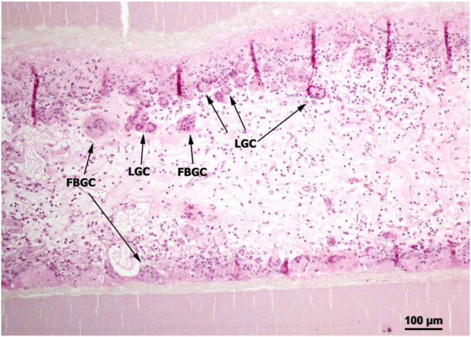
Foreign body multinucleated giant cells (FBGC) and Langhans type giant cells (LGC) adjacent to the inner surface of the nerve conduit in light microscopic H&E staining (scale bars, 100 μm, n = 10).
The histology of the tibial nerve distal to the conduit in both groups revealed myelinated nerve fibers on H&E staining (Figure 5), proved by monoclonal antibody TUJ1 immunohistochemistry staining (Figure 6) and transmission electron microscopy (TEM) (Figures 7 and 8).
Figure 5.
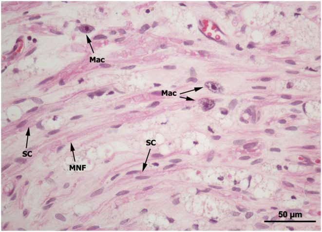
Schwann cells (SCs), myelinated nerve fibers (MNFs), and macrophage (Mac) of the tibial nerve distal to the nerve conduit in H&E staining (scale bars, 50 μm, n = 10).
Figure 6.

Monoclonal antibody TUJ1 immunohistochemistry was used to stain the tibial nerve (PLCL group). (A) Proximal (P) part of the nerve conduit (NT). (B) Middle part of the nerve conduit. (C) Distal (D) part of the nerve conduit (scale bars, 100 μm). (D) Myelinated nerve fiber (MNFs) stained by TUJ1 (brown color) and Schwann cells (SCs) in high-power magnification (scale bars, 50 μm, n = 10).
Figure 7.

Semithin cross section (A–D) and transmission electron microscopy (TEM) (E–F) of the tibial nerve of the experimental side sample of the PLCL-Col group. Round shape of the nerve and regular axon distribution in the proximal portion of the tibial nerve on low (A) and moderate (B) power magnification. Irregular shape and axon distribution in the distal portion of the tibial nerve on low (C) and moderate (D) power magnification (scale bars, 100 μm). Mast cells (arrow) were found in the distal portion as the regeneration process of the axon. TEM shows degeneration of the axon in some part ((E), arrow) (scale bars, 3000 nm) and myelinated nerve fibers ((F), MNFs) (scale bars, 2000 m, n = 10).
Figure 8.

Semithin cross section (A, B) and transmission electron microscopy (TEM) (C, D) of the tibial nerve of the experimental side sample of the PLCL group. Irregular shape and axon distribution in the distal portion of the tibial nerve on low (A) (Scale bars, 100 μm) and moderate (B) power magnification (scale bars, 100 μm). Mast cells (arrow) were found in the distal portion as the regeneration process of the axon. TEM shows degeneration of the axon in some part ((C), arrow), myelinated nerve fibers (C, MNFs) (scale bars, 3000 nm), and (D) Schwann cells (SCs) surrounding the axon (Ax) (scale bars, 600 nm, n = 10).
The available quantitative data of histomorphometric study comparing the two groups are shown in Table 2. There was no statistically significant difference in any parameter on comparing between the PLCL-Col and PLCL groups.
Table 2. Parameters in Quantitative Histomorphometric Study (n = 10).
| perimeter (mm) | diameter (mm) | cross-sectional area (mm2) | number of fibers | density (count/mm2) | thickness of myelin (μm) | |
|---|---|---|---|---|---|---|
| PLCL-Col | 0.76 ± 0.13 | 0.24 ± 0.04 | 0.05 ± 0.02 | 1667 ± 658 | 3.95 ± 1.81 | 0.42 ± 0.06 |
| PLCL | 0.76 ± 0.11 | 0.24 ± 0.03 | 0.05 ± 0.01 | 1975 ± 833 | 4.40 ± 1.97 | 0.43 ± 0.05 |
| autograft | 1.44 ± 0.00 | 0.46 ± 0.00 | 0.17 ± 0.00 | 8299 ± 707 | 1.15 ± 0.15 | 0.49 ± 0.01 |
| t value | 0.42 | 0.42 | 0.09 | –0.80 | –0.47 | –0.24 |
| p-value | 0.97 | 0.97 | 0.93 | 0.44 | 0.64 | 0.81 |
As presented in Table 2, axon count measures the total number of axons. However, an increased number of axons may not necessarily correlate with improved functional outcomes as an overproduction of axons may result from impaired axon growth. Consequently, the axon count may not be a reliable indicator of overall nerve function. The diameter of axons is influenced by both the origin of the axon and maturation of the nerve, which may or may not be directly related to the functional performance. The degree of myelination, while indicative of axon maturity, may not be directly associated with function as myelination occurs prior to the axon reaching its target organ. Furthermore, small axons typically exhibit a thin myelin sheath, making the myelin thickness an unreliable measure of functional capacity. While these morphometric parameters may provide insights into nerve maturity, they do not necessarily reflect functional outcomes due to factors such as improper reinnervation or inadequate integration within the central nervous system. Muscle weight is commonly assumed to correlate with the extent of muscle innervation with the presumption that an innervated muscle weighs more than a denervated one. However, the presence of fibrosis and an increased fat content in denervated tissues may undermine this assumption. Additionally, improper reinnervation, such as the incorrect innervation of a flexor motor nerve to an extensor muscle, may also impact the muscle weight. Therefore, any single measurement will not be able to evaluate nerve recovery well and multiple measurements will be required to make conclusions.
All parameters of nerve regeneration were observed in every animal of each study group. There was evidence of nerve regeneration across the gap from the histomorphometric study. The TFI showed sequential improvements with implantation time in every group (p < 0.05). However, at 12 weeks post operation, the TFI and GMW measurements revealed that the standard nerve graft group showed superior results to the PLCL-Col and PLCL nerve guide groups with a statistical significance of p < 0.05. Our results clearly showed that reinnervation and return of function in both groups. Although the fibrous PLCL presented a good biocompatibility in in vitro cell proliferation study and many studies reported that collagen has low antigenicity, very good cell compatibility, and the ability to promote tissue regeneration, unfortunately, none of the measurements was able to show the superiority of nerve regeneration in adding collagen with polymer in this animal model. Meanwhile, the previous in vitro biocompatible study showed that the use of low loadings of collagen-blend electrospun scaffolds promoted the initial proliferation of cells and reduced necrosis.21 Not surprisingly, the control autografts contained more axons than the PLCL-Col and PLC. Published works from previous studies show that the high number of axons in the autografts may be attributed to the existence of the native axonal extracellular matrix (ECM) structure in the autograft tissue, which facilitates the migration of neural cells and promotes efficient neuronal sprouting.45 Furthermore, collagen conduits did not produce the same functional recovery as nerve autografts. This could be due to the improper target reinnervation caused by the dispersion of misdirected axons inside the collagen conduit.46 This may address the unnecessary collagen component of the conduit wall. This may also have been caused by the short 7 mm distance gap, which is not critical enough to stretch the difference between both groups. From this limitation, we can conclude that the increased collagen loading or the collagen intraluminal filler should be designed with a long-distance gap in the long-term animal study. This may support the fluid phase and the matrix phase of regeneration, or on Schwann cells, which support the cellular migration phase, the axonal phase, and the myelination phase.47
4. Conclusions
The in vivo study in rats used multiple measurements to evaluate nerve regeneration and functional results because the best indicator of nerve function remains unproved in the experimental animals. All parameters of nerve regeneration were observed in every animal of the study groups. Our results clearly showed that there are reinnervation and return of function in both fibrous PLCL and PLCL-Col groups similarly to the autograft. The measurements rarely showed any superiority of nerve regeneration in PLCL-Col in this model. This may be addressed by the porous conduit and the low loading of collagen component in PLCL. It may also have been caused by the short 7 mm distance gap, which is not critical enough to stretch the difference between both groups.
Acknowledgments
The authors thank Thailand’s National Science and Technology Development Agency (NSTDA) and the Commission on Higher Education (CHE) under the National Research University (NRU) Project for financial support. This project also received funding from the Center of Excellence in Materials Science and Technology, Chiang Mai University. This research has received funding support from the NSRF via the Program Management Unit for Human Resources & Institutional Development, Research and Innovation (PMU-B) [Grant No. B13F660056]. This research and innovation activity was funded by the National Research Council of Thailand (NRCT) under the “Hub of Talents in Bioplastics for Use in Medical Applications” (Grant No. N34E670071).
The authors declare no competing financial interest.
References
- Faroni A.; Mobasseri S. A.; Kingham P. J.; Reid A. J. Peripheral nerve regeneration: experimental strategies and future perspectives. Adv. Drug Delivery Rev. 2015, 82–83, 160–167. 10.1016/j.addr.2014.11.010. [DOI] [PubMed] [Google Scholar]
- Gu X.; Ding F.; Williams D. F. Neural tissue engineering options for peripheral nerve regeneration. Biomaterials 2014, 35, 6143–6156. 10.1016/j.biomaterials.2014.04.064. [DOI] [PubMed] [Google Scholar]
- Muheremu A.; Ao Q. Past, present, and future of nerve conduits in the treatment of peripheral nerve injury. BioMed Res. Int. 2015, 2015, 237507 10.1155/2015/237507. [DOI] [PMC free article] [PubMed] [Google Scholar]
- Manoukian O. S.; Baker J.; Rudraiah S.; Arul M.; Vella A.; Domb A.; Kumbar S. G. Functional polymeric nerve guidance conduits and drug delivery strategies for peripheral nerve repair and regeneration. J. Control Release 2020, 317, 78–95. 10.1016/j.jconrel.2019.11.021. [DOI] [PubMed] [Google Scholar]
- Meek M. F.; Coert J. H. Clinical Use of Nerve Conduits in Peripheral-Nerve Repair: Review of the Literature. J. Reconstr. Microsurg. 2002, 18, 097–110. 10.1055/s-2002-19889. [DOI] [PubMed] [Google Scholar]
- Habre S. B.; Bond G.; Jing X. L.; Kostopoulos E.; Wallace R. D.; Konofaos P. The surgical management of nerve gaps: present and future. Ann. Plast. Surg. 2018, 80, 252–261. 10.1097/sap.0000000000001252. [DOI] [PubMed] [Google Scholar]
- Farzamfar S.; Naseri-Nosar M.; Vaez A.; et al. Neural tissue regeneration by a gabapentinloaded cellulose acetate/gelatin wet-electrospun scaffold. Cellulose 2018, 25, 1229–1238. 10.1007/s10570-017-1632-z. [DOI] [Google Scholar]
- Jiang H.; Qian Y.; Fan C.; Ouyang O. Polymeric Guide Conduits for Peripheral Nerve Tissue Engineering. Front. Bioeng. Biotechnol. 2020, 8, 582646 10.3389/fbioe.2020.582646. [DOI] [PMC free article] [PubMed] [Google Scholar]
- Büeler H. Impaired mitochondrial dynamics and function in the pathogenesis of Parkinson’s disease. Exp. Neurol. 2009, 218, 235–246. 10.1016/j.expneurol.2009.03.006. [DOI] [PubMed] [Google Scholar]
- Freed L. E.; Vunjak-Novakovic G.; Biron R. J.; Eagles D. B.; Lesnoy D. C.; Barlow S. K.; Langer R. Biodegradable polymer scaffolds for tissue engineering. Nat. Biotechnol. 1994, 12, 689–693. 10.1038/nbt0794-689. [DOI] [PubMed] [Google Scholar]
- Evans G. R. D.; Brandt K.; Widmer M. S.; Lu L.; Meszlenyi R. K.; Gupta P. K.; Mikos A. G.; Hodges J.; Williams J.; Gürlek A.; Nabawi A.; Lohman R.; Patrick C. W. Jr In vivo evaluation of poly(l-lactic acid) porous conduits for peripheral nerve regeneration. Biomaterials 1999, 20, 1109–1115. 10.1016/S0142-9612(99)00010-1. [DOI] [PubMed] [Google Scholar]
- Nakamura T.; Inada Y.; Fukuda S.; Yoshitani M.; Nakada A.; Itoi S.-i.; Kanemaru S.-i.; Endo K.; Shimizu Y. Experimental study on the regeneration of peripheral nerve gaps through a polyglycolic acid–collagen (PGA–collagen) tube. Brain Res. 2004, 1027, 18–29. 10.1016/j.brainres.2004.08.040. [DOI] [PubMed] [Google Scholar]
- Panseri S.; Cunha C.; Lowery J.; Carro U. D.; Taraballi F.; Amadio S.; Vescovi A.; Gelain F. Electrospun micro-and nanofiber tubes for functional nervous regeneration in sciatic nerve transections. BMC Biotechnol. 2008, 8, 39 10.1186/1472-6750-8-39. [DOI] [PMC free article] [PubMed] [Google Scholar]
- Farzamfar S.; Salehi M.; Tavangar S. M.; Verdi J.; Mansouri K.; Ai A.; Malekshahi Z. V.; Ai J. A novel polycaprolactone/carbon nanofiber composite as a conductive neural guidance channel: an in vitro and in vivo study. Prog. Biomater. 2019, 8, 239–248. 10.1007/s40204-019-00121-3. [DOI] [PMC free article] [PubMed] [Google Scholar]
- Wang X.; Hu W.; Cao Y.; Yao J.; Wu J.; Gu X. Dog sciatic nerve regeneration across a 30-mm defect bridged by a chitosan/PGA artificial nerve graft. Brain 2005, 128, 1897–1910. 10.1093/brain/awh517. [DOI] [PubMed] [Google Scholar]
- Ramesh P. A.; Sethuraman S.; Subramanian A. Fabrication of Anatomically Equivalent Pectin-Based Multifilament Nerve Conduits. ACS Appl. Bio Mater. 2024, 7 (10), 6706–6719. 10.1021/acsabm.4c00872. [DOI] [PubMed] [Google Scholar]
- Shlapakova L. E.; Pryadko A. S.; Zharkova I. I.; Volkov A.; Kozadaeva M.; Chernozem R. V.; Mukhortova Y. R.; Chesnokova D.; Zhuikov V. A.; Zeltser A.; Dudun A. A.; Makhina T.; Bonartseva G. A.; Voinova V. V.; Shaitan K. V.; Romanyuk K.; Kholkin A. L.; Bonartsev A. P.; Surmeneva M. A.; Surmenev R. A. Osteogenic Potential and Long-Term Enzymatic Biodegradation of PHB-based Scaffolds with Composite Magnetic Nanofillers in a Magnetic Field. ACS Appl. Mater. Interfaces 2024, 16 (42), 56555–56579. 10.1021/acsami.4c06835. [DOI] [PubMed] [Google Scholar]
- Bini T. B.; Gao S.; Xu X.; Wang S.; Ramakrishna S.; Leong K. W. Peripheral nerve regeneration by microbraided poly(L-lactide-co-glycolide) biodegradable polymer fibers. J. Biomed. Mater. Res., Part A 2004, 68A, 286–295. 10.1002/jbm.a.20050. [DOI] [PubMed] [Google Scholar]
- Bini T. B.; Shujun G.; Tan T. C.; Shu W.; Aymeric L.; Hai L. B.; Ramakrishna S. Electrospun poly(L-lactide- co -glycolide) biodegradable polymer nanofibre tubes for peripheral nerve regeneration. Nanotechnology 2004, 15, 1459 10.1088/0957-4484/15/11/014. [DOI] [Google Scholar]
- Bini T. B.; Gao S.; Wang S.; Ramakrishna S. Poly(l-lactide-co-glycolide) biodegradable microfibers and electrospun nanofibers for nerve tissue engineering: an in vitro study. J. Mater. Sci. 2006, 41, 6453–6459. 10.1007/s10853-006-0714-3. [DOI] [Google Scholar]
- Daranarong D.; Thapsukhon B.; Wanandy N. S.; Molloy R.; Punyodom W.; Foster L. J. R. Application of low loading of collagen in electrospun poly[(l-lactide)-co-(ε-caprolactone)] nanofibrous scaffolds to promote cellular biocompatibility. Polym. Int. 2014, 63, 1254–1262. 10.1002/pi.4631. [DOI] [Google Scholar]
- Hiljanen-Vainio M.; Karjalainen T.; Seppälä J. Biodegradable lactone copolymers. I. Characterization and mechanical behavior of ε-caprolactone and lactide copolymers. J. Appl. Polym. Sci. 1996, 59, 1281–1288. 10.1002/(SICI)1097-4628(19960222)59:8<1281::AID-APP11>3.0.CO;2-9. [DOI] [Google Scholar]
- Jeong S. I.; Kim B.-S.; Kang S. W.; Kwon J. H.; Lee Y. M.; Kim S. H.; Kim Y. H. In vivo biocompatibilty and degradation behavior of elastic poly(l-lactide-co-ε-caprolactone) scaffolds. Biomaterials 2004, 25, 5939–5946. 10.1016/j.biomaterials.2004.01.057. [DOI] [PubMed] [Google Scholar]
- Xie J.; Ihara M.; Jung Y.; Jung Y.; Kwon I. K.; Kim S. H.; Kim Y. H.; Matsuda T. Mechano-Active Scaffold Design Based on Microporous Poly(L-lactide-co-ε-caprolactone) for Articular Cartilage Tissue Engineering: Dependence of Porosity on Compression Force-Applied Mechanical Behaviors. Tissue Eng. 2006, 12, 449–458. 10.1089/ten.2006.12.449. [DOI] [PubMed] [Google Scholar]
- Hudson T. W.; Evans G. R.; Schmidt C. E. Engineering strategies for peripheral nerve repair. Clin. Plast. Surg. 1999, 26, 617–628. 10.1016/S0094-1298(20)32661-4. [DOI] [PubMed] [Google Scholar]
- Sarker M.; Naghieh S.; McInnes A. D.; Schreyer D. J.; Chen X. Strategic design and fabrication of nerve guidance conduits for peripheral nerve regeneration. Biotechnol. J. 2018, 13 (7), 1700635 10.1002/biot.201700635. [DOI] [PubMed] [Google Scholar]
- Shi Y.; Rittman L.; Vesely I. Novel Geometries for Tissue-Engineered Tendonous Collagen Constructs. Tissue Eng. 2006, 12, 2601–2609. 10.1089/ten.2006.12.2601. [DOI] [PubMed] [Google Scholar]
- Matthews J. A.; Wnek G. E.; Simpson D. G.; Bowlin G. L. Electrospinning of Collagen Nanofibers. Biomacromolecules 2002, 3, 232–238. 10.1021/bm015533u. [DOI] [PubMed] [Google Scholar]
- Prabhakaran M. P.; Venugopal J. R.; Ramakrishna S. Mesenchymal stem cell differentiation to neuronal cells on electrospun nanofibrous substrates for nerve tissue engineering. Biomaterials 2009, 30, 4996–5003. 10.1016/j.biomaterials.2009.05.057. [DOI] [PubMed] [Google Scholar]
- Kanaya F.; Firrell J. C.; Breidenbach W. C. Sciatic function index, nerve conduction tests, muscle contraction, and axon morphometry as indicators of regeneration. Plast. Reconstr. Surg. 1996, 98, 1264–1271. 10.1097/00006534-199612000-00023. [DOI] [PubMed] [Google Scholar]
- Nalampang K.; Molloy R.; Punyodom W. Synthesis and characterization of poly(L-lactide-co-ε- caprolactone) copolymers: influence of sequential monomer addition on chain microstructure. Polym. Adv. Technol. 2007, 18, 240–248. 10.1002/pat.880. [DOI] [Google Scholar]
- Baimark Y.; Molloy R.; Molloy N.; Siripitayananon J.; Punyodom W.; Sriyai M.. Synthesis, characterization and melt spinning of a block copolymer of L-lactide and ε -caprolactone for potential use as an absorbable monofilament surgical suture J. Mater. Sci.: Mater. Med. 16699–707. 10.1007/s10856-005-2605-6. [DOI] [PubMed] [Google Scholar]
- Thapsukhon B.; Meepowpan P.; Molloy R.; Inthanon K.; Wongkham W.; Punyodom W. Effects of Copolymer Microstructure on the Properties of Electrospun Poly(L-lactide-co-caprolactone) Absorbable Nerve Guide Tubes. J. Appl. Polym. Sci. 2013, 130, 4357–436. 10.1002/app.39675. [DOI] [Google Scholar]
- Thapsukhon B.; Daranarong D.; Meepowpan P.; Suree N.; Molloy R.; Inthanon K.; Wongkham W.; Punyodom W. Effect of topology of poly(L-lactide-co-ε-caprolactone) scaffolds on the response of cultured human umbilical cord Wharton’s jelly-derived mesenchymal stem cells and neuroblastoma cell lines. J. Biomater. Sci., Polym. Ed. 2014, 25, 1028–1044. 10.1080/09205063.2014.918457. [DOI] [PubMed] [Google Scholar]
- Bain J. R.; Mackinnon S. E.; Hunter D. A. Functional Evaluation of Complete Sciatic, Peroneal, and Posterior Tibial Nerve Lesions in the Rat. Plast. Reconstr. Surg. 1989, 83, 129–136. 10.1097/00006534-198901000-00024. [DOI] [PubMed] [Google Scholar]
- Thomson R. C.; Wake M. C.; Yaszemski M. J.; Mikos A. G.. Biodegradable polymer scaffolds to regenerate organs. In Biopolymers II; Peppas N.; Langer R., Eds.; Advances in Polymer Science; Springer Berlin Heidelberg, 1995; Vol. 122, pp 245–274. [Google Scholar]
- Parry D. A. D.; Craig A. S.. Collagen; CRC Press: Boaca Raton, FL, 1988. [Google Scholar]
- Titorencu I.; Jinga V.; Lungu A.; Trandafir V.; Albu M. G.; Rau I.; Iovu H. New Scaffold Structure Based on Collagen. Fabrication and Biocompatibility Evaluation. Mol. Cryst. Liq. Cryst. 2008, 486, 147–189. 10.1080/15421400801917932. [DOI] [Google Scholar]
- Dornseifer U. L. F.; Fichter A. M.; Leichtle S.; Wilson A.; Rupp A.; Rodenacker K.; Ninkovic M.; Biemer E.; Machens H.-G.; Matiasek K.; Papadopulos N. A. Peripheral nerve reconstruction with collagen tubes filled with denatured autologous muscle tissue in the rat model. Microsurgery 2011, 31, 632–641. 10.1002/micr.20926. [DOI] [PubMed] [Google Scholar]
- Giusti G.; Willems W. F.; Kremer T.; Friedrich P. F.; Bishop A. T.; Shin A. Y. Return of Motor Function After Segmental Nerve Loss in a Rat Model: Comparison of Autogenous Nerve Graft, Collagen Conduit, and Processed Allograft (AxoGen). J. Bone Jt. Surg 2012, 94, 410–417. 10.2106/JBJS.K.00253. [DOI] [PubMed] [Google Scholar]
- Xu L.; Zhou S.; Feng G.-Y.; Zhang L.-P.; Zhao D.-M.; Sun Y.; Liu Q.; Huang F. Neural Stem Cells Enhance Nerve Regeneration after Sciatic Nerve Injury in Rats. Mol. Neurobiol. 2012, 46, 265–274. 10.1007/s12035-012-8292-7. [DOI] [PubMed] [Google Scholar]
- Ichihara S.; Inada Y.; Nakamura T. Artificial nerve tubes and their application for repair of peripheral nerve injury: an update of current concepts. Injury 2008, 39, 29–39. 10.1016/j.injury.2008.08.029. [DOI] [PubMed] [Google Scholar]
- ZENG X.; Sun Z.; Jia W.; et al. Recovery from rat sciatic nerve injury in vivo through the use of differentiated MDSCs in vitro. Exp. Ther. Med. 2013, 5, 193–196. 10.3892/etm.2012.785. [DOI] [PMC free article] [PubMed] [Google Scholar]
- Deumens R.; Bozkurt A.; Meek M. F.; Marcus M. A. E.; Joosten E. A. J.; Weis J.; Brook G. A. Repairing injured peripheral nerves: Bridging the gap. Prog. Neurobiol. 2010, 92, 245–276. 10.1016/j.pneurobio.2010.10.002. [DOI] [PubMed] [Google Scholar]
- Adams A. M.; VanDusen K. W.; Kostrominova T. Y.; Mertens J. P.; Larkin L. M. Scaffoldless tissue-engineered nerve conduit promotes peripheral nerve regeneration and functional recovery after tibial nerve injury in rats. Neural Regener. Res. 2017, 12 (9), 1529–1537. 10.4103/1673-5374.215265. [DOI] [PMC free article] [PubMed] [Google Scholar]
- Long Q.; Wu B.; Yang Y.; Wang S.; Shen Y.; Bao Q.; Xu F. Nerve guidance conduit promoted peripheral nerve regeneration in rat. Artif. Organs 2021, 45, 616–624. 10.1111/aor.13881. [DOI] [PubMed] [Google Scholar]
- Daly W.; Yao L.; Zeugolis D.; Windebank A.; Pandit A. A biomaterials approach to peripheral nerve regeneration: bridging the peripheral nerve gap and enhancing functional recovery. J. R. Soc. Interface 2012, 9 (67), 202–221. 10.1098/rsif.2011.0438. [DOI] [PMC free article] [PubMed] [Google Scholar]