Abstract
Bunyamwera virus (BUNV) is the prototype of the Bunyaviridae family of RNA viruses. BUNV genomic strands are templates for both replication and transcription, whereas the antigenomic RNAs serve only as templates for replication. By mutagenesis of model templates, we showed that the BUNV transcription promoter comprises elements within both the 3′ and the 5′ nontranslated regions. Using this information, we designed a model ambisense BUNV segment that transcribed BUNV S mRNA from the genomic strand and green fluorescent protein (GFP) mRNA from the antigenome. Demonstration of GFP expression showed that this ambisense strategy represents a viable approach for generating BUNV segments able to express additional proteins.
The Bunyaviridae family of RNA viruses comprises five genera, namely, Orthobunyavirus, Hantavirus, Nairovirus, Phlebovirus, and Tospovirus. Many bunyaviruses cause life-threatening human disease and consequently are described by the Centers for Disease Control and Prevention as category A, B, and C priority pathogens. The prototype of the Bunyaviridae family is Bunyamwera virus (BUNV), which serves as a model for the many pathogens within this family.
The BUNV genome comprises three segments of negative-sense RNA, designated small (S), medium (M), and large (L). The S segment encodes the nucleocapsid (N) and nonstructural proteins (NSs) expressed from overlapping open reading frames (ORFs) on the same mRNA (8, 11, 12). The M segment encodes a polyprotein that is cleaved to yield Gn, Gc, and NSm proteins (6, 12, 13). The L segment encodes the RNA-dependent RNA polymerase (RdRp) (7).
The ORFs of each segment are flanked by nontranslated regions (NTRs) that direct the RdRp to perform two distinct RNA synthesis activities: (i) transcription to generate a single mRNA and (ii) replication to generate antigenomes that are replicated to generate further genomes. The BUNV S-segment genome is a template for both replication and transcription, whereas the antigenome serves only as a template for replication (5, 14). This division of template activity, in which a functional mRNA is transcribed only from the genomic strand, is not a universal feature of all bunyavirus members. Many viruses within this group perform ambisense transcription, in which both genome and antigenomes are transcriptionally active. The ambisense strategy of gene expression is a feature of the segmented arenaviruses and recently has been artificially bestowed upon several nonsegmented negative-sense RNA viruses (9, 18).
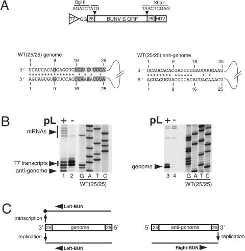
The fundamental difference in activity of the BUNV genomic and antigenomic RNAs suggests that these strands are recognized differently by the BUNV RdRp, which is likely due to critical nucleotide differences within the 3′ and 5′ NTRs of these RNAs. For the BUNV S segment, the 3′ and 5′ NTRs are 85 and 170 nucleotides in length, respectively. To facilitate our search for nucleotides responsible for the different strand activities, we constructed WT(25/25), which comprises nucleotides 1 to 25 of 3′ and 5′ NTRs surrounding the intact BUNV S-segment ORF (Fig. 1A). To analyze the RNA synthesis activity of WT(25/25), the corresponding cDNA was transfected together with BUNV S- and L-segment ORF containing plasmids into BHK-21 cells previously infected with recombinant vaccinia virus vTF7-3, which expresses T7 RNA polymerase. Primer extension of end-labeled oligonucleotides Left-BUN (5′-GCGACCTCTGGGTCAAAAGTACTGC-3′) and Right-BUN (5′-GGAAGAAAACCAATGTTAGTGCAGC-3′), performed as described previously (3-5), showed that the genomic and antigenomic strands of WT(25/25) exhibited the same RNA-synthetic characteristics as their full-length counterparts such that the genome signaled both transcription and replication (Fig. 1B, lane 1, and C), whereas the antigenome signaled only replication (Fig. 1B, lane 3, and C).
FIG. 1.
Schematic representation of the genomic and antigenomic strands of model template WT(25/25) and analysis of their RNA synthesis activity. (A) Schematic of T7 transcription plasmid pWT(25/25) designed to express the antigenomic strand of template WT(25/25). The resulting primary transcript contains nucleotides 1 to 25 of the BUNV 3′ and 5′ S-segment NTRs and nucleotides representing BglII and XhoI restriction sites surrounding the BUNV S-segment ORF. The primary transcript also possesses two additional G residues at the 5′ end and the Hepatitis delta virus ribozyme (HDV) at the 3′ end. The BUNV-specific nucleotides within 3′ and 5′ NTRs of the genome and antigenome of template WT(25/25) are shown. Differences between the two strands are shaded, and the cases in which there is the potential to form canonical Watson-Crick pairings (*) or noncanonical pairs (•) are shown. (B) The RNA synthesis activities of WT(25/25) genomic and antigenomic strands was determined using primer extension analysis. A cDNA expressing the model BUNV template WT(25/25) was transfected into vTF7-3-infected BHK-21 cells together with BUNV S and L plasmids (+) or BUNV S plasmid alone (-). Positive-sense RNAs generated from the genomic strand of WT(25/25) were detected by primer extension using 33P end-labeled negative-sense oligonucleotide Left-BUN. The products were separated by polyacrylamide gel electrophoresis (PAGE) and visualized by autoradiography. Initial T7 RNA polymerase transcripts and antigenomic replication products are marked with an arrow, and the BUNV mRNAs characterized by the host-cell-derived and variably sized 5′ extensions are marked with a vertical bar. (B) Negative-sense RNAs generated by the antigenomic strand of WT(25/25) were analyzed by use of 33P end-labeled positive-sense oligonucleotide Right-BUN. For each panel, a sequence ladder is provided as a marker. (C). The ability of WT(25/25) genome or antigenome strands to transcribe and replicate is schematically summarized below each corresponding autoradiograph.
There are 18 nucleotide differences between these strands, and one or more must be a critical component of the transcription signal. These nucleotides comprise mismatched nucleotides at 3′ and 5′ position 9 and eight further nucleotides within each NTR (positions 16 to 18, 20, and 22 to 25 [Fig. 1A]). The goal of this study is to identify which of these nucleotides are essential for transcription.
We previously showed that the U residue at 3′ position 9 was critical for transcription, whereas corresponding 5′ position 9 could be changed with no effect (5). We also showed that this U residue was not the sole component of the transcription promoter, as it did not signal transcription when within the transcriptionally silent antigenome (5). In addition, we determined that the remaining eight nucleotide differences between the 3′ NTRs of genomic and antigenomic strands played no role in transcription signaling (5). Together, these findings imply that an essential transcription signal likely resides within two elements located at opposite ends of the template: the U at genomic 3′ position 9 and the eight nucleotides located at genomic 5′ positions 16 to 18, 20, and 22 to 25. To test this hypothesis, we introduced these elements into the nontranscribing antigenome of WT(25/25). If our hypothesis was correct, transfer of both elements would confer transcription signaling ability to this strand.
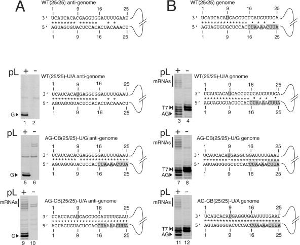
We first inserted each element individually to show that neither was sufficient alone to signal transcription. First, the U residue was incorporated at position 9 within the WT(25/25) antigenome to generate WT(25/25)-U/A (Fig. 2A, top schematic). Primer extension analysis using oligonucleotide Right-BUN showed that the WT(25/25)-U/A antigenome signaled replication but not transcription (Fig. 2A, lane 1). We next placed the eight-nucleotide element within the antigenomic 5′ NTR of WT(25/25) to generate AG-CB(25/25)-U/G (Fig. 2A, middle schematic). Again, primer extension analysis using oligonucleotide Right-BUN showed that the antigenome signaled replication but not transcription (Fig. 2A, lane 5). Finally, we incorporated both elements into WT(25/25) to generate AG-CB(25/25)-U/A (Fig. 2A, bottom schematic). Primer extension analysis using oligonucleotide Right-BUN showed that the antigenome strand signaled both replication and transcription (Fig. 2A, lane 9), thus confirming our hypothesis that nucleotides essential for transcription were located at 3′ position 9 and also 5′ positions 16 to 18, 20, and 22 to 25.
FIG. 2.
Schematic representation of the genomic and antigenomic strands of WT(25/25) and its derivatives and analysis of their RNA synthesis activities. (A) Nucleotides hypothesized to be essential for signaling BUNV transcription (shaded) were incorporated into the transcriptionally inactive antigenomic strand of template WT(25/25) to generate templates WT(25/25)-U/A, AG-CB(25/25)-U/G, and AG-CB(25/25)-U/A. The abilities of these antigenomic strands to signal transcription were investigated by primer extension analysis. Plasmids expressing the model BUNV templates WT(25/25)-U/A, AG-CB(25/25)-U/G, and AG-CB(25/25)-U/A were transfected into vTF7-3 infected BHK-21 cells together with BUNV S and L plasmids (+) or BUNV S plasmid alone (−). Negative-sense RNAs generated from these antigenomes were analyzed by primer extension using 33P end-labeled positive-sense oligonucleotide Right-BUN. The products were separated by PAGE and visualized by autoradiography. Genomic replication products are marked with an arrow, and the variably sized negative-sense BUNV transcripts are marked with a vertical bar. (B) The RNA synthesis ability of the genomic strands of templates WT(25/25)-U/A, AG-CB(25/25)-U/G, and AG-CB(25/25)-U/A was investigated by primer extension analysis using negative-sense oligonucleotide Left-BUN. Initial T7 RNA polymerase transcripts and antigenomic replication products are marked with an arrow and the variably sized BUNV mRNAs are marked with a vertical bar. G, genome; AG, antigenome.
Based on our hypothesis, all alterations made to the antigenomic strands of WT(25/25)-U/A, AG-CB(25/25)-U/G, and AG-CB(25/25)-U/A should not affect the existing transcription signal within the corresponding genomic RNAs. Primer extension analysis using oligonucleotide Left-BUN showed these genomic strands maintained transcription signaling ability (Fig. 2B, lanes 3, 7, and 11), demonstrating that both genomic and antigenomic strands of AG-CB(25/25)-U/A are transcriptionally competent. Template AG-CB(25/25)-U/A is thus capable of ambisense transcription.
These results show the transcription promoter involves nucleotides located at 3′ and 5′ ends of the template. We recently determined that BUNV RNA replication required complementarity between nucleotides within 3′ and 5′ NTRs, and their signaling ability depended on base-pairing potential rather than specific identity (3). In contrast, nucleotides required for transcription are predicted to be unpaired in the wild-type template (Fig. 1A), and so their signaling ability may depend on nucleotide identity rather than base-pairing potential with nucleotides at the opposite end of the template. However, despite these possible differences, the common requirement for 3′ and 5′ nucleotides suggests that both replication and transcription require template circularization. It is well established that Influenza virus segments also require terminal interaction for RNA synthesis (1, 2, 10, 15-17, 19-21), and so these results establish functional similarities between RNA synthesis signals of bunyaviruses and orthomyxoviruses.
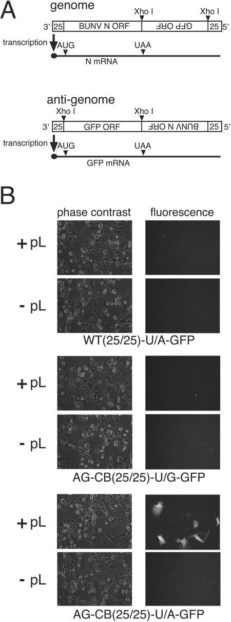
We next investigated whether transcripts made from the AG-CB(25/25)-U/A antigenome could be translated. To test this, we inserted the green fluorescent protein (GFP) ORF downstream of the antigenomic 3′ NTR in templates WT(25/25)-U/A, AG-CB(25/25)-U/G, and AG-CB(25/25)-U/A (Fig. 3A). The ability of these templates to express functional GFP mRNAs was analyzed by using fluorescence microscopy.
FIG. 3.
Construction of an ambisense BUNV model template able to express an additional foreign protein. (A) A cDNA encompassing the entire GFP ORF was inserted at the XhoI site directly downstream of the antigenomic 3′ NTR of templates WT(25/25)-U/A, AG-CB(25/25)-U/G, and AG-CB(25/25)-U/A. (B) Plasmids expressing the antigenomic strand of templates WT(25/25)-U/A-GFP, AG-CB(25/25)-U/G-GFP, and AG-CB(25/25)-U/A-GFP were transfected into vTF7-3-infected BHK-21 cells together with plasmids expressing BUNV S and L support plasmids (+pL) or BUNV S support plasmid alone (−pL). The ability of the antigenomic strands of these templates to express GFP was analyzed using a Nikon Eclipse TE2000-E fluorescence microscope 20 h posttransfection at a magnification of 60×.
Only cells transfected with AG-CB(25/25)-U/A-GFP and both S and L plasmids generated fluorescence (Fig. 3B), indicating that the GFP mRNA was translatable. As predicted from our earlier results (Fig. 2A, lanes 1 and 5), fluorescence was not detected in dishes containing WT(25/25)-U/A-GFP and AG-CB(25/25)-U/G-GFP or dishes lacking the L plasmid. Interestingly, the AG-CB(25/25)-U/A-GFP antigenome does not contain the signal that terminates BUNV S-segment transcription upstream of the genomic 5′ end (J. N. Barr, J. W. Rodgers, and G. W. Wertz, submitted for publication). This signal could place sequences or secondary structures at the mRNA 3′ end to functionally replace poly(A) tails that BUNV mRNAs lack. However, translation of the GFP mRNAs suggests that BUNV-specific sequences or structures are not essential for translation.
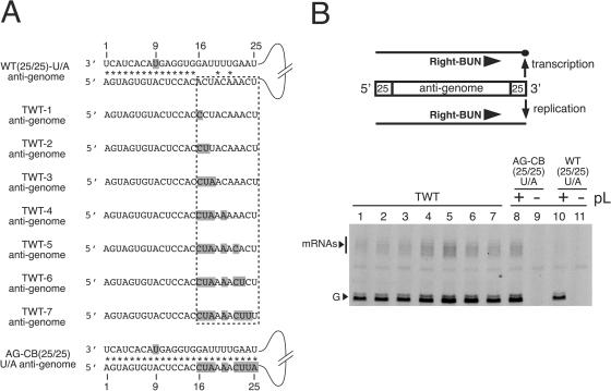
We next wanted to determine which nucleotides within 5′ positions 16 to 18, 20, and 22 to 25 were essential for transcription. To achieve this, we individually altered each of these nucleotides within the nontranscribing antigenome of WT(25/25)-U/A to the identity of the corresponding nucleotide from the transcriptionally active antigenome of AG-CB(25/25)-U/A (Fig. 4A). We anticipated that stepwise alteration of these nucleotides would confer transcription ability to the WT(25/25)-U/A antigenome only when nucleotides critical for transcription signaling were present.
FIG. 4.
Identification of nucleotides within the 5′ NTR that are essential for BUNV transcription. (A). Schematic showing the eight nucleotide differences within the 5′ NTR of the antigenomic strands of WT(25/25)-U/A and AG-CB(25/25)-U/A. These eight nucleotides were altered in a stepwise manner such that the nontranscribing antigenome of WT(25/25)-U/A was converted to the transcribing antigenome of AG-CB(25/25)-U/A in seven increments to give templates TWT1 to TWT7. The shaded positions represent nucleotides normally found within the 5′ NTR of AG-CB(25/25)-U/A. (B). Plasmids expressing the model BUNV templates TWT1 to TWT7, WT(25/25)-U/A, and AG-CB(25/25)-U/A were transfected into vTF7-3-infected BHK-21 cells together with BUNV S and L support plasmids (+) or BUNV S plasmid alone (−). The abilities of the antigenomic strands of these model templates to perform transcription were analyzed by primer extension by use of 33P end-labeled positive-sense oligonucleotide Right-BUN. The products were separated by PAGE and visualized by autoradiography. Genomic replication products are marked with an arrow, and the variably sized negative-sense BUNV transcripts are marked with a vertical bar. G, genome.
The ability of the resulting antigenomes (TWT1 to TWT7) to signal transcription was analyzed by primer extension using oligonucleotide Right-BUN. Our results show that all antigenomes from TWT1 to TWT7 (Fig. 4B, lanes 1 to 7) signaled increased transcription compared to the nontranscribing WT(25/25)-U/A antigenome (Fig. 4B, lane 10). Transcript abundance increased through the series as follows: TWT1 < TWT2 < TWT3 < TWT4 (Fig. 4B, lanes 1 to 4), such that the transcription signaling ability of TWT4 was indistinguishable from that of AG-CB(25/25)-U/A (Fig. 4B, lane 8). This shows that corresponding nucleotides 16, 17, 18, and 20 of the 5′ NTR form a critical element that is required for abundant BUNV transcription.
In conclusion, we have identified nucleotides within BUNV 3′ and 5′ NTRs that are essential for signaling transcription. We used this information to generate a BUNV segment capable of ambisense expression, and we suggest that this mode of transcription represents a potential strategy for generating recombinant infectious BUNV variants containing additional transcriptional units.
Acknowledgments
We thank members of the G. W. Wertz laboratory for helpful suggestions during the preparation of the manuscript. We also thank R. M. Elliott (Glasgow, United Kingdom) for the continued use of cDNAs expressing BUNV S- and L-segment ORFs.
This work was supported by National Institutes of Health grant AI 59174 to J.N.B. and an unrestricted infectious diseases research award to G.W.W.
REFERENCES
- 1.Azzeh, M., R. Flick, and G. Hobom. 2001. Functional analysis of the influenza A virus cRNA promoter and construction of an ambisense transcription system. Virology 289:400-410. [DOI] [PubMed] [Google Scholar]
- 2.Bae, S. H., H. K. Cheong, J. H. Lee, C. Cheong, M. Kainosho, and B. S. Choi. 2001. Structural features of an influenza virus promoter and their implications for viral RNA synthesis. Proc. Natl. Acad. Sci. USA 98:10602-10607. [DOI] [PMC free article] [PubMed] [Google Scholar]
- 3.Barr, J. N., R. M. Elliott, E. F. Dunn, and G. W. Wertz. 2003. Segment-specific terminal sequences of Bunyamwera bunyavirus regulate genome replication. Virology 311:326-338. [DOI] [PubMed] [Google Scholar]
- 4.Barr, J. N., and G. W. Wertz. 2004. Bunyamwera bunyavirus RNA synthesis requires cooperation of 3′- and 5′-terminal sequences. J. Virol. 78:1129-1138. [DOI] [PMC free article] [PubMed] [Google Scholar]
- 5.Barr, J. N., and G. W. Wertz. 2005. Role of the conserved nucleotide mismatch within the 3′ and 5′ terminal regions of Bunyamwera virus in signaling transcription. J. Virol. 79:3586-3594. [DOI] [PMC free article] [PubMed] [Google Scholar]
- 6.Elliott, R. M. 1985. Identification of nonstructural proteins encoded by viruses of the Bunyamwera serogroup (family Bunyaviridae). Virology 143:119-126. [DOI] [PubMed] [Google Scholar]
- 7.Elliott, R. M. 1989. Nucleotide sequence analysis of the large (L) genomic RNA segment of Bunyamwera virus, the prototype of the family Bunyaviridae. Virology 173:426-436. [DOI] [PubMed] [Google Scholar]
- 8.Elliott, R. M. 1989. Nucleotide sequence analysis of the small (S) RNA segment of Bunyamwera virus, the prototype of the family Bunyaviridae. J. Gen. Virol. 70:1281-1285. [DOI] [PubMed] [Google Scholar]
- 9.Finke, S., and K. K. Conzelmann. 1997. Ambisense gene expression from recombinant rabies virus: random packaging of positive- and negative-strand ribonucleoprotein complexes into rabies virions. J. Virol. 71:7281-7288. [DOI] [PMC free article] [PubMed] [Google Scholar]
- 10.Flick, R., and G. Hobom. 1999. Interaction of influenza virus polymerase with viral RNA in the ‘corkscrew’ conformation. J. Gen. Virol. 80:2565-2572. [DOI] [PubMed] [Google Scholar]
- 11.Fuller, F., A. S. Bhown, and D. H. Bishop. 1983. Bunyavirus nucleoprotein, N, and a non-structural protein, NSS, are coded by overlapping reading frames in the S RNA. J. Gen. Virol. 64:1705-1714. [DOI] [PubMed] [Google Scholar]
- 12.Fuller, F., and D. H. Bishop. 1982. Identification of virus-coded nonstructural polypeptides in bunyavirus-infected cells. J. Virol. 41:643-648. [DOI] [PMC free article] [PubMed] [Google Scholar]
- 13.Gentsch, J. R., and D. L. Bishop. 1979. M viral RNA segment of bunyaviruses codes for two glycoproteins, G1 and G2. J. Virol. 30:767-770. [DOI] [PMC free article] [PubMed] [Google Scholar]
- 14.Jin, H., and R. M. Elliott. 1993. Characterization of Bunyamwera virus S RNA that is transcribed and replicated by the L protein expressed from recombinant vaccinia virus. J. Virol. 67:1396-1404. [DOI] [PMC free article] [PubMed] [Google Scholar]
- 15.Leahy, M. B., H. C. Dobbyn, and G. G. Brownlee. 2001. Hairpin loop structure in the 3′ arm of the influenza A virus virion RNA promoter is required for endonuclease activity. J. Virol. 75:7042-7049. [DOI] [PMC free article] [PubMed] [Google Scholar]
- 16.Leahy, M. B., G. Zecchin, and G. G. Brownlee. 2002. Differential activation of influenza A virus endonuclease activity is dependent on multiple sequence differences between the virion RNA and cRNA promoters. J. Virol. 76:2019-2023. [DOI] [PMC free article] [PubMed] [Google Scholar]
- 17.Lee, M. T., K. Klumpp, P. Digard, and L. Tiley. 2003. Activation of influenza virus RNA polymerase by the 5′ and 3′ terminal duplex of genomic RNA. Nucleic Acids Res. 31:1624-1632. [DOI] [PMC free article] [PubMed] [Google Scholar]
- 18.Le Mercier, P., D. Garcin, S. Hausmann, and D. Kolakofsky. 2002. Ambisense sendai viruses are inherently unstable but are useful to study viral RNA synthesis. J. Virol. 76:5492-5502. [DOI] [PMC free article] [PubMed] [Google Scholar]
- 19.Pritlove, D. C., L. L. Poon, E. Fodor, J. Sharps, and G. G. Brownlee. 1998. Polyadenylation of influenza virus mRNA transcribed in vitro from model virion RNA templates: requirement for 5′ conserved sequences. J. Virol. 72:1280-1286. [DOI] [PMC free article] [PubMed] [Google Scholar]
- 20.Rao, P., W. Yuan, and R. M. Krug. 2003. Crucial role of CA cleavage sites in the cap-snatching mechanism for initiating viral mRNA synthesis. EMBO J. 22:1188-1198. [DOI] [PMC free article] [PubMed] [Google Scholar]
- 21.Zheng, H., P. Palese, and A. Garcia-Sastre. 1996. Nonconserved nucleotides at the 3′ and 5′ ends of an influenza A virus RNA play an important role in viral RNA replication. Virology 217:242-251. [DOI] [PubMed] [Google Scholar]


