Abstract
Three species of Bacillus were evaluated as potential surrogates for Bacillus anthracis for determining the sporicidal activity of chlorination as commonly used in drinking water treatment. Spores of Bacillus thuringiensis subsp. israelensis were found to be an appropriate surrogate for spores of B. anthracis for use in chlorine inactivation studies.
The use of spores of Bacillus anthracis as a bioterrorist weapon has prompted renewed interest in the study of the inactivation of Bacillus spores by chemical disinfectants. Information regarding the resistance of B. anthracis spores to chlorination is of particular interest in reference to drinking water treatment. There has been a growing awareness, due to restrictions of working with select agents, of the need to evaluate the resistance to disinfectants of spores of attenuated strains of B. anthracis as well as other closely related species of Bacillus which might serve as surrogates for the overt pathogenic agent. The three species evaluated as surrogate organisms in this study were chosen based upon their genetic homogeneity (9). Indeed, some investigators have suggested that the three organisms comprise a single species (5).
The sporicidal effectiveness of free available chlorine was determined at two pH levels and two temperatures. The inactivation experiments were conducted under chlorine-demand-free (CDF) conditions. CT values (C is the concentration of chlorine in mg/liter, and T is the exposure time in minutes) were determined for each Bacillus species for the various experimental conditions. The sporulation and purification procedures were performed in the same manner for each species.
Three species of Bacillus were used in this study: B. anthracis Sterne (34F2; Colorado Serum Co., Denver, CO), Bacillus cereus (ATCC 7039), and Bacillus thuringiensis subsp. israelensis (ATCC 35646). B. anthracis Sterne is an attenuated strain of B. anthracis. Endospores were produced in a broth sporulation medium (3). The cultures were grown at 35°C with agitation on a rotary shaker for 5 days. Spores were purified by gradient separation using RenoCal-76 (Bracco Diagnostics, Princeton, NJ) and washed three times by centrifugation with distilled water, as previously described (7). Purified spore preparations (approximately 1 × 10⁷ CFU/ml) were examined using phase-contrast microscopy and stored in 40% (vol/vol) ethanol at 5°C until the time of use. Microscopic examination of the purified spore preparations exhibited <0.1% vegetative cells.
Sterile CDF buffer (0.05 M KH2PO4) was used in the experiments. The buffer was made chlorine demand free by adding reagent-grade sodium hypochlorite (4 to 6%) to the buffer to achieve a free-chlorine residual of approximately 3.0 mg/liter. The pH of the buffer was adjusted by the addition of 10 M sodium hydroxide. The buffer was boiled for 5 minutes, transferred to 4-liter beakers, and exposed to short-wave UV irradiation in a biological-safety cabinet for 48 hours to remove the chlorine. The buffer was then sterilized by autoclaving and stored in a sealed container for no longer than 3 weeks. An appropriate volume of a 1:200 (vol/vol) aqueous solution of reagent-grade sodium hypochlorite was added to the CDF buffer to obtain the desired free-chlorine level.
Inactivation experiments were conducted at 5°C in a recirculating, refrigerated water bath and at ambient room temperature (22 to 23°C) at pH 7.0 and 8.0. These parameters were chosen as representative of conditions which might normally be encountered in drinking water treatment facilities. All temperatures in the reaction vessels were verified using a mercury-filled glass thermometer. Borosilicate glass beakers (1,000 ml) containing 500 ml of buffer served as the reaction vessels. Reaction vessels were continuously stirred using a magnetic stirring apparatus. The vessels were inoculated with the various spore preparations to yield an initial level of approximately 1 × 10⁴ CFU/ml. Chlorine concentrations were determined at each exposure time using the N,N-diethyl-p-phenylenediamine colorimetric method (1). Controls for these experiments consisted of CDF buffer without a chlorine residual. Samples were withdrawn from the reaction vessels at the various exposure times, and any residual chlorine was immediately neutralized by the addition of 0.1 ml of a 10% (wt/vol) sodium thiosulfate solution. Control and test samples were treated in the same manner throughout the experiments. Triplicate experiments were performed for each Bacillus species for each experimental condition. The numbers of spores present in the control and chlorine-exposed samples were determined by culture count using a membrane filtration procedure, as previously described (10).
Levels of inactivation were determined by plotting the log10 ratio of survivors against the exposure time (minutes) for each experimental condition. Covariance analysis was performed to compare the inactivation rates. The analysis was performed separately for each of the four experimental conditions of pH and temperature. The Chick-Watson model was used for determining CT values (6). CT values were calculated based upon a first-order exponential relationship for chlorine decay. The chlorine decay rate was calculated from the slope of the line plotted on the basis of the ratio of chlorine concentrations at each exposure time and at time zero (Ct/C0) against the exposure time. The mean chlorine decay rate was 0.0012 min−1 (range, 0.00023 to 0.00259 min−1).
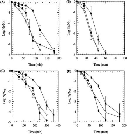
The results of the inactivation experiments are shown in Fig. 1. Each data point represents the mean of the results of three experiments. Under the chlorine-demand-free conditions, the chlorine concentrations averaged 2.0 ± 0.2 mg/liter during the course of the experiments. The standard error of the mean ranged from 0 to 0.58, with the average standard error being 0.14 for the log10 microbial counts for the individual data points (Fig 1). As expected, in all cases the rates of inactivation were greater at the higher temperature (23°C). Inactivation also occurred more rapidly, as anticipated, at the lower pH level (pH 7.0). These results concur with established observations regarding the role of temperature and pH in chlorine inactivation studies (6).
FIG. 1.
Inactivation of Bacillus species spores exposed to 2.0 mg/liter free chlorine under different conditions. (A) pH 7, 5°C; (B) pH 7, 23°C; (C) pH 8, 5°C; (D) pH 8, 23°C. Symbols: •, B. anthracis Sterne; ▿, B. cereus; ▪, B. thuringiensis subsp. israelensis.
Differences were observed for the inactivation of the different Bacillus species. B. thuringiensis subsp. israelensis spores were consistently more resistant than spores of either B. anthracis Sterne or B. cereus. Spores of B. anthracis Sterne and B. cereus behaved similarly but did not exhibit consistent patterns of resistance under the various conditions. Spores of B. anthracis Sterne were more resistant than spores of B. cereus at pH 7.0, but spores of B. cereus were more resistant than spores of B. anthracis Sterne at pH 8.0 at both temperatures (Fig. 1). The covariance analysis indicated that the rate of inactivation for spores of B. thuringiensis was significantly lower (P < 0.05) than the rate of inactivation for spores of B. anthracis or B. cereus. There was no significant difference (P < 0.05) between the inactivation rates for spores of B. anthracis Sterne or B. cereus.
The calculated CT values (mg · min/liter) for inactivation of spores of the three Bacillus spp. at 2, 3, and 4 orders of magnitude are given in Table 1. The CT values derived from the model illustrate the differences in inactivation observed for the three species of Bacillus. Also shown in Table 1 are CT values which have recently been reported for 2 and 3 log10 levels of inactivation at pH 7.0 for spores of a virulent strain of anthrax, B. anthracis Ames (11).
TABLE 1.
CT values for inactivation of spores of Bacillus spp. exposed to 2.0 mg/liter free chlorine
| Temp (°C) | pH | Log10 inactivation |
CT (mg · min/liter)
|
|||
|---|---|---|---|---|---|---|
| B. anthracis Amesa | B. anthracis Sterne | B. cereus | B. thuringiensis | |||
| 23 | 7 | 2 | 79 | 45 | 41 | 66 |
| 3 | 102 | 68 | 62 | 99 | ||
| 4 | ND | 90 | 82 | 132 | ||
| 8 | 2 | ND | 127 | 132 | 246 | |
| 3 | ND | 191 | 199 | 369 | ||
| 4 | ND | 254 | 264 | 492 | ||
| 5 | 7 | 2 | 220 | 140 | 117 | 229 |
| 3 | 339 | 210 | 175 | 344 | ||
| 4 | ND | 280 | 233 | 458 | ||
| 8 | 2 | ND | 319 | 340 | 481 | |
| 3 | ND | 478 | 510 | 721 | ||
| 4 | ND | 637 | 680 | 961 | ||
Data from reference 11. ND, not determined.
Chlorine is the most widely used disinfectant for water treatment in the United States, but there is a limited amount of data on the effectiveness of chlorination in inactivating spores of B. anthracis under conditions used in drinking water treatment (2,4). The CT values for two of the surrogate organisms used in this study (B. anthracis Sterne and B. cereus) were substantially lower than the CT values for spores of B. thuringiensis subsp. israelensis and for the virulent B. anthracis Ames strain. The CT values which most closely approximated those for the virulent strain were seen for spores of B. thuringiensis subsp. israelensis. Based upon these findings, spores of B. thuringiensis subsp. israelensis would be an appropriate surrogate to use in place of B. anthracis in chlorine inactivation studies.
Methodological variations can substantially alter the measured activities of sporicidal agents (12). In a recent report on UV inactivation of Bacillus species spores (8), it was noted that the most reliable method for testing intrinsic differences between strains requires parallel testing, using identical conditions for sporulation, purification, and survival determinations. The present study meets these criteria. Future studies directly comparing the inactivation of spores of other surrogate species and other strains of the proposed surrogate organisms with that of other virulent strains will be beneficial in evaluating the use of surrogate Bacillus spp. as an alternative to B. anthracis in disinfection studies.
Acknowledgments
We thank Anna Yu and Christy M. Frietch for their assistance in this project.
REFERENCES
- 1.American Public Health Association. 1998. Standard methods for the examination of water and wastewater, 20th ed. American Public Health Association, Washington, D.C.
- 2.Brazis, A. R., J. E. Leslie, P. W. Kabler, and R. L. Woodward. 1958. The inactivation of spores of Bacillus globigii and Bacillus anthracis by free available chlorine. Appl. Microbiol. 6:338-342. [DOI] [PMC free article] [PubMed] [Google Scholar]
- 3.Coroller, L., I. Leguérinel, and P. Mafart. 2001. Effect of water activities of heating and recovery media on apparent heat resistance of Bacillus cereus spores. Appl. Environ. Microbiol. 67:317-322. [DOI] [PMC free article] [PubMed] [Google Scholar]
- 4.Fair, G. M., J. C. Morris, and S. L. Chang. 1947. The dynamics of water chlorination. J. N. Engl. Water Works Assoc. 61:285-301. [Google Scholar]
- 5.Helgason, E., O. A. Økstad, D. A. Caugant, H. A. Johansen, A. Fouet, M.Mock, I. Hegna, and A.-B. Kolstø. 2000. Bacillus anthracis, Bacillus cereus and Bacillus thuringiensis—one species on the basis of genetic evidence. Appl. Environ. Microbiol. 66:2627-2630. [DOI] [PMC free article] [PubMed] [Google Scholar]
- 6.Hoff, J. C. 1986. Inactivation of microbial agents by chemical disinfectants. EPA/600/2- 86/067. U.S. Environmental Protection Agency, Cincinnati, Ohio.
- 7.Nicholson, W. L., and P. Setlow. 1990. Sporulation, germination and outgrowth, p. 391-429. In C. R. Harwood and S. M. Cutting (ed.), Molecular biology methods for Bacillus. John Wiley and Sons, New York, N.Y.
- 8.Nicholson, W. L., and B. Galeano. 2003. UV resistance of Bacillus anthracis spores revisited: validation of Bacillus subtilis spores as UV surrogates for spores of B. anthracis Sterne. Appl. Environ. Microbiol. 69:1327-1330. [DOI] [PMC free article] [PubMed] [Google Scholar]
- 9.Radnedge, L., P. G. Agron, K. K. Hill, P. J. Jackson, L. O. Ticknor, P. Keim, and G. L. Andersen. 2003. Genome differences that distinguish Bacillus anthracis from Bacillus cereus and Bacillus thuringiensis. Appl. Environ. Microbiol. 69:2755-2764. [DOI] [PMC free article] [PubMed] [Google Scholar]
- 10.Rice, E. W., K. R. Fox, R. J. Miltner, D. A. Lytle, and C. H. Johnson. 1996. Evaluating plant performance using endospores. J. Am. Water Works Assoc. 88:122-130. [Google Scholar]
- 11.Rose, L. J., E. W. Rice, B. Jensen, R. Murga, A. Peterson, R. M. Donlan, and M. J. Arduino. 2005. Chlorine inactivation of bacterial bioterrorism agents. Appl. Environ. Microbiol. 71:566-568. [DOI] [PMC free article] [PubMed] [Google Scholar]
- 12.Sagripanti, J.-L., and A. Bonifacino. 1996. Comparative sporicidal effects of liquid chemical agents. Appl. Environ. Microbiol. 62:545-551. [DOI] [PMC free article] [PubMed] [Google Scholar]


