

Abstract
Background:
Dental amalgam is a popular restorative material used in posterior teeth. Hair dryers can emit electromagnetic fields (EMFs) that may affect the microleakage of the amalgam-tooth interface.
Objective:
The aim of this experimental study was to investigate whether the EMFs produced by commercial hair dryers could cause microleakage in amalgam restorations
Material and Methods:
In this experimental study, a total of 100 human extracted teeth without cavities were selected and prepared for class V preparations on their buccal aspects. The teeth were divided into five groups (G1–G5), each containing 20 teeth. Group 1 served as the control group and was not subjected to any treatment. Groups 2 to 5 were exposed to EMFs of a hair dryer (2000 W, 220 V, and 50 Hz). Groups 2 and 3 were exposed to “EMFs +Hot Air” for 20 min at 10 cm and 30 min at 5 cm, respectively. Groups 4 and 5 were exposed to “EMFs +Cool Air” for 20 min at 10 cm and 30 min at 5cm, respectively. After preparation, the sectioned teeth were evaluated for microleakage using dye penetration measurement.
Results:
The microleakage scores showed a significant difference among the three exposure groups (G2, G3, and G5) and the control group (P=0.001, 0.002, and 0.01, respectively). However, there was no significant difference between G4 and G1. The microleakage score in G2 was higher than that in G4.
Conclusion:
This study suggests that the common use of hair dryers can lead to damage in amalgam restorations
Keywords: Electromagnetic Fields, Dental Leakage, Dental Amalgams
Introduction
The most commonly used dental material is a dental amalgam for posterior teeth restorations. This filling material was widely used for restoring posterior teeth because of its convenience of manipulation, low technique sensitivity, high wear-resistant, more affordable than alternative materials, durability, and being insoluble in oral fluids [ 1 - 4 ]. However, as a cause of microleakage, the absence of chemical adhesion to the dentin and enamel is one of the most significant disadvantages of amalgam filling materials. As a definition, the entry of microbial germs and their secretions in intervals of the amalgam material and the prepared dental wall is known as microleakage. In addition to tooth discoloration at the restoration’s margins, microleakage may cause subsequent decay, failure of filling materials, sensitivity, pulpal injury, and partial or complete restoration loss [ 1 , 5 , 6 ].
Extremely Low-frequency Electromagnetic Fields (ELF-EMF) and Radiofrequency Electromagnetic Fields (RF-EMF) are just two types of electromagnetic fields. However, electrical power sources and appliances create ELF-EMFs (3 Hz to 3,000 kHz) and wireless gadgets, including mobile phones and other communication devices like radars emit RF-EMFs (10 MHz to 300 GHz) .
Hair dryers are common household devices used to quicken the evaporation of water and dry hair, by directing a stream of cool, warm, or hot air toward humid hair [ 9 ]. The majority of hairdryers include a label with a power output to show their maximum power (Wattage), which varies between 800 and 1800 Watts. The mode, in which a hair dryer runs, determines how much energy it uses. Typically, hair dryers consume electricity in the range of 1,500 to 2,000 watts (W), which is dependent on the specific model. These devices usually use 15 to 20 amps and require a 120/220 volt outlet for connection [ 10 ].
Every electrical appliance in our home emits electromagnetic fields. An electrical appliance that is plugged in produces an electric field even during turned off. An electrical device also emits a magnetic field during turned on (the electrical current is flowing) [ 11 ].
Since a lot of power is used, and the motor or heater is typically held quite close to the user’s head, the Electromagnetic Fields (EMFs) of electrical devices, like hair dryers and wireless signals, may cause public anxiety because of their negative consequences possibilities [ 12 ]. As reported by WHO, even similar devices produce different levels of magnetic fields. In this light, the WHO states that the strength of the magnetic fields produced by some hair dryers can reach very high levels [ 13 ]. In recent years, attention has extended to examining the health threats of various tools that EMFs, like smartphones. [ 14 - 22 ], stations for mobile devices [ 23 ], cellular phone jammer equipment [ 23 ], laptops [ 24 , 25 ], radars [ 26 ], cavitron instruments in dental offices [ 27 ], and magnetic resonance imaging devices [ 28 , 29 ].
Keshavarz et al. recently conducted a study on the effects of various physical stresses on microleakage and mercury released in harmful amounts in amalgam materials and also investigated the effect of a broad range of stressors, including Magnetic Resonance Imaging (MRI) as Static Magnetic Fields (SMF) and mobile phones as Electromagnetic Fields (EMF) producing devices, ionizing radiation, like X-rays, and lasers as non-ionizing radiation [ 30 ].
The present study is the first investigation of the effect of the magnetic fields emitted by commercial hair dryers on the microleakage of dental amalgam restorations.
Material and Methods
Teeth Samples
In this experimental study, one hundred non-carious extracted premolars and molars were selected without any fractured or damaged teeth. Following debridement and washing of the teeth with distilled water, they were immersed in a saline solution and kept there for two months. Class V restorative cavities with standard size (3 mm length, 5 mm width, and 2 mm depth) were prepared on the buccal aspects of the teeth just in their Cement-enamel Junction (CEJs) using a template by carbide burs (SS White Burs, Lakewood, NJ). It’s worth mentioning, after every six cavity preparation, a fresh bur was used to ensure cutting effectiveness. Next, the high copper spherical amalgam (Cinalux, Faghihi Dental, Tehran, Iran) was used to fill the preparative cavities in all samples. The amalgams were incrementally applied and condensed, by using small condensers, against the preparation walls after being triturated following the manufacturer’s instructions. Burnishing was then completed by an ovoid shaped burnisher. The identic dentist carried out the all steps of restoration. The restored teeth were kept for seven days in distilled water at 37 °C.
Exposure of the samples
All restored teeth were divided into 5 groups; G1 to G5 with 20 restored teeth in each group. In addition, the control group as G1 group was not exposed to EMFs, while all samples in G2 to G5 groups were subjected to electromagnetic fields created by a commercial hair dryer (2000 W, 220 V, and 50 Hz) under the following hairdryer’s modes: G2 and G3 for hot air mode for 20 and 30 min at a distance of 10 and 5 cm, respectively; G4 for cold air mode for 20 min at a distance of 10 cm, and G5 for cold air mode for 30 teeth.
A calibrated EMF meter was used to control the exposure setup. During exposure, the environment temperature was controlled by a calibrated thermometer. The teeth at the end of 5 ml Eppendorf were placed in the circular pattern surrounding the hairdryer device to ensure uniform irradiation. Further, a microleakage was evaluated.
Microleakage evaluation
Excluding the amalgam fillings and their 1-mm surrounding, all of the teeth surfaces received two coats of nail varnish. In all groups, the restored teeth were soaked in 2% basic fuchsin dye solution (Merck, Germany) for 24 h at 25 °C, washed with tap water, and dried. Following that, each tooth was buccolingually cut into two sections using a slow-moving saw with air and water-cooling.
An interpreter, who was unaware of the groups, examined the gingival, axial, and occlusal margins of the segment corresponding to the central area of the tooth restoration using a stereomicroscope (Olympus, Tokyo, Japan). The degree of microleakage was assessed using a standardized ranking method [ 6 ], in which 0, 1, and 3 denote no dye infiltration, dye passage anywhere along enamel, dye entry on the Dentine-enamel Junction (DEJ), but not across the axial wall, respectively. Moreover, 3 shows dye permeability along an axial wall (Figures 1-2).
Figure 1.
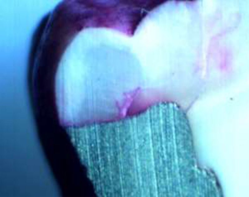
Microleakage evaluation under stereomicroscope shows a dye penetrating the enamel and spreading through the dentine-enamel junction (DEJ) to the dentin (score 3)
Figure 2.
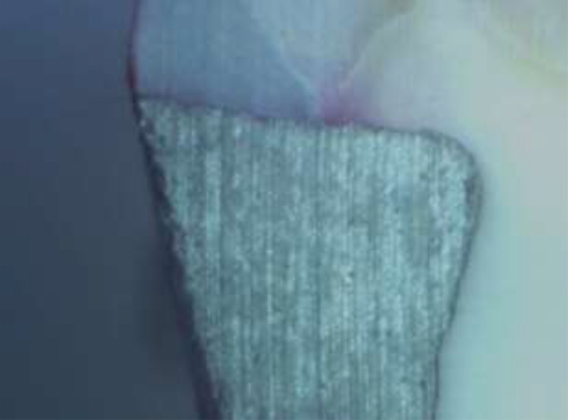
A sample control tooth under stereomicroscope microleakage evaluation reveals no dye penetration (Score 0)
At the statistical significance of 0.95 (P-value<0.05), the Kruskal-Wallis test and the Mann-Whitney U-test were conducted to assess the data and identify any statistically significant association between the experimental and control groups for microleakage.
Results
Table 1 displays the distribution of each group’s microleakage scores. A total of 12.5% of teeth in G2 and 5.0% in G3 had a grade 3 score, while the percentage of teeth with a grade 3 microleakage score was zero in the control group, G4, and G5. Also, the microleakage with grade 2 in G2, G3, and G5 groups was 5.0%, 5.0%, and 3.6%, respectively, while it was 0% in G4 and the control group.
Table 1.
The summary of the grades in the control and radiofrequency heating and electromagnetic exposure groups
| Group | EMF | Heat | Distance (cm) | Exposure Time (min) | Percent (%) of the scores | Mean rank | *P-value | |||
|---|---|---|---|---|---|---|---|---|---|---|
| 0 | 1 | 2 | 3 | |||||||
| G1 | 90.0 | 10.0 | 0.0 | 0.0 | 71.90 | 0.003 | ||||
| G2 | √ | √ | 20 | 10 | 57.7 | 25.0 | 5.0 | 12.5 | 104.06 | |
| G3 | √ | √ | 30 | 5 | 60.0 | 30.0 | 5.0 | 5.0 | 99.75 | |
| G4 | √ | No | 20 | 10 | 25.0 | 7.0 | 0.0 | 0.0 | 81.88 | |
| G5 | √ | No | 30 | 5 | 18.0 | 9.0 | 1.0 | 0.0 | 94.34 | |
*Kruskal-Walis test
EMF: Electromagnetic Field
Significantly more microleakage occurred in the G2, G3, and G5 groups than in the control group (P-value=0.001, 0.002, and 0.01). However, the difference between the scores of microleakage in G4 and the control group was not statistically significant (P-value=0.167).
The microleakage scores in G2 were significantly higher than in G4 (P-value=0.033). However, these scores were not substantially higher than G3 and G5 (P-value=0.623, 0.338). The microleakage degree of G3 was not significant compared with those of G4 and G5 (P-value=0.71 and 0.591, respectively). The microleakage scores between G4 and G5 didn’t have a significant difference statistically (P-value=0.215) (Table 2).
Table 2.
The P-values in comparing all of the groups according to the Mann-Whitney U-test
| Groups | G1 | G2 | G3 | G4 | G5 |
|---|---|---|---|---|---|
| G1 | 0.001 | 0.002 | 0.167 | 0.01 | |
| G2 | 0.623 | 0.033 | 0.338 | ||
| G3 | 0.71 | 0.591 | |||
| G4 | 0.215 |
Discussion
The current study showed that amalgam restorations cause greater microleakage when exposed to EMFs produced by commercial hair dryers. Some previous studies showed an increase in microleakage of dental amalgam restoration after exposure to the MRI’s electromagnetic fields [ 31 , 32 ]. On the contrary, Akgun OA et al. could not detect any significant difference between whether the dental amalgam samples were exposed to MRI’s electromagnetic fields or not, in the scores of microleakage [ 33 ].
Amalgam microleakage was significantly higher in the exposed group to a hairdryer than in the control group. According to Shahidi et al. [ 32 ], the thermoelectromagnetic convection brought on by exposure to EMFs can increase microleakage after MRI. The intensification of the diffusion process, grain boundary migration, and void formation resulting in microleakage were all attributed to this convection. However, the rate of rising temperature induced by EMFs was insufficient to support their theory [ 34 ].
To the best of our knowledge, this is the first study that investigates the effect of exposure to electromagnetic fields of commercial hair dryers on the microleakage of dental amalgam restorations. It should be noted that hair dryers are widely used in hair salons and homes. On the other hand, many people have some amalgam dental fillings in their oral cavities. Therefore, this present study can clarify some of the foggy aspects of the complicated questions about the rise in microleakage from dental amalgam restorations. Regarding the results of this study, it can be suggested that populations with wide amalgam restorations should limit hairdryer use.
Although it can be concluded that frequent everyday use of hairdryers can lead to damaged amalgam restorations, providing the significance of these findings, additional ex-vivo and in vivo research is required to fully understand the mechanisms of EMFs-induced damages.
Conclusion
According to this study, the frequent use of hair dryers may cause damage to amalgam restorations. Although the findings suggest that individuals with wide amalgam restorations should limit their use of hair dryers, more research, both ex-vivo and in vivo, is needed to fully understand the mechanisms of damage caused by EMFs. Therefore, clinicians should be mindful of the potential impact of hair dryers on dental amalgam restorations and advise their patients accordingly. While the study concludes that everyday use of hair dryers can be detrimental to amalgam restorations, further research is required to confirm the significance of these findings.
Acknowledgment
The Vice-Chancellory of Research at the Shiraz University of Medical Science provided funding for the present study, which is acknowledged by the authors. For the statistical analysis, the researchers of this study additionally thank Dr. Vosoughi of the Dental School’s Dental Research Development Center.
Authors’ Contribution
The study was conceived and designed by SMJ. Mortazavi and M. Paknahad. Data collection and analysis were performed by I. Khaleghi and M. Eghlidespour. The article was written by M. Paknahad and SMJ. Mortazavi. All authors reviewing, revising, and approving the final manuscript.
Ethical Approval
This work was approved by the Shiraz University of Medical Sciences Ethics Committee (IR.SUMS.REC.1395.S402).
Funding
The funding for this present study was provided by the Vice-Chancellory of Research at the Shiraz University of Medical Science (Grant# 10027).
Conflict of Interest
SMJ. Mortazavi, as the Editorial Board Member, was not involved in the peer-review and decision-making processes for this manuscript.
References
- 1.Bembi S, Bembi NN, Sood A, Gambhir A. To Evaluate the Effect of Different Adhesive Materials on the Microleakage of Bonded Amalgam Restorations: An in vitro Study. Int J Clin Pediatr Dent. 2012;5(3):185–9. doi: 10.5005/jp-journals-10005-1163. [ PMC Free Article ] [DOI] [PMC free article] [PubMed] [Google Scholar]
- 2.Opdam NJ, Bronkhorst EM, Loomans BA, Huysmans MC. 12-year survival of composite vs. amalgam restorations. J Dent Res 2010;89(10):1063–7. doi: 10.1177/0022034510376071. [DOI] [PubMed] [Google Scholar]
- 3.Cenci MS, Piva E, Potrich F, Formolo E, Demarco FF, Powers JM. Microleakage in bonded amalgam restorations using different adhesive materials. Braz Dent J. 2004;15(1):13–8. doi: 10.1590/s0103-64402004000100003. [DOI] [PubMed] [Google Scholar]
- 4.Mitchell RJ, Koike M, Okabe T. Posterior amalgam restorations--usage, regulation, and longevity. Dent Clin North Am. 2007;51(3):573–89. doi: 10.1016/j.cden.2007.04.004. [DOI] [PubMed] [Google Scholar]
- 5.Morrow LA, Wilson NH, Setcos JC, Watts DC. Microleakage of amalgam cavity treatment systems: an in vitro evaluation. Am J Dent. 2002;15(4):262–7. [PubMed] [Google Scholar]
- 6.Burgess JO, Walker R, Davidson JM. Posterior resin-based composite: review of the literature. Pediatr Dent. 2002;24(5):465–79. [PubMed] [Google Scholar]
- 7.Odaci E, Bas O, Kaplan S. Effects of prenatal exposure to a 900 MHz electromagnetic field on the dentate gyrus of rats: a stereological and histopathological study. Brain Res. 2008;1238:224–9. doi: 10.1016/j.brainres.2008.08.013. [DOI] [PubMed] [Google Scholar]
- 8.Orendácová J, Raceková E, Orendác M, Martoncíková M, Saganová K, Lievajová K, et al. Immunohistochemical study of postnatal neurogenesis after whole-body exposure to electromagnetic fields: evaluation of age- and dose-related changes in rats. Cell Mol Neurobiol. 2009;29(6):981–90. doi: 10.1007/s10571-009-9385-3. [DOI] [PMC free article] [PubMed] [Google Scholar]
- 9.Toothman J, Meeker-O’Connell A. How Hair Dryers Work. Howstuffworks; 2023. Available from: https://home.howstuffworks.com/hair-dryer.htm.
- 10.EnergySage. How many watts does a hair dryer use? EnergySage; 2023. Available from: https://news.energysage.com/how-many-watts-does-a-hair-dryer-use/
- 11.Garrido C, Otero AF, Cidras J. Low-frequency magnetic fields from electrical appliances and power lines. IEEE Transactions on Power Delivery. 2003;18(4):1310–9. doi: 10.1109/TPWRD.2003.817744. [DOI] [Google Scholar]
- 12.Hardell L, Sage C. Biological effects from electromagnetic field exposure and public exposure standards. Biomed Pharmacother. 2008;62(2):104–9. doi: 10.1016/j.biopha.2007.12.004. [DOI] [PubMed] [Google Scholar]
- 13.WHO. Electromagnetic fields (EMF): What are electromagnetic fields? World Health Organization; 2016. Available from: https://www.who.int/news-room/questions-and-answers/item/radiation-electromagnetic-fields.
- 14.Mortazavi SMJ, Motamedifar M, Namdari G, Taheri M, Mortazavi SAR, Shokrpour N. Non-linear adaptive phenomena which decrease the risk of infection after pre-exposure to radiofrequency radiation. Dose Response. 2013;12(2):233–45. doi: 10.2203/dose-response.12-055.Mortazavi. [ PMC Free Article ] [DOI] [PMC free article] [PubMed] [Google Scholar]
- 15.Mortazavi SMJ, Rouintan MS, Taeb S, Dehghan N, Ghaffarpanah AA, Sadeghi Z, Ghafouri F. Human short-term exposure to electromagnetic fields emitted by mobile phones decreases computer-assisted visual reaction time. Acta Neurol Belg. 2012;112(2):171–5. doi: 10.1007/s13760-012-0044-y. [DOI] [PubMed] [Google Scholar]
- 16.Mortazavi SMJ, Mosleh-Shirazi MA, Tavassoli A, Taheri M, Bagheri Z, Ghalandari R, et al. A comparative study on the increased radioresistance to lethal doses of gamma rays after exposure to microwave radiation and oral intake of flaxseed oil. Int J Radiat Res. 2011;9(1):9–14. [Google Scholar]
- 17.Mortazavi SMJ, Daiee E, Yazdi A, Khiabani K, Kavousi A, Vazirinejad R, et al. Mercury release from dental amalgam restorations after magnetic resonance imaging and following mobile phone use. Pak J Biol Sci. 2008;11(8):1142–6. doi: 10.3923/pjbs.2008.1142.1146. [DOI] [PubMed] [Google Scholar]
- 18.Mortazavi SMJ, Ahmadi J, Shariati M. Prevalence of subjective poor health symptoms associated with exposure to electromagnetic fields among university students. Bioelectromagnetics. 2007;28(4):326–30. doi: 10.1002/bem.20305. [DOI] [PubMed] [Google Scholar]
- 19.Mortazavi SMJ. Safety Issue of Mobile Phone Base Stations. J Biomed Phys Eng. 2013;3(1):1–2. [Google Scholar]
- 20.Mortavazi S, Habib A, Ganj-Karami A, Samimi-Doost R, Pour-Abedi A, Babaie A. Alterations in TSH and Thyroid Hormones following Mobile Phone Use. Oman Med J. 2009;24(4):274–8. doi: 10.5001/omj.2009.56. [ PMC Free Article ] [DOI] [PMC free article] [PubMed] [Google Scholar]
- 21.Mortazavi SMJ, Mosleh-Shirazi MA, Tavassoli A, Taheri M, Mehdizadeh AR, Namazi SAS, et al. Increased Radioresistance to Lethal Doses of Gamma Rays in Mice and Rats after Exposure to Microwave Radiation Emitted by a GSM Mobile Phone Simulator. Dose Response. 2012;11(2):281–92. doi: 10.2203/dose-response.12-010.Mortazavi. [DOI] [PMC free article] [PubMed] [Google Scholar]
- 22.Shirbandi K, Khalafi M, J Bevelacqua J, Sadeghian N, Adiban S, Bahaeddini Zarandi F, et al. Exposure to Low Levels of Radiofrequency Electromagnetic Fields Emitted from Cell-phones as a Promising Treatment of Alzheimer’s Disease: A Scoping Review Study. J Biomed Phys Eng. 2023;13(1):3–16. doi: 10.31661/jbpe.v0i0.2109-1398. [ PMC Free Article ] [DOI] [PMC free article] [PubMed] [Google Scholar]
- 23.Mortazavi SMJ, Parsanezhad M, Kazempour M, Ghahramani P, Mortazavi SAR, Davari M. Male reproductive health under threat: Short term exposure to radiofrequency radiations emitted by common mobile jammers. J Hum Reprod Sci. 2013;6(2):124–8. doi: 10.4103/0974-1208.117178. [ PMC Free Article ] [DOI] [PMC free article] [PubMed] [Google Scholar]
- 24.Mortazavi SMJ, Tavassoli A, Ranjbar F, Moammaiee P. Effects of Laptop Computers’ Electromagnetic Field on Sperm Quality. J Reprod Infertil. 2010;11(4):251–8. [Google Scholar]
- 25.Parsanezhad ME, Mortazavi SMJ, Doohandeh T, Jahromi BN, Mozdarani H, Zarei A, et al. Exposure to Radiofrequency Radiation Emitted from Mobile Phone Jammers Adversely Affects the Quality of Human Sperm. Int J Radiat Res. 2017;15(1):63–70. doi: 10.18869/acadpub.ijrr.15.1.63. [DOI] [Google Scholar]
- 26.Mortazavi SMJ, Taeb S, Dehghan N. Alterations of visual reaction time and short term memory in military radar personnel. Iran J Public Health. 2013;42(4):428–35. [ PMC Free Article ] [PMC free article] [PubMed] [Google Scholar]
- 27.Mortazavi SMJ, Vazife-Doost S, Yaghooti M, Mehdizadeh S, Rajaie-Far A. Occupational exposure of dentists to electromagnetic fields produced by magnetostrictive cavitrons alters the serum cortisol level. J Nat Sci Biol Med. 2012;3(1):60–4. doi: 10.4103/0976-9668.95958. [ PMC Free Article ] [DOI] [PMC free article] [PubMed] [Google Scholar]
- 28.28. Fcc Enforcement Bureau Steps up Education and Enforcement Efforts Against Cellphone and Gps Jamming. NEWS; Washington : Federal Communications Commission; 2011. [Google Scholar]
- 29.Mortazavi SMJ, Neghab M, Anoosheh SM, Bahaeddini N, Mortazavi G, Neghab P, Rajaeifard A. High-field MRI and mercury release from dental amalgam fillings. Int J Occup Environ Med. 2014;5(2):101–5. [ PMC Free Article ] [PMC free article] [PubMed] [Google Scholar]
- 30.Keshavarz M, Eslami J, Abedi-Firouzjah R, Mortazavi SAR, Abbasi S, Mortazavi G. How Do Different Physical Stressors’ Affect the Mercury Release from Dental Amalgam Fillings and Microleakage? A Systematic Review. J Biomed Phys Eng. 2022;12(3):227–36. doi: 10.31661/jbpe.v0i0.2009-1175. [ PMC Free Article ] [DOI] [PMC free article] [PubMed] [Google Scholar]
- 31.Yilmaz S, Misirlioglu M. The effect of 3 T MRI on microleakage of amalgam restorations. Dentomaxillofac Radiol. 2013;42(8):20130072. doi: 10.1259/dmfr.20130072. [ PMC Free Article ] [DOI] [PMC free article] [PubMed] [Google Scholar]
- 32.Shahidi SH, Bronoosh P, Alavi AA, Zamiri B, Sadeghi AR, Bagheri MH, Javadpour S. Effect of magnetic resonance imaging on microleakage of amalgam restorations: an in vitro study. Dentomaxillofac Radiol. 2009;38(7):470–4. doi: 10.1259/dmfr/30077669. [DOI] [PubMed] [Google Scholar]
- 33.Akgun OM, Polat GG, Turan Illca A, Yildirim C, Demir P, Basak F. Does magnetic resonance imaging affect the microleakage of amalgam restorations? Iran J Radiol. 2014;11(3):e15565. doi: 10.5812/iranjradiol.15565. [ PMC Free Article ] [DOI] [PMC free article] [PubMed] [Google Scholar]
- 34.Mortazavi SMJ, Paknahad M. Effect of magnetic resonance imaging on microleakage of amalgam restorations: an in vitro study (Letter) . Dentomaxillofac Radiol. 2016;45(1):20150187. doi: 10.1259/dmfr.20150187. [ PMC Free Article ] [DOI] [PMC free article] [PubMed] [Google Scholar]