Abstract
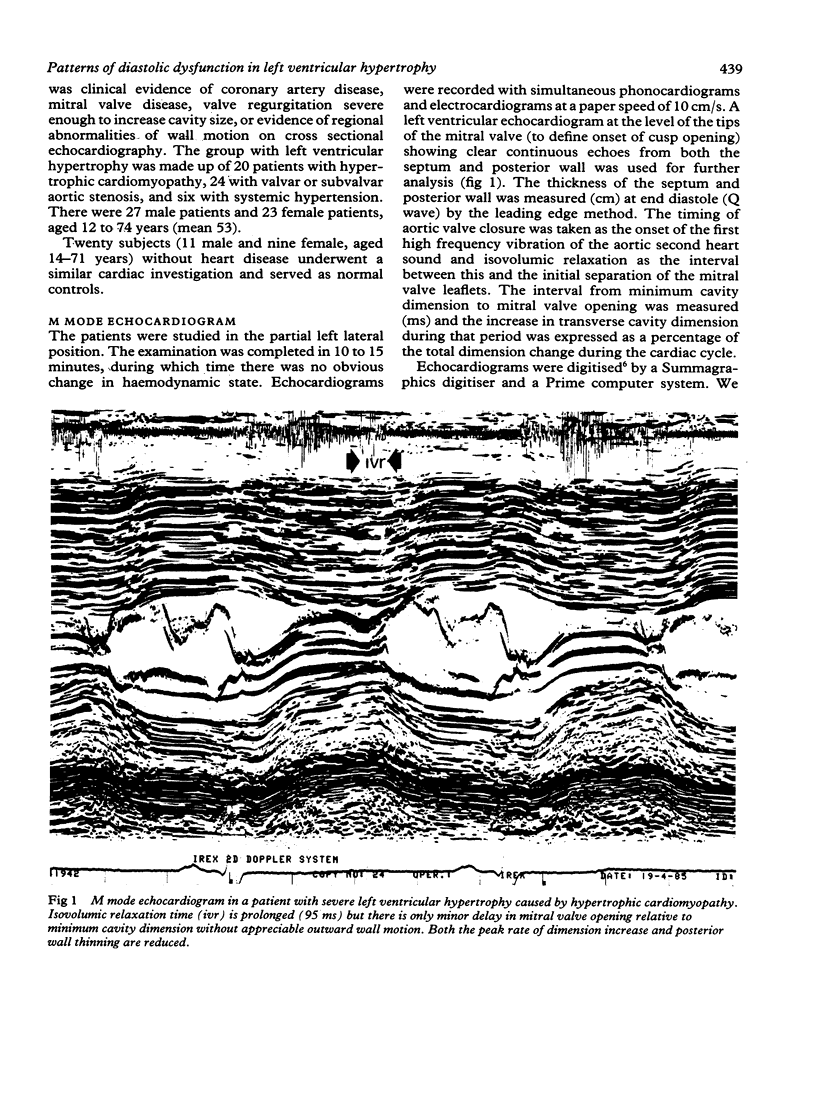
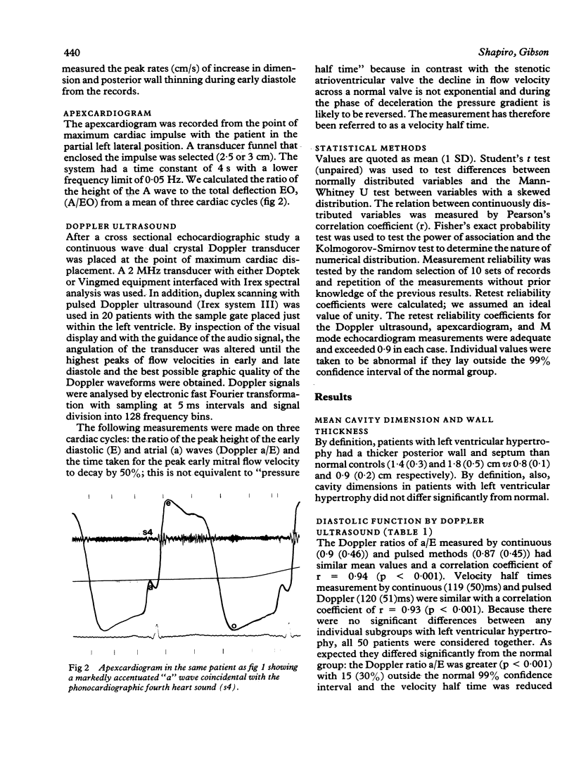
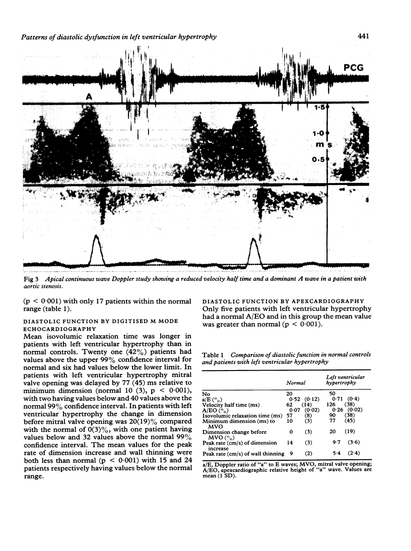
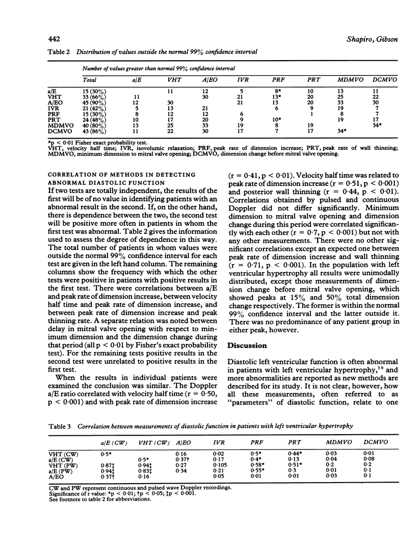
The relative sensitivities of and interrelations between different measurements of diastolic function were studied in 50 patients with left ventricular hypertrophy diagnosed on anatomical grounds. Isovolumic relaxation time, the interval from minimum cavity dimension to mitral valve opening and relative dimension increase during this period, and the peak rate of dimension increase and wall thinning during rapid ventricular filling were measured by digitised M mode echocardiography. The relative heights of peak early diastolic and atrial velocities (a/E) and the time for decline of early diastolic velocity to half its peak value (velocity half time) were measured on continuous wave and pulsed Doppler and the relative height of the "a" wave was measured by apexcardiogram. All sets of values except those of the interval from minimum dimension to mitral opening were unimodally distributed, and all differed significantly from those in 20 age matched controls. The relative height of the "a" wave on the apexcardiogram (90% values were abnormal) was the most sensitive method of studying left ventricular diastolic function and peak rate of dimension increase was the least sensitive. Though none of the correlations was high, there were individual associations between peak rate of dimension increase, a/E, peak wall thinning rate, and velocity half time, and independently between delay in mitral valve opening and dimension change during this period. Other values seemed to be independent of one another, suggesting a different physiological basis. It is concluded that these various abnormal values do not reflect a single underlying disturbance of diastolic function. There are at least four possible discrete abnormalities: prolongation of isovolumic relaxation; incoordination during isovolumic relaxation; reduced rate of rapid filling; and an increase in the relative amplitude of the "a" wave probably caused by increased passive stiffness. These may be present singly or in combination in any patient.
Full text
PDF
Images in this article
Selected References
These references are in PubMed. This may not be the complete list of references from this article.
- Bonow R. O., Frederick T. M., Bacharach S. L., Green M. V., Goose P. W., Maron B. J., Rosing D. R. Atrial systole and left ventricular filling in hypertrophic cardiomyopathy: effect of verapamil. Am J Cardiol. 1983 May 1;51(8):1386–1391. doi: 10.1016/0002-9149(83)90317-x. [DOI] [PubMed] [Google Scholar]
- Brutsaert D. L., Housmans P. R., Goethals M. A. Dual control of relaxation. Its role in the ventricular function in the mammalian heart. Circ Res. 1980 Nov;47(5):637–652. doi: 10.1161/01.res.47.5.637. [DOI] [PubMed] [Google Scholar]
- Denef B., Popeye R., Geest H. D., Kesteloot H. On the clinical value of calibrated displacement apexcardiography. Circulation. 1975 Mar;51(3):541–551. doi: 10.1161/01.cir.51.3.541. [DOI] [PubMed] [Google Scholar]
- Gibson D. G., Brown D. Measurement of instantaneous left ventricular dimension and filling rate in man, using echocardiography. Br Heart J. 1973 Nov;35(11):1141–1149. doi: 10.1136/hrt.35.11.1141. [DOI] [PMC free article] [PubMed] [Google Scholar]
- Gibson D. G., Prewitt T. A., Brown D. J. Analysis of left ventricular wall movement during isovolumic relaxation and its relation to coronary artery disease. Br Heart J. 1976 Oct;38(10):1010–1019. doi: 10.1136/hrt.38.10.1010. [DOI] [PMC free article] [PubMed] [Google Scholar]
- Gibson D. G., Traill T. A., Hall R. J., Brown D. J. Echocardiographic features of secondary left ventricular hypertrophy. Br Heart J. 1979 Jan;41(1):54–59. doi: 10.1136/hrt.41.1.54. [DOI] [PMC free article] [PubMed] [Google Scholar]
- Grossman W., Stefadouros M. A., McLaurin L. P., Rolett E. L., Young D. T. Quantitative assessment of left ventricular diastolic stiffness in man. Circulation. 1973 Mar;47(3):567–574. doi: 10.1161/01.cir.47.3.567. [DOI] [PubMed] [Google Scholar]
- Hammermeister K. E., Warbasse J. R. The rate of change of left ventricular volume in man. II. Diastolic events in health and disease. Circulation. 1974 Apr;49(4):739–747. doi: 10.1161/01.cir.49.4.739. [DOI] [PubMed] [Google Scholar]
- Hanrath P., Mathey D. G., Siegert R., Bleifeld W. Left ventricular relaxation and filling pattern in different forms of left ventricular hypertrophy: an echocardiographic study. Am J Cardiol. 1980 Jan;45(1):15–23. doi: 10.1016/0002-9149(80)90214-3. [DOI] [PubMed] [Google Scholar]
- Henry W. L., Gardin J. M., Ware J. H. Echocardiographic measurements in normal subjects from infancy to old age. Circulation. 1980 Nov;62(5):1054–1061. doi: 10.1161/01.cir.62.5.1054. [DOI] [PubMed] [Google Scholar]
- Ishida Y., Meisner J. S., Tsujioka K., Gallo J. I., Yoran C., Frater R. W., Yellin E. L. Left ventricular filling dynamics: influence of left ventricular relaxation and left atrial pressure. Circulation. 1986 Jul;74(1):187–196. doi: 10.1161/01.cir.74.1.187. [DOI] [PubMed] [Google Scholar]
- Mattheos M., Shapiro E., Oldershaw P. J., Sacchetti R., Gibson D. G. Non-invasive assessment of changes in left ventricular relaxation by combined phono-, echo-, and mechanocardiography. Br Heart J. 1982 Mar;47(3):253–260. doi: 10.1136/hrt.47.3.253. [DOI] [PMC free article] [PubMed] [Google Scholar]
- Miyatake K., Okamoto M., Kinoshita N., Owa M., Nakasone I., Sakakibara H., Nimura Y. Augmentation of atrial contribution to left ventricular inflow with aging as assessed by intracardiac Doppler flowmetry. Am J Cardiol. 1984 Feb 1;53(4):586–589. doi: 10.1016/0002-9149(84)90035-3. [DOI] [PubMed] [Google Scholar]
- Sanderson J. E., Gibson D. G., Brown D. J., Goodwin J. F. Left ventricular filling in hypertrophic cardiomyopathy. An angiographic study. Br Heart J. 1977 Jun;39(6):661–670. doi: 10.1136/hrt.39.6.661. [DOI] [PMC free article] [PubMed] [Google Scholar]
- Shapiro L. M., Moore R. B., Logan-Sinclair R. B., Gibson D. G. Relation of regional echo amplitude to left ventricular function and the electrocardiogram in left ventricular hypertrophy. Br Heart J. 1984 Jul;52(1):99–105. doi: 10.1136/hrt.52.1.99. [DOI] [PMC free article] [PubMed] [Google Scholar]
- Spirito P., Maron B. J., Bonow R. O. Noninvasive assessment of left ventricular diastolic function: comparative analysis of Doppler echocardiographic and radionuclide angiographic techniques. J Am Coll Cardiol. 1986 Mar;7(3):518–526. doi: 10.1016/s0735-1097(86)80461-2. [DOI] [PubMed] [Google Scholar]


