Abstract
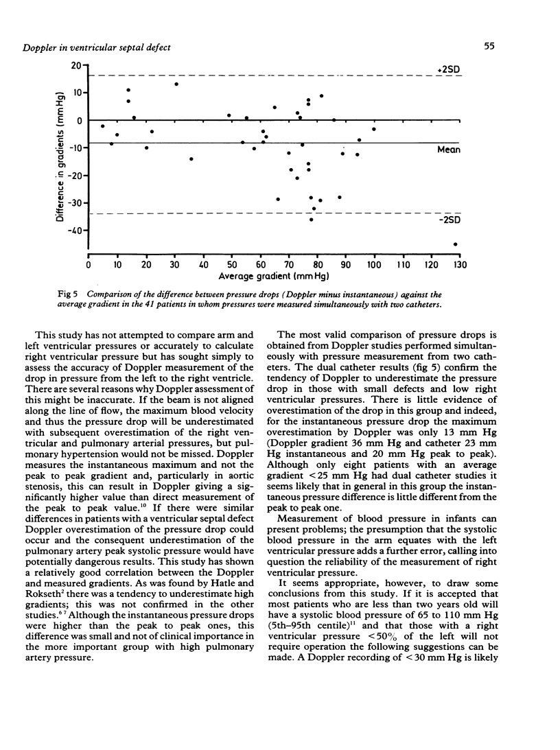
Doppler ultrasound was used to assess the pressure drop between the ventricles in 109 infants and children (61 less than two years old) with a ventricular septal defect who underwent cardiac catheterisation. The pressure in both ventricles was measured at catheterisation in 103 patients either simultaneously through two catheters (41) or with a single catheter withdrawn across the septum or removed from one ventricle to the other (62). When pressure was measured simultaneously with two catheters (41 patients) the peak to peak and instantaneous gradients showed a maximum difference of 20 mm Hg with levels within 10 mm Hg of each other in 36. Comparison of the difference in the gradients with the average of the measurements demonstrated a tendency for Doppler to underestimate the difference when it was high (greater than 50 mm Hg) and overestimate it when it was low. A Doppler estimate of a low pressure difference between the ventricles indicates pulmonary arterial hypertension and a high one low pulmonary artery pressure, but in the intermediate group Doppler is as yet not sufficiently sensitive to allow selection of those patients who require further investigation and possible operation. Doppler ultrasound was found to be a sensitive method of detecting a very small ventricular septal defect. Thus although Doppler is a very useful means of assessing and following patients with a ventricular septal defect, further studies are required to determine its exact place in clinical practice.
Full text
PDF
Images in this article
Selected References
These references are in PubMed. This may not be the complete list of references from this article.
- Bland J. M., Altman D. G. Statistical methods for assessing agreement between two methods of clinical measurement. Lancet. 1986 Feb 8;1(8476):307–310. [PubMed] [Google Scholar]
- Currie P. J., Hagler D. J., Seward J. B., Reeder G. S., Fyfe D. A., Bove A. A., Tajik A. J. Instantaneous pressure gradient: a simultaneous Doppler and dual catheter correlative study. J Am Coll Cardiol. 1986 Apr;7(4):800–806. doi: 10.1016/s0735-1097(86)80339-4. [DOI] [PubMed] [Google Scholar]
- Currie P. J., Seward J. B., Chan K. L., Fyfe D. A., Hagler D. J., Mair D. D., Reeder G. S., Nishimura R. A., Tajik A. J. Continuous wave Doppler determination of right ventricular pressure: a simultaneous Doppler-catheterization study in 127 patients. J Am Coll Cardiol. 1985 Oct;6(4):750–756. doi: 10.1016/s0735-1097(85)80477-0. [DOI] [PubMed] [Google Scholar]
- Hatle L., Brubakk A., Tromsdal A., Angelsen B. Noninvasive assessment of pressure drop in mitral stenosis by Doppler ultrasound. Br Heart J. 1978 Feb;40(2):131–140. doi: 10.1136/hrt.40.2.131. [DOI] [PMC free article] [PubMed] [Google Scholar]
- Hatle L., Rokseth R. Noninvasive diagnosis and assessment of ventricular septal defect by Doppler ultrasound. Acta Med Scand Suppl. 1981;645:47–56. doi: 10.1111/j.0954-6820.1981.tb02600.x. [DOI] [PubMed] [Google Scholar]
- Londe S., Goldring D. High blood pressure in children: problems and guidelines for evaluation and treatment. Am J Cardiol. 1976 Mar 31;37(4):650–657. doi: 10.1016/0002-9149(76)90410-0. [DOI] [PubMed] [Google Scholar]
- Murphy D. J., Jr, Ludomirsky A., Huhta J. C. Continuous-wave Doppler in children with ventricular septal defect: noninvasive estimation of interventricular pressure gradient. Am J Cardiol. 1986 Feb 15;57(6):428–432. doi: 10.1016/0002-9149(86)90766-6. [DOI] [PubMed] [Google Scholar]
- Remington J. W., O'Brien L. J. Construction of aortic flow pulse from pressure pulse. Am J Physiol. 1970 Feb;218(2):437–447. doi: 10.1152/ajplegacy.1970.218.2.437. [DOI] [PubMed] [Google Scholar]
- Silbert D. R., Brunson S. C., Schiff R., Diamant S. Determination of right ventricular pressure in the presence of a ventricular septal defect using continuous wave Doppler ultrasound. J Am Coll Cardiol. 1986 Aug;8(2):379–384. doi: 10.1016/s0735-1097(86)80054-7. [DOI] [PubMed] [Google Scholar]
- Sutherland G. R., Godman M. J., Smallhorn J. F., Guiterras P., Anderson R. H., Hunter S. Ventricular septal defects. Two dimensional echocardiographic and morphological correlations. Br Heart J. 1982 Apr;47(4):316–328. doi: 10.1136/hrt.47.4.316. [DOI] [PMC free article] [PubMed] [Google Scholar]


